. 2025 Jul 9;22(1):177.
doi: 10.1186/s12974-025-03487-3.

Liver-specific expression of ANGPTL8 promotes Alzheimer's disease progression through activating microglial pyroptosis

Affiliations

Liver-specific expression of ANGPTL8 promotes Alzheimer's disease progression through activating microglial pyroptosis

Jiarui Wei et al. J Neuroinflammation. .

Abstract

Introduction: Liver dysfunction contributes to Alzheimer's disease (AD) pathogenesis, and evidence suggests that the liver is involved in amyloid β (Aβ) clearance, and regulates Aβ deposition in the brain. However, the specific regulatory mechanism remains elusive.

Objectives: Angiopoietin-like protein 8 (ANGPTL8), a high expression of liver-specific secreted proinflammatory factor, crosses the blood‒brain barrier from the bloodstream to abnormally activate microglia and promote AD progression.

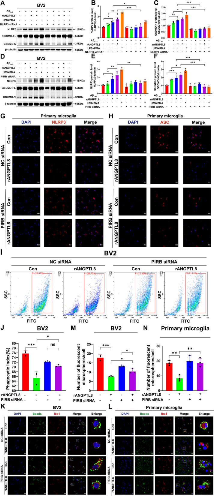
Methods: The ANGPTL8-/- mice and 5 × FAD mice were crossed mutated and subjected to the Morris water maze test and novel object recognition test to assess cognitive ability in different cohorts. Thioflavin-S, NeuN, and Nissl staining were used to assess Aβ deposition and neuron loss. The number of phagocytic microglia was evaluated with Fitc latex beads. Adeno-associated virus 8 (AAV8) hydrodynamically injected restored the liver ANGPTL8 levels of ANGPTL8-/- 5 × FAD mice for further experiments. Single-cell RNA sequencing, bulk RNA sequencing and transmission electron microscopy were used to explore the role of ANGPTL8 in regulating AD progression, and drug screening was carried out to identify an effective inhibitor of ANGPTL8.

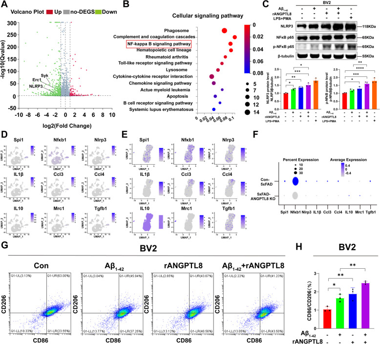
Results: ANGPTL8 knockout improved cognitive function and reduced Aβ deposition by reducing microgliosis and microglial activation in 5xFAD mice. Mechanistically, ANGPTL8 crossed the blood‒brain barrier and interacted with the microglial membrane receptor PirB/LILRB2. This interaction subsequently activated the downstream NLRP3 inflammasome, leading to microglial pyroptosis and exacerbating the Aβ-induced release of inflammatory factors, thereby accelerating AD progression. Furthermore, the administration of metformin, an ANGPTL8 inhibitor, improved learning and memory deficits in 5 × FAD mice by negating microglial pyroptosis and neuroinflammation.

Conclusions: ANGPTL8 aggravates microglial pyroptosis via the PirB/NLRP3 pathway to accelerate the pathogenesis of AD. Targeting high expression of ANGPTL8 in the liver may hold potential for developing therapies for AD.

Keywords: ANGPTL8; Alzheimer's disease; Liver‒brain axis; Microglial pyroptosis; Neuroinflammation.


Conflict of interest statement

Declarations. Ethics approval and consent to participate: All experiments involving animals were conducted according to ethical policies and procedures approved by the ethics committee of Hubei University of Medicine, China. The ethical committee number for the study was 202016. Consent for publication: Not applicable. Competing interests: The authors declare no competing interests.

Figures

Fig. 1
Fig. 1
ANGPTL8 KO improved the cognitive function of 5xFAD mice. A Representative images of the tracks of 9-month-old WT and ANGPTL8 KO mice in the Morris water maze (MWM) test. B-C Statistical results of the escape latency (B) and number of platform crossings (C) of 9-month-old mice (n = 8) on d 7. D Schematics of the experimental procedures for the four groups of mice (WT, ANGPTL8 KO, 5 × FAD, and ANGPTL8 KO:5xFAD; every group included half male and half female mice). E Representative plot of the MWM test results of 3-month-old mice in the four groups (n = 8–10). F The time needed to reach the hidden platform (escape latency) was plotted across training days. Statistical analysis revealed a difference in 3-month-old ANGPTL8 KO:5 × FAD mice compared with WT, ANGPTL8 KO and 5 × FAD mice on d 7 (n = 8–10). GI. Escape latency, number of platform crossings and swimming speed (mm/s) were analysed during the MWM test (d 7) in 3-month-old mice. JK The escape latency and number of platform crossings were analysed during the MWM test (d 7) in 6-month-old mice (n = 6‒8). LM Recognition indices of the four groups of 3- and 6-month-old mice were detected via a novel object recognition experiment (n = 6–8). NP. IHC staining (N) and statistical analysis (OP) of Aβ plaques per pixel area in the hippocampus and cortex of four groups of mice (WT, ANGPTL8 KO, 5xFAD, and ANGPTL8 KO:5xFAD). (n = 7—11 slices from three mice per group). *P < 0.05, ** P < 0.01, *** P < 0.001, ns = not significant
Fig. 2
Fig. 2
ANGPTL8 promotes AD progression by inducing microgliosis and microglial activation. A Changes in cortical cells in the brains of 6-month-old 5xFAD: ANGPTL8-KO and Con-5xFAD mice were analysed via single-nucleus RNA sequencing (snRNA-seq). Joint uniform manifold approximation and projection (UMAP), colored by total major cell type (17 main clusters) in mice. B-C. Global breakdown, region composition, enrichment and number of nuclei for 17 main cluster subtypes in the Con-5xFAD mouse group (B) and 5xFAD:ANGPTL8-KO mouse group (C). D-F. Subpopulations of microglia. UMAP embeddings of single nuclei profiled via snRNA-seq, colored according to clusters, and annotated for each subpopulation, indicating the proportion of each subpopulation in the Con-5xFAD mouse group (E) and the 5xFAD:ANGPTL8-KO mouse group (F). G. Immunostaining for Iba-1 and DAPI in the CA1 area of the hippocampus. Microglia were labelled with an Iba-1 antibody. H. Colocalization of Aβ and Iba-1 in the hippocampus of the mice in the indicated groups. Aβ plaques and microglia were labelled with anti-Aβ (red) and anti-Iba-1 antibodies (green), respectively
Fig. 3
Fig. 3
Liver-derived ANGPTL8 activated microglia. A-D. ANGPTL8 mRNA was detected by qPCR in liver of 1, 3, 6 and 9 months old WT and 5xFAD mice. E. The effect of Aβ stimulation on ANGPTL8 expression was analyzed by qPCR in LO2 cells. F-I. Serum ANGPTL8 levels were detected by ELISA in 1-, 3-, 6-, and 9-month-old WT and 5xFAD mice (n ≥ 3). J-K. ANGPTL8 protein levels in the hippocampal and cortical homogenates of 6-month-old 5xFAD and WT mice were determined via ELISA. *P < 0.05, **P < 0.01, ***P < 0.001, ns = not significant. L. Schematic of the establishment of a model for long-term complementation of the liver expression of ANGPTL8 in ANGPTL8 KO:5xFAD mice via hydrodynamic tail vein injection of AAV8. M. Representative images of the tracks of 6-month-old ANGPTL8 KO:5xFAD-AAV8-con and ANGPTL8 KO:5xFAD-AAV8-ANGPTL8 mice in the Morris water maze (MWM) test. NO. Statistical analysis of the number of platform crossings (N) and escape latency (O) of 6-month-old ANGPTL8 KO:5xFAD-AAV8-con and ANGPTL8 KO:5xFAD-AAV8-ANGPTL8 mice (n = 8) on d 7. P‒R. IHC staining (P) and statistical analysis (QR) of Aβ plaques per pixel area in the hippocampus and cortex of two groups of mice (ANGPTL8 KO:5xFAD-AAV8-con and ANGPTL8 KO:5xFAD-AAV8-ANGPTL8; n = 7–11 slices from three mice per group). SU. Thioflavin-S (S) and statistical analysis (TU) of Aβ plaques per pixel area in the hippocampus and cortex of two groups of mice (ANGPTL8 KO:5xFAD-AAV8-con and ANGPTL8 KO:5xFAD-AAV8-ANGPTL8; n = 7–11 slices from three mice per group). *P < 0.05, **P < 0.01, ***P < 0.001, ns = not significant
Fig. 4
Fig. 4
ANGPTL8 reduces microglial phagocytosis to inhibit the clearance of Aβ. A Morphological changes (resting ramified microglia and activated amoeboid microglia) in BV2 cells treated with rANGPTL8 (600 ng/ml) for 6 h under a bright field. Scale bar: 50 μm. B-E. Flow cytometry analysis of the phagocytic ability of fluorescence microspheres in BV2 cells or primary microglia after treatment with Aβ1-42 (400 ng/ml) and/or ANGPTL8 (600 ng/ml) for 6 h. Representative scatter plots showing the frequencies of the cells containing fluorescent microspheres (B, C) and summary bar graphs (D, E). FI. Microscopic analysis of the phagocytosis of fluorescent microspheres by BV2 cells or primary microglia after treatment with Aβ1‒42 (400 ng/ml) and/or ANGPTL8 (600 ng/ml) for 6 h. Representative images of fluorescent microspheres (green) engulfed by BV2 cells (F) or primary microglia (G) (marked with anti-Iba1 (red) and DAPI for the nucleus (blue)) and a summary bar graph (HI). Scale bar: 50 μm. *P < 0.05, ** P < 0.01, *** P < 0.001, ****P < 0.0001
Fig. 5
Fig. 5
ANGPTL8 induced inflammatory factor expression and microglial polarization. A. Volcano plot visualizing the differential expression of the genes of the heart transcriptome (n = 3 in each group). B. Kyoto Encyclopedia of Genes and Genomes pathway enrichment analysis of the identified differentially expressed genes on the basis of the RNA-seq dataset from the hippocampi of 6-week-old 5 × FAD (WT) and ANGPTL8-KO:5 × FAD mice (n = 3 in each group). C. Representative immunoblots probed with antibodies against NLRP3, p-NF-κBp65, NF-κBp65, and β-tubulin and the statistical results of Western blotting. D-F. UMAP gene expression patterns of NF-κB pathway markers in 17 labelled clusters (D). Gene expression patterns of NF-κB pathway markers in UMAPs of microglial subpopulations (E). Distinct gene expression patterns of NF-κB pathway markers in microglial subpopulations in 5 × FAD (WT) and ANGPTL8-KO:5 × FAD mice. The dot color shows the mean expression in expressing cells (column scale); the dot size shows the percentage of cells expressing the gene (F). G. Representative images of CD86 and CD206 expression detected by flow cytometry in the indicated groups. H. Quantification of CD86/CD206 in BV2 cells in the indicated groups. LPS (500 ng/ml) + PMA (500 ng/ml) was used as a positive control. *P < 0.05, ** P < 0.01, *** P < 0.001, ****P < 0.0001
Fig. 6
Fig. 6
ANGPTL8-induced microglial pyroptosis. A. Representative morphological images of pyroptosis in BV2 cells treated with Aβ1-42 (400 ng/ml) and/or rANGPTL8 (600 ng/ml) for 36 h. B. Statistical analysis of pyroptotic cell numbers/mm.2 in each group. C. Representative morphological images of the indicated groups observed via transmission electron microscopy. LPS (500 ng/ml) + PMA (500 ng/ml) was used as a positive control. D. Quantification of the levels of GSDMD-N normalized to those of β‐tubulin. Experiments were performed in the indicated groups (n = 3 mice/group) E. Representative western blot bands of GSDMD-FL, GSDMD-N, and β-tubulin in 3-month-old mice in each group. F-I. IHC staining (F&H) and statistical analysis (G&I) of NLRP3 and ASC in the hippocampus and cortex of four groups of mice (WT, ANGPTL8 KO, 5xFAD, and ANGPTL8 KO:5xFAD). N = 7 to 11 slices from three mice per group, *P < 0.05, ** P < 0.01, *** P < 0.001, ns = not significant. JK. Colocalization of ASC and Iba-1 (J) or NRLP3 and Iba-1 (K) in the hippocampus of the mice in the indicated groups. L. Representative immunoblots probed with antibodies against GSDMD-FL, GSDMD-N, cleaved caspase-1, ASC and β-tubulin in BV2 cells treated with Aβ1-42 (400 ng/ml) and/or rANGPTL8 (600 ng/ml) for 36 h. MO. The levels of GSDMD-N, cleaved caspase-1, and ASC were quantified and normalized to those of β-tubulin. GSDMD-FL represents full-length GSDMD, and c-caspase-1 represents cleaved caspase-1. *P < 0.05, ** P < 0.01, *** P < 0.001, ****P < 0.0001
Fig. 7
Fig. 7
ANGPTL8 promoted microglial pyroptosis via the PirB/NLRP3 pathway. A. To analyse the role of NLRP3 in BV2 cell pyroptosis after treatment with ANGPLT8 and/or Aβ1-42, a siRNA strategy was used to manipulate the expression of NLRP3 in BV2 cells. Immunoblots of NLRP3, GSDMD-FL, GSDMD-N, and β-tubulin (loading control) in BV2 cells and NLRP3-knockdown cells treated with Aβ1-42 (400 ng/ml) and/or rANGPTL8 (600 ng/ml) for 6 h. LPS (500 ng/ml) + PMA (500 ng/ml) was used as a positive control. B-C. The levels of NLRP3 and GSDMD-N were quantified and normalized to those of β-tubulin. D. To analyse the role of PirB in BV2 cell pyroptosis after treatment with ANGPLT8 and/or Aβ1-42 by western blotting, a siRNA strategy was used to manipulate the expression of PirB. EF. Quantification of NLRP3 and GSDMD-N in BV2 and PirB-knockdown BV2 cells treated with Aβ1-42 with or without rANGPTL8. G-H. Representative images of NLRP3 and ASC expression detected by immunofluorescence in the indicated groups. IJ. Flow cytometry analysis of the phagocytic ability of fluorescent microspheres in BV2 and PirB-knockdown BV2 cells after treatment with ANGPTL8 (600 ng/ml) for 6 h. Representative scatter plots showing the frequencies of the cells containing fluorescent microspheres (I) and summary bar graphs (J). KN. Confocal microscopic examination of the phagocytosis of fluorescent microspheres by BV2 cells or primary microglia and PirB-knockdown BV2 cells or primary microglia after treatment with ANGPTL8 for 6 h. Representative images (K-L) of fluorescent microspheres (green) engulfed by BV2 cells or primary microglia (marked with anti-Iba1 (red) and DAPI for nuclei (blue)) and a summary bar graph (MN) are shown
Fig. 8
Fig. 8
Metformin improved cognitive dysfunction in 5xFAD mice by inhibiting ANGPTL8. A. Schematics of the experimental procedures for the eight groups of mice. B. A representative locus plot of the MWM test results of 4-month-old WT, ANGPTL8KO, 5 × FAD, ANGPTL8KO: 5 × FAD mice that received without or with Met (300 mg/kg, i.g.) for 4 weeks is shown. CE. The swimming speed, number of platform crossings, and escape latency of the mice in the indicated groups were analysed during the Morris water maze test. F. The recognition index was detected by a novel object recognition experiment in 4-month-old mice in the indicated groups. G. Serum ANGPTL8 levels were detected by ELISA in 4-month-old 5 × FAD mice without or with Met (300 mg/kg, i.g.) treatment for 4 weeks. H. Amyloid plaques were detected by thioflavin-S staining (scale bars, 100 μm) in the cortex and hippocampus of the brains of 4-month-old mice from the indicated groups. IJ. Quantitative analysis of amyloid plaques in the cortex (I) and hippocampus (J) in each group (the average value of 8‒10 slices from each mouse, n = 3 mice/group), *P < 0.05, ** P < 0.01, ns = not significant. K. Schematic representation of the proposed mechanism by which ANGPTL8, as a liver-secreted inflammatory trigger, aggravates microglial pyroptosis via the PirB/NLRP3 pathway to accelerate the pathogenesis of AD. The elevated Aβ levels in the AD model of 5 × FAD mice upregulated liver ANGPTL8 expression, and secreted ANGPTL8 acted as an inflammatory trigger and directly interacted with the microglial membrane receptor PirB to activate downstream PirB/NLRP3 signalling. Furthermore, the NLRP3 inflammasome initiates cleaved GSDMD to release a GSDMD-N fragment that forms pores on microglia, leading to the extracellular release of inflammatory factors such as IL-1β. Metformin inhibited the expression of ANGPTL8 and decreased Aβ deposition in 5 × FAD mice

References

    1. Porsteinsson AP, Isaacson RS, Knox S, Sabbagh MN, Rubino I. Diagnosis of Early Alzheimer’s Disease: Clinical Practice in 2021. J Prev Alzheimers Dis. 2021;8:371–86. - PubMed
    1. Scheltens P, De Strooper B, Kivipelto M, Holstege H, Chételat G, Teunissen CE, Cummings J, van der Flier WM. Alzheimer’s disease. Lancet. 2021;397:1577–90. - PMC - PubMed
    1. Meier-Stephenson FS, Meier-Stephenson VC, Carter MD, Meek AR, Wang Y, Pan L, Chen Q, Jacobo S, Wu F, Lu E, et al. Alzheimer’s disease as an autoimmune disorder of innate immunity endogenously modulated by tryptophan metabolites. Alzheimers Dement (N Y). 2022;8:e12283. - PMC - PubMed
    1. Chen X, Firulyova M, Manis M, Herz J, Smirnov I, Aladyeva E, Wang C, Bao X, Finn MB, Hu H, et al. Microglia-mediated T cell infiltration drives neurodegeneration in tauopathy. Nature. 2023;615:668–77. - PMC - PubMed
    1. Moonen S, Koper MJ, Van Schoor E, Schaeverbeke JM, Vandenberghe R, von Arnim CAF, Tousseyn T, De Strooper B, Thal DR. Pyroptosis in Alzheimer’s disease: cell type-specific activation in microglia, astrocytes and neurons. Acta Neuropathol. 2023;145:175–95. - PubMed

Substances

LinkOut - more resources