. 2025 Jul 9;32(1):63.
doi: 10.1186/s12929-025-01159-1.

Goblet cell breakdown: transcriptomics reveals Acinetobacter baumannii early and robust inflammatory response in differentiated human bronchial epithelial cells

Affiliations

Goblet cell breakdown: transcriptomics reveals Acinetobacter baumannii early and robust inflammatory response in differentiated human bronchial epithelial cells

Daniela Scribano et al. J Biomed Sci. .

Abstract

Background: The airway epithelium represents the first line of defense of the lungs, functioning both as a physical barrier as well as an active immune modulator. However, in the last years, pneumonia caused by the opportunistic pathogen Acinetobacter baumannii have become difficult to treat due to the increase of the number of extensively drug resistant strains. In this study, we report for the first time the use of an ex vivo air-liquid interface (ALI) model of differentiated human bronchial epithelial cells to unravel the early response to A. baumannii infection.

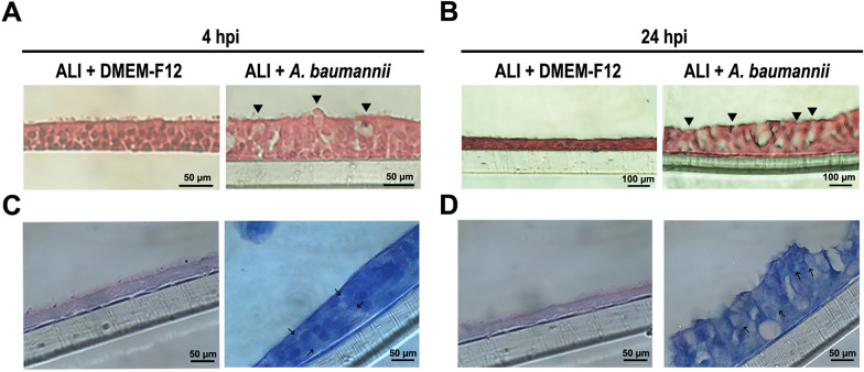
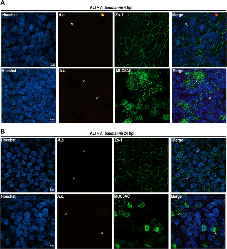
Methods: Epithelial integrity, tissue architecture, and goblet cell function were assessed through FITC-dextran permeability assays, hematoxylin and eosin staining, and indirect immunofluorescence. Transcriptomic profiling was performed to characterize host gene expression changes.

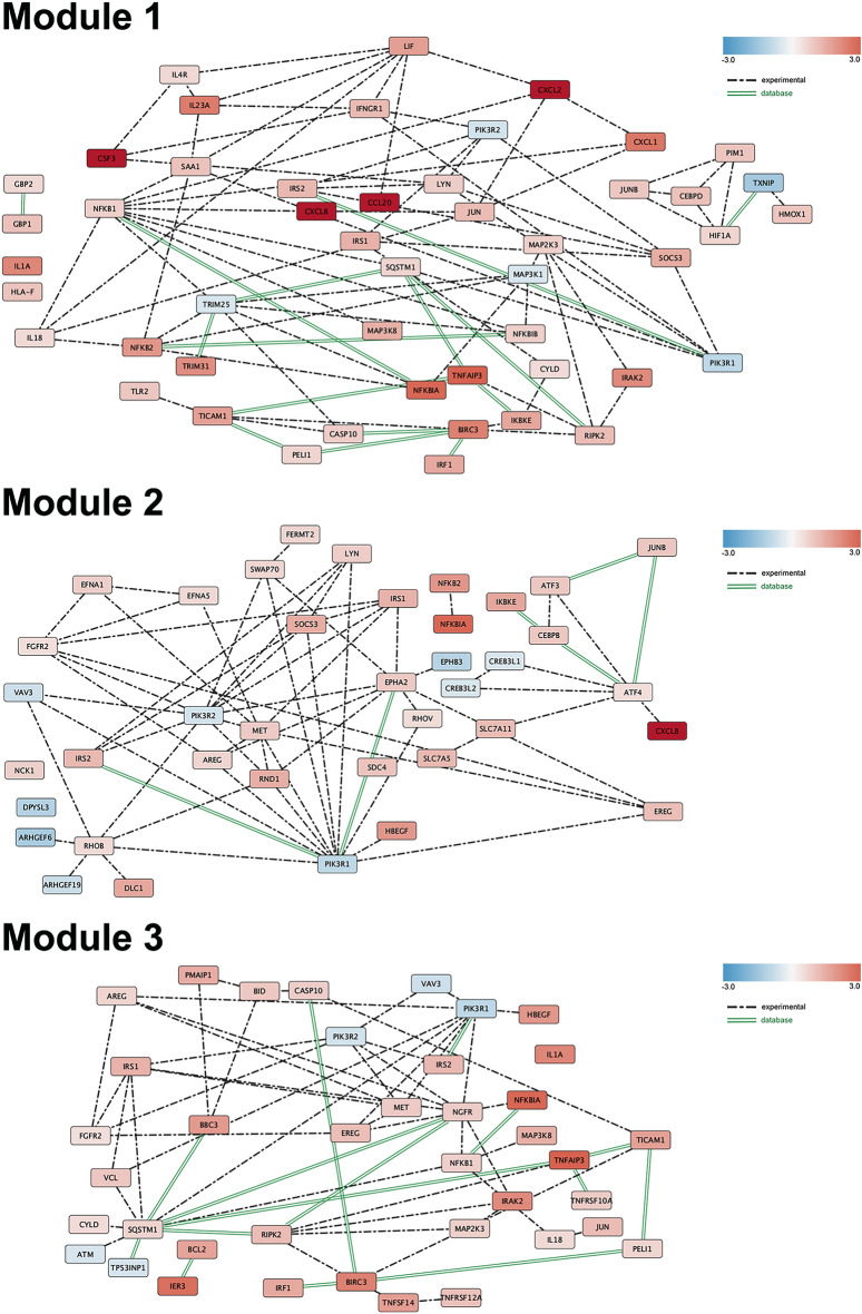
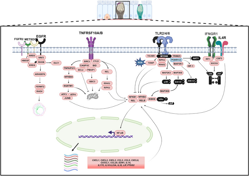
Results: Initial tissue damage began as early as at 4 h post-infection (hpi); at 24 hpi, goblet cell hypertrophy, reduced mucin secretion, and compromised epithelial integrity were highly evident. Transcriptomic data at 4 hpi revealed 668 differentially expressed genes (441 upregulated, 227 downregulated), mainly involved in a strong pro-inflammatory response and characterized by IL-8/CCL20-driven neutrophil recruitment and type 2 cytokine activation (IL-4, IL-13). Noteworthy, genes related to cytoskeletal organization, adhesion, and extracellular matrix remodeling were significantly altered, suggesting a bacterial mechanism to enhanced tissue dissemination. The PI3K-Akt survival pathway was inhibited, with downregulation of PIK3R1 and PIK3R2 genes, implying the induction of apoptosis/cell death and epithelial damage. Our findings are in agreement with previous in vivo studies, further strengthening the value of our ALI model in mimicking the early infection response of bronchial cells to A. baumannii infection.

Conclusion: Our data highlight the early molecular mechanisms underlying A. baumannii pathogenesis and open new avenues for future investigations for therapeutic interventions.

Keywords: Acinetobacter baumannii; 2D co-culture; Air liquid interface epithelium; Host–pathogen interaction; Infection model; Transcriptome.


Conflict of interest statement

Declarations. Ethics approval and consent to participate: Not applicable. Consent for publication: Not applicable. Competing interests: The authors declare that they have no competing interests.

Figures

Fig. 1
Fig. 1
Characterization of differentiated NHBE cells cultured at the air–liquid interface (ALI). 2D ALI cultures were grown for 28 days, before being analyzed. A After 28 days of air exposure, the pseudostratified epithelium was stained with specific antibodies to visualize ciliated cells with well-developed apical projections (acetylated tubulin, red), and sparse goblet cells secreting mucin-5 subtype AC- (MUC5AC, green). Nuclei were counterstained with Hoechst 33342 (blue). B An image illustrating the spatial organization and differentiation of ciliated and goblet cells within the epithelium. C, D Representative histological image of tangential sections of the epithelium stained with H&E and PAS, respectively. Three independent experiments using cells from the same donor were performed. Scale bar sizes are indicated in the images
Fig. 2
Fig. 2
Histology of ALI infected with A. baumannii. A total of 20 µl of bacteria resuspended in DMEM-F12 was used for infection. Non-infected ALI cultures were treated with an equivalent volume of DMEM-F12 as a negative control. A, B Representative images of infected and unifected ALI cultures stained with H&E at 4 h post-infection (hpi) and 24 hpi, respectively, as indicated. C, D Representative images of the same sections shown in panels (A and B), stained with Giemsa. Three independent experiments using cells from the same donor were performed. Scale bar sizes are indicated in the images
Fig. 3
Fig. 3
Immunofluorescence of ALI infected with A. baumannii. Representative images of infected ALI cultures stained with antibodies against A. baumannii (A.b., red) and either MUC5AC (green) or ZO-1 (green) at 4 and 24 h post-infection (hpi). Nuclei were counterstained with Hoechst 33342 (blue). Non-infected ALI cultures were treated with an equivalent volume of DMEM-F12 as a negative control. Three independent experiments were performed. Scale bar sizes are indicated in the images
Fig. 4
Fig. 4
A. baumannii infection increases paracellular permeability in ALI cultures. Differentiated 2D ALI bronchial epithelial cultures were infected apically with strain AB507. At 24 h post-infection (hpi), barrier integrity was assessed by measuring the paracellular flux of 2 mg/ml of FITC-dextran (4 kDa) from the apical to the basolateral compartment. After 2 h of further incubation at 37 °C and 5% CO2, fluorescence measurement (excitation at 492 nm and emission at 520 nm) were recorded using a CLARIOStar microplate reader (BMG Labtech, Offenburg, Germany) in a 96-well black microtiter. The Adjust Gain setting was calibrated using a 2 mg/ml solution to standardize all sample readings. Data are expressed as mean ± standard deviation of three independent experiments. Statistical analysis was performed using Student’s t-test
Fig. 5
Fig. 5
Global transcriptiomic response of bronchial epithelium to A. baumannii AB5075 infection at 4 hpi. A Principal Component Analysis (PCA) plot illustrating the clustering of unexposed (group A) and A. baumannii-exposed (group B) bronchial cells based on transcriptomic profiles. B Heatmap and hierarchical clustering dendrogram of the top differentially expressed genes (DEGs) in unexposed and exposed bronchial cells (FDR P value < 0.05). Warmer colors indicate upregulated genes, while cooler colors indicate downregulated genes. C Tree plot of the top 30 Gene Ontology (GO) terms related to the biological process of DEGs. The size of each node represents the level of enrichment significance, and the hierarchical arrangement reflects functional similarities between terms. D Description of the top 30 pathway enrichment analysis within the DEGs list, obtained through the use of a hypergeometric model. E Chord diagram illustrating the log2 fold changes of genes involved in the selected GO terms. F Volcano plot depicting the distribution of DEGs, with the most significantly upregulated (green) and downregulated (red) genes highlighted. The x-axis represents log2 fold change, and the y-axis represents -log10 (FDR P value), with a threshold of FDR < 0.05 used to identify significant DEGs
Fig. 6
Fig. 6
Protein–protein interaction network. The three modules represent the protein–protein interaction in the main pathways idendified constructed by Reactome functional interactions Cytoscape Plugin. Module 1, DEGs involved in inflammation and signaling regulation, module 2 DEGs involved in cell–cell interaction, module 3 DEGs in cell stress, survival and apoptosis. The color code indicates up o downregulation. Dotted lines reports experimentally demonstrated interactions, solid green lines interactions reported in the databases
Fig. 7
Fig. 7
Proposed regulatory networks of human bronchial epithelium upon exposure to A. baumannii. Based on the results of this study, three main pathways highly interconneced were triggered by bacteria: inflammation and signaling regulation, cell–cell interaction, and autophagy/cell death. Black-filled proteins were not found among DEGs. Orange and light blue proteins represent upregulated and downregulated genes, respectively. Solid lines indicate previously demonstrated interactions, while dotted lines refer to missing interactors. Additional details are provided in the main text. The figure was created with BioRender.com

References

    1. Cain AK, Hamidian M. Portrait of a killer: uncovering resistance mechanisms and global spread of Acinetobacter baumannii. PLoS Pathog. 2023;19(8): e1011520. - PMC - PubMed
    1. Sarshar M, Behzadi P, Scribano D, Palamara AT, Ambrosi C. Acinetobacter baumannii: an ancient commensal with weapons of a pathogen. Pathogens. 2021. 10.3390/pathogens10040387. - PMC - PubMed
    1. Shields RK, Paterson DL, Tamma PD. Navigating available treatment options for carbapenem-resistant Acinetobacterbaumannii-calcoaceticus complex infections. Clin Infectious Diseas. 2023;76(S2):S179–93. - PMC - PubMed
    1. Shadan A, Pathak A, Ma Y, Pathania R, Singh RP. Deciphering the virulence factors, regulation, and immune response to Acinetobacter baumannii infection. Front Cell Infect Microbiol. 2023;13:156. - PMC - PubMed
    1. Pompilio A, Scribano D, Sarshar M, Di Bonaventura G, Palamara AT, Ambrosi C. Gram-negative bacteria holding together in a biofilm: the Acinetobacter baumannii way. Microorganisms. 2021;9(7):1353. - PMC - PubMed

LinkOut - more resources