. 2025 Jul 9;22(1):176.
doi: 10.1186/s12974-025-03507-2.

Pharmacological inhibition of the cGAS-STING pathway suppresses microglia pyroptosis in sepsis-associated encephalopathy

Affiliations

Pharmacological inhibition of the cGAS-STING pathway suppresses microglia pyroptosis in sepsis-associated encephalopathy

Qing-Quan Zeng et al. J Neuroinflammation. .

Abstract

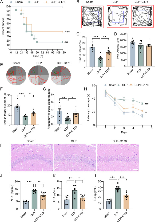
Sepsis-associated encephalopathy (SAE) presents significant challenges in clinical management due to its association with cognitive impairments and high mortality rates. This study aims to elucidate the molecular mechanisms underlying microglial pyroptosis and the stimulator of interferon genes (STING) signaling pathway in SAE, with a focus on identifying potential therapeutic targets. Employing the cecal ligation and puncture (CLP) model - the gold-standard polymicrobial sepsis model that faithfully replicates human sepsis progression - in C57BL/6J mice, we assessed the effects of various pharmacological agents, including the pyroptosis inhibitor dimethyl fumarate (DMF), the STING inhibitor C176, and the mitochondrial protectant idebenone, on cognitive and behavioral outcomes in SAE mice. Results indicated that DMF significantly prevented microglial pyroptosis, reduced inflammatory cytokine levels in the hippocampus, thereby enhancing survival rates and cognitive function. Additionally, inhibition of microglial pyroptosis through C176 effectively inhibited the STING signaling pathway, consequently reducing microglial pyroptosis and ameliorating behavioral symptoms associated with SAE. Furthermore, idebenone was observed to exert mitochondrial protection and inhibit STING-mediated pyroptosis of microglia, leading to an improvement in behavioral symptoms in the SAE model. In conclusion, our findings underscore the pivotal roles of pyroptosis and the STING signaling pathway in the pathophysiology of SAE, suggesting that targeting these mechanisms may provide promising therapeutic avenues for improving cognitive recovery in sepsis patients. Future studies should focus on elucidating the underlying molecular mechanisms and exploring their clinical viability in the management of SAE.

Graphical Abstract:

Supplementary Information: The online version contains supplementary material available at 10.1186/s12974-025-03507-2.

Keywords: Microglia; Pyroptosis; STING; Sepsis; Sepsis-associated encephalopathy.


Conflict of interest statement

Declarations. Ethical approval: All experimental protocols complied with international guidelines and were conducted under the supervision of the Ethics Committee for Animal Experiments of Fujian Medical University (Approval No. IACUC FJMU 2022 − 0880, approved on Dec. 21, 2022). Consent for publication: Not applicable. Competing interests: The authors declare no competing interests.

Figures

Fig. 1
Fig. 1
Pyroptosis inhibitor improved survival rate, alleviated cognitive impairment and neuroinflammation after SAE. (A) DMF improved survival rates in CLP-induced septic mice (n = 9 per group). (B-D) Open field test demonstrated the effects of DMF on the behavior of septic mice (n = 9 per group). (E-H) Morris water maze test illustrated the impact of DMF on the behavior of septic mice (n = 9 per group). (I) HE staining assessed hippocampal tissue pathology in septic mice treated with DMF. (J-L) ELISA analysis of TNF-α, IL-1β, and IL-6 levels in the hippocampus of mice (n = 6 per group). Kaplan–Meier survival curves were employed for survival rate analysis. Behavioral test results were analyzed using repeated-measures ANOVA for intergroup comparisons, alongside one-way ANOVA followed by Dunnett T3 tests. Statistical significance: ##P < 0.01; ###P < 0.001 compared to the Sham group; *P < 0.05; **P < 0.01; ***P < 0.001 compared to the CLP group
Fig. 2
Fig. 2
Pyroptosis inhibitor prevented microglia pyroptosis after SAE in the hippocampal of CLP mice. (A-D) Quantitative PCR analysis of NLRP3, Caspase-1, GSDMD, and IL-1β mRNA in the hippocampus (n = 6). (E) Representative western blot showed the protein levels of NLRP3, Pro-Caspase-1, Caspase-1, GSDMD-N, Pro-IL-1β. The quantity of protein levels (F-K). (L) Co-localization of IBA-1 with GSDMD-N in in hippocampus (scale = 100 μm). Data are presented as the mean ± S.E.M. Separate one-way ANOVA followed by Dunnett T3 test. *P < 0.05; **P < 0.01; ***P < 0.001
Fig. 3
Fig. 3
STING inhibitor C176 improved survival rate, alleviated cognitive impairment and neuroinflammation after SAE. (A) C176 improved survival rates in CLP-induced septic mice (n = 9 per group). (B-D) Open field test demonstrated the effects of C176 on the behavior of septic mice (n = 9 per group). (E-H) Morris water maze test illustrated the impact of C176 on the behavior of septic mice (n = 9 per group). (I) HE staining assessed hippocampal tissue pathology in septic mice treated with C176. (J-L) ELISA analysis of TNF-α, IL-1β, and IL-6 levels in the hippocampus of mice (n = 6 per group). Kaplan–Meier survival curves were employed for survival rate analysis. Behavioral test results were analyzed using repeated-measures ANOVA for intergroup comparisons, alongside one-way ANOVA followed by Dunnett T3 tests. Statistical significance: ###P < 0.001 compared to the Sham group; *P < 0.05; **P < 0.01; ***P < 0.001 compared to the CLP group
Fig. 4
Fig. 4
STING inhibitor C176 prevented microglia pyroptosis after SAE in the hippocampal of CLP mice. (A-G) Quantitative PCR analysis of STING, IFN-α, IFN-β, NLRP3, Caspase-1, IL-1β, and GSDMD in the hippocampus (n = 6). (H) Representative western blot showed the protein levels of STING, p-STING, IRF3, p-IRF3, TBK1, p-TBK1, NLRP3, pro-Caspase-1, GSDMD-N, and pro-IL-1β proteins. The quantity of protein levels (I-T). (U) Co-localization of IBA-1 with GSDMD-N in in hippocampus (scale = 100 μm). Data are presented as the mean ± S.E.M. Separate one-way ANOVA followed by Dunnett T3 test. *P < 0.05; **P < 0.01; ***P < 0.001
Fig. 5
Fig. 5
STING knockout alleviated CLP-induced SAE and inhibited microglia pyroptosis in hippocampal microglia. (A) STING KO improved survival rates in the CLP sepsis model. (B-D) Open field test results demonstrating the effects of STING KO on the behavior of septic mice. (E-H) Morris water maze test results illustrating the impact of STING KO on the behavior of septic mice. (I) Representative western blot showed the protein levels of NLRP3, pro-Caspase-1, Caspase-1, GSDMD-N, pro-IL-1β. (J-P) The quantity of protein levels. (Q) Co-localization of IBA-1 with GSDMD-N in in hippocampus (scale = 100 μm). Kaplan–Meier survival curves were used for survival rate analysis. Behavioral test results were analyzed using repeated-measures ANOVA for intergroup comparisons, along with one-way ANOVA followed by LSD or Dunnett T3 tests. Statistical significance: ###P < 0.001 compared to the Sham group; *P < 0.05; **P < 0.01 compared to the CLP group
Fig. 6
Fig. 6
Idebenone exerted mitochondrial protection in SAE mice. (A) Representative electron microscopy images of microglia in the hippocampus of mice, with white arrows indicating mitochondria in microglia. The expression levels of genes Dloop1, Dloop2, and Dloop3 in the cytoplasm were detected by qPCR. (n = 6). (C) Co-localization of NEUN with ds-DNA in in hippocampus (scale = 100 μm). Statistical significance was determined via one-way ANOVA followed by the LSD test or Dunnett’s T3 test. **P < 0.01; ***P < 0.001 compared to the CLP group
Fig. 7
Fig. 7
Idebenone reduced hippocampal STING expression and attenuated microglial pyroptosis markers in SAE mice. (A-G) Quantitative PCR analysis of STING, IFN-α, IFN-β, GSDMD, IL-1β, NLRP3, Caspase-1 in the hippocampus (n = 6). (H) Representative western blot showed the protein levels of STING, p-STING, IRF3, p-IRF3, TBK1, p-TBK1, NLRP3, pro-Caspase-1, GSDMD-N, and pro-IL-1β proteins. (I-T) The quantity of protein levels. (U) Co-localization of IBA-1 with GSDMD-N in in hippocampus (scale = 100 μm). Data are presented as the mean ± S.E.M. Separate one-way ANOVA followed by Dunnett T3 test. *P < 0.05; **P < 0.01; ***P < 0.001
Fig. 8
Fig. 8
Idebenone improved behavioral symptoms of sepsis encephalopathy induced by CLP in mice. (A) Idebenone improved survival rates in CLP-induced septic mice (n = 9 per group). (B-D) Open field test demonstrated the effects of idebenone on the behavior of septic mice (n = 9 per group). (E-H) Morris water maze test illustrated the impact of idebenone on the behavior of septic mice (n = 9 per group). (I) HE staining assessed hippocampal tissue pathology in septic mice treated with idebenone. (J-L) ELISA analysis of TNF-α, IL-1β, and IL-6 levels in the hippocampus of mice (n = 6 per group). Kaplan–Meier survival curves were employed for survival rate analysis. Behavioral test results were analyzed using repeated-measures ANOVA for intergroup comparisons, alongside one-way ANOVA followed by Dunnett T3 tests. Statistical significance: ###P < 0.001 compared to the Sham group; *P < 0.05; **P < 0.01; ***P < 0.001 compared to the CLP group
Fig. 9
Fig. 9
Mechanistic insights into how cGAS-STING pathway inhibition attenuates microglial pyroptosis in sepsis-associated encephalopathy

References

    1. Mazeraud A, Righy C, Bouchereau E, Benghanem S, Bozza FA, Sharshar T. Septic-Associated encephalopathy: a comprehensive review. Neurotherapeutics. 2020;17:392–403. - PMC - PubMed
    1. Sonneville R, Benghanem S, Jeantin L, de Montmollin E, Doman M, Gaudemer A, Thy M, Timsit JF. The spectrum of sepsis-associated encephalopathy: a clinical perspective. Crit Care. 2023;27:386. - PMC - PubMed
    1. Hong Y, Chen P, Gao J, Lin Y, Chen L, Shang X. Sepsis-associated encephalopathy: from pathophysiology to clinical management. Int Immunopharmacol. 2023;124:110800. - PubMed
    1. Zhu DD, Huang YL, Guo SY, Li N, Yang XW, Sui AR, Wu Q, Zhang Y, Kong Y, Li QF, et al. AQP4 aggravates cognitive impairment in Sepsis-Associated encephalopathy through inhibiting Na(v) 1.6-Mediated astrocyte autophagy. Adv Sci (Weinh). 2023;10:e2205862. - PMC - PubMed
    1. Fu Q, Zhang Y-B, Shi C-X, Jiang M, Lu K, Fu Z-H, Ruan J-P, Wu J. Gu X-P: GSDMD/Drp1 signaling pathway mediates hippocampal synaptic damage and neural Oscillation abnormalities in a mouse model of sepsis-associated encephalopathy. J Neuroinflammation. 2024;21:96. - PMC - PubMed

LinkOut - more resources