. 2025 Jul;24(7):e70292.
doi: 10.1111/jocd.70292.

Innate and Adaptive Immune Responses to Clinical Hyaluronic Acid Fillers

Affiliations

Innate and Adaptive Immune Responses to Clinical Hyaluronic Acid Fillers

Joshua S T Hooks et al. J Cosmet Dermatol. 2025 Jul.

Abstract

Background: Crosslinked hyaluronic acid (HA)-based hydrogels are commonly used as dermal fillers where they interact with surrounding tissues including host stromal and immune cells. HA fillers are widely used for aesthetic applications, with products designed with varying properties depending on their indication. Although HA fillers have been demonstrated to have a strong biocompatibility profile, a small subset of patients' experiences delayed-onset adverse events hypothesized to be inflammatory and allergy-related outcomes such as delayed-onset hypersensitivity.

Aims: The overall goal of this study was to evaluate the innate and adaptive immune response to two clinically available HA filler formulations.

Methods: Using multiparametric flow cytometry, we characterized the immune response to Juvèderm Volbella (VYC-15 L) and Juvèderm Ultra 3 (SGD-30XP) in a murine quadricep muscle resection that enables implantation of larger volumes and exposure to muscle and adipose.

Results: Presence of the implanted HA filler increased recruitment of immune cells, specifically antigen presenting macrophages, eosinophils, and gamma-delta (γδ) T cells to the injury site compared to no implant (saline) controls. Comparing the two materials, VYC-15 L increased interleukin 17a (IL17a) production by lymphocyte subsets at the injury site and induced higher levels of circulating IgE relative to SGD-30XP and saline controls.

Conclusion: Overall, these results provide insights into the immune response to HA fillers and how different formulations may alter the immune outcomes.

Keywords: biomaterials; flow cytometry; hyaluronic acid; immune response.


Conflict of interest statement

Allergan funded this study and provided the materials for this study. J.H.E. is an inventor on intellectual property related to biological scaffolds and inhibiting fibrosis. J.H.E. holds equity in Unity Biotechnology and Aegeria Soft Tissue. J.H.E. is a member of the scientific advisory boards of Tessera Therapeutics, HapInScience, and Font Bio. J.H.E. is a consultant for Vericel. C.K.H. is an employee of Allergan Aesthetics, an AbbVie company, and owns stock and stock options in AbbVie.

Figures
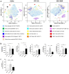
FIGURE 1
FIGURE 1
HA fillers polarize immune profile toward an allergy‐like response at 6 weeks following VML. Mice underwent volumetric muscle loss (VML) surgery and VYC‐15 L or SGD‐30XP HA fillers were implanted in the resection. Control mice were treated with a physiological saline. (a) Immune profiles as tSNE plots from spectral flow cytometry. (b) Bar plots of key immune cell populations shifted by HA filler implant compared to saline control. (c) Increased serum IgE detected in VYC‐15 L condition via ELISA. (Statistics) Data are mean ± SD, n = 4, ****p < 0.0001, ***p < 0.001, **p < 0.01, and *p < 0.05 by one‐way ANOVA with Tukey's multiple comparisons test.
FIGURE 2
FIGURE 2
HA fillers enhance immune cell recruitment and increased macrophage antigen presentation markers. (a) HA fillers increase immune cell recruitment to the wound at all follow‐up time points. (b) Macrophages as a percent of immune cells increase over time across all conditions. No consistent shifts in M1 vs. M2 polarization (CD206/CD86 MFI) in either HA filler. Markers for antigen presentation (CD11c & MHCII) are upregulated on macrophages in HA filler conditions at the 6‐week time point. (c) H&E staining reveals residual filler at the wound site at both 3 and 6 weeks following implant. VYC‐15 L filler had less remaining material compared to SGD‐30XP at both follow‐up time points. Scale bar, 1 mm. (Statistics) Data are mean ± SD, n = 4.
FIGURE 3
FIGURE 3
Differential cytokine production by local T cells following HA filler implant over course of wound healing. (a) At 6 weeks following VML, T cells were upregulated in the VYC‐15 L condition. T cells in the VYC‐15 L condition at 6 weeks following VML significantly increased IL17a expression. Both HA fillers downregulated T cell production of IFNγ. (b) There was not a difference in expression of overall CD4+ T helper cells throughout wound healing, but T helper cell subsets did shift significantly. IFNγ producing T helper cells (Th1) were significantly downregulated by HA fillers and FoxP3+ T regulatory cells (Tregs) were upregulated by SGD‐30XP at 6 weeks following VML. (c) VYC‐15 L upregulated IL17a+ γδ T cells, seen significantly at the 6‐week time point. (Statistics) Data are mean ± SD, n = 4, ***p < 0.001, **p < 0.01, and *p < 0.05 by one‐way ANOVA with Tukey's multiple comparisons test.
FIGURE 4
FIGURE 4
Immune expression of the draining inguinal lymph node (iLN) immune populations following VML and filler implantation. (a) Expression of immune cell populations was not significantly shifted by HA filler implant in the iLN. IL17a and IFNγ producing subsets were not significantly changed across all CD3+ cells. (b) Immune cell subsets reveal an increase in IL17a+ T helper cells (Th17) in the VYC‐15 L filler condition and a general downward trend in IFNγ producing lymphocytes (Th1, CD8+, and NKTs) in the SGD‐30XP filler condition compared to VYC‐15 L. (Statistics) Data are mean ± SD, n = 3–4, one‐way ANOVA with Tukey's multiple comparisons test.

References

    1. Fraser J. R. E., Laurent T. C., and Laurent U. B. G., “Hyaluronan: Its Nature, Distribution, Functions and Turnover,” Journal of Internal Medicine 242 (1997): 27–33. - PubMed
    1. Huerta‐ángeles G., Nešporová K., Ambrožová G., Kubala L., and Velebný V., “An Effective Translation: The Development of Hyaluronan‐Based Medical Products From the Physicochemical, and Preclinical Aspects,” Frontiers in Bioengineering and Biotechnology 6 (2018): 1–13. - PMC - PubMed
    1. La Gatta A., Salzillo R., Catalano C., et al., “Hyaluronan‐Based Hydrogels as Dermal Fillers: The Biophysical Properties That Translate Into a “Volumetric” Effect,” PLoS One 14 (2019): 1–17, e0218287. - PMC - PubMed
    1. Alijotas‐Reig J., Hindié M., Kandhaya‐Pillai R., and Miro‐Mur F., “Bioengineered Hyaluronic Acid Elicited a Nonantigenic T Cell Activation: Implications From Cosmetic Medicine and Surgery to Nanomedicine,” Journal of Biomedical Materials Research. Part A 95 (2010): 180–190. - PubMed
    1. Slevin M., Kumar S., and Gaffney J., “Angiogenic Oligosaccharides of Hyaluronan Induce Multiple Signaling Pathways Affecting Vascular Endothelial Cell Mitogenic and Wound Healing Responses *,” Journal of Biological Chemistry 277 (2002): 41046–41059. - PubMed

Grants and funding