. 2025 Jun 25:16:1617146.
doi: 10.3389/fimmu.2025.1617146. eCollection 2025.

Organ of Corti macrophages: a distinct group of cochlear macrophages with potential roles in supporting cell degeneration and survival

Affiliations

Organ of Corti macrophages: a distinct group of cochlear macrophages with potential roles in supporting cell degeneration and survival

Mengxiao Ye et al. Front Immunol. .

Abstract

Macrophages are the primary immune cells in the cochlea, essential for maintaining cochlear homeostasis and orchestrating inflammatory responses to pathological events. Although these cells have been found in various parts of the cochlea, their presence in the organ of Corti, a critical structure for acoustic sensing, remains poorly understood. The present study was designed to examine macrophage responses to ototoxic drug-induced cochlear damage and age-related cochlear degeneration, with a particular focus on the pathological conditions that trigger macrophage recruitment into the organ of Corti. We used a model of ototoxicity induced by cyclodextrin, a cyclic oligosaccharide known for its ability to induce rapid sensory cell damage at high doses. Cochlear tissues were collected for macrophage assessment. Multiple protein markers, including CD45, Iba1, galectin-3, CD68, myosin 7α, and IFIT3, were used to label sensory cells, macrophages, and supporting cells in cochlear sensory epithelia. Consistent with previous reports, our study confirms that macrophages are absent from the organ of Corti in mouse cochleae under normal conditions and during the acute phase of cochlear damage. However, macrophages enter the organ of Corti during the chronic phase of cochlear pathogenesis. These macrophages exhibit an activated state and display a distinct functional profile compared to macrophages outside the organ of Corti. Importantly, we demonstrate that the organ of Corti macrophage activity is not directly related to the process of sensory cell degradation. Instead, their activity is associated with supporting cell pathogenesis. Moreover, our study shows that the organ of Corti macrophages are also present in aging mouse cochleae. Collectively, our findings reveal the conditions that lead to macrophage recruitment into the organ of Corti and their involvement in supporting cell survival and degeneration. These findings provide valuable insights for future strategies focused on modulating macrophage activity to reduce tissue damage and promote repair in the cochlea following injury.

Keywords: aging; cochlea; galectin-3; inflammation; macrophage; organ of Corti; ototoxicity; supporting cells.


Conflict of interest statement

The authors declare that the research was conducted in the absence of any commercial or financial relationships that could be construed as a potential conflict of interest. The author(s) declared that they were an editorial board member of Frontiers, at the time of submission. This had no impact on the peer review process and the final decision.

Figures

Figure 1
Figure 1
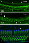
Cyclodextrin treatment induces rapid sensory cell death in mouse cochleae. (A–D) Sensory cells in a control cochlea stained for F-actin (A), Myosin-7α (B), and DAPI (C). Both OHCs and IHCs are intact. (E–H) Typical image of hair cells in the apical region of the cochlea (25% from the apex) examined at one day post-cyclodextrin treatment. Most hair cells are present. (I–L) Significant hair cell death was observed at the basal end of the cochlea at one day post-cyclodextrin treatment. Arrows in (I) point to regions of OHC cuticular plates that lack F-actin staining, suggesting cell loss. Arrows in (J) indicate fragmented OHC bodies illustrated by Myosin-7α staining. Arrows in (K) show condensed or fragmented nuclei in DAPI staining.
Figure 2
Figure 2
Hair cell loss at the chronic phases of cyclodextrin-induced ototoxicity. (A-C) Typical image of the apical region of the cochlea (approximately 25% from the apex), showing areas of hair cell loss (indicated by arrows) in a cochlea examined 6 weeks after cyclodextrin treatment. (D-F) Representative image of the basal region of the cochlea (approximately 85% from the apex), taken 6 weeks after cyclodextrin treatment. The image shows complete hair cell loss, with areas of supporting cell loss also evident (indicated by the arrow). (G, H) Quantification of missing OHCs (G) and IHCs (H) at the acute phase (3–4 days) and chronic phase (6–10 weeks) of cyclodextrin treatment. OHC loss spans the middle and basal regions (41–100% from the apex), while IHC loss is confined to the basal region (86–100% from the apex). The severity of OHC loss remains consistent between the acute and chronic phases. However, IHC loss significantly increases in the transition area between the mild and severe IHC loss regions during the chronic phase. **** in indicates p < 0.0001.
Figure 3
Figure 3
Macrophages in the cochlea under normal conditions. (A) A schematic illustration of a cross-sectional view of the cochlea, showing the organ of Corti and its surrounding tissues. The red dotted box highlights the regions of macrophage observation: the organ of Corti and the basilar membrane. (B) An enlarged histological view of the areas of macrophage observation. The dark space marked by the white asterisks indicates the basement membrane, which separates the OC macrophages from the BM macrophages. (C) Iba1 immunostaining in the middle region of the cochlea from a whole-mount preparation, showing the regions of the osseous spiral lamina (OSL), basilar membrane (BM), and lateral wall (LW). The two dotted lines mark the boundaries of the basilar membrane. Note that Iba1-positive immune cells are present in these regions (arrows). (D) Double staining of CD45 in the same tissue as shown in panel (C, E) Merged view of (C, D) showing that CD45-positive cells also exhibit Iba1 immunoreactivity, indicating that these cells are macrophages. (F–H) A cross-sectional view of the whole-mount tissue confirms that the macrophages observed in the basilar membrane (double arrows) are located on the scala tympani side of the basilar membrane. The organ of Corti is devoid of macrophages. The dark space marked by the asterisks indicates the basement membrane, which separates the OC macrophages from the BM macrophages. BM: basilar membrane, OC, organ of Corti; OSL, osseous spiral lamina; SL, spiral ligament; LW, lateral wall.
Figure 4
Figure 4
Macrophage activity during the acute phase of cyclodextrin ototoxicity. (A) A typical image of BM macrophages stained with Iba1 in the middle section of the cochlea at 1 day post-treatment. At this early stage, BM macrophages retain their normal elongated morphology (arrows). (B) A representative image of BM macrophages observed 4 days post-treatment. Numerous small, round Iba1-positive cells are present on the basilar membrane (arrows). These cells are infiltrating monocytes. (C) Quantification of Iba1-positive cells reveals a significant increase in BM macrophages during the acute phase of cyclodextrin ototoxicity. ** indicates p < 0.01. (D–F) A side view of the sensory epithelium shows that Iba1-positive cells (double arrows) are confined to the basilar membrane. Macrophages remain absent in the organ of Corti (arrow) at this acute stage of the damage.
Figure 5
Figure 5
Galectin-3 expression in BM macrophages under normal conditions and during the acute phase of cochlear damage. (A) Galectin-3 immunoreactivity in the middle section of a normal cochlea. Macrophages in the basilar membrane region (marked by the dotted line) exhibit little, if any, galectin-3 immunoreactivity. (B) A few galectin-3 positive macrophages (arrows) are present in the basilar membrane (marked by the dotted line) 1 day post-treatment. (C) A significant increase in the number of galectin-3 positive macrophages (arrows) on the basilar membrane 4 days after cyclodextrin treatment. Notably, many galectin-3-positive cells are small and round, suggesting they are infiltrating monocytes. (D) A side view of the sensory epithelium shows that all galectin-3 and Iba1 double-positive cells (arrow) are confined to the basilar membrane. BM: basilar membrane.
Figure 6
Figure 6
Regression of macrophage activity on the basilar membrane during the chronic phase of cyclodextrin ototoxicity. (A) Comparison of the number of BM macrophages among the control, the acute (3–4 days post-treatment), and the chronic phase (6–10 weeks post-treatment) shows that the macrophage number returns to baseline level at the chronic phase (** indicates p < 0.01, *** indicates p < 0.001). (B) Typical morphology of BM macrophages in the middle section of the cochlea at 6 weeks post-treatment. Most BM macrophages exhibit a branched or elongated shape (arrows). The small, round macrophages commonly observed during the acute phase are now rarely present. (C, D) Double immunostaining for galectin-3 and Iba1 in the middle section of a cochlea examined six weeks after cyclodextrin treatment. Galectin-3 immunoreactivity was barely detectable in the basilar membrane region (indicated by the area between the two dotted lines in panel (C), despite the presence of Iba1-positive macrophages in this region (arrows in panel (D). Notably, cells medial to the inner hair cells and a subset of macrophages in the lateral wall (indicated by the double arrow) exhibit galectin-3 immunoreactivity. ns, not significant.
Figure 7
Figure 7
Identification of OC macrophages during the chronic phase of cyclodextrin ototoxicity. (A) A side view of the sensory epithelium stained for CD45 and Iba1 from a cochlea examined 6 weeks post-treatment. The two downward arrows at the top indicate OC macrophages, and the upward arrow at the bottom indicates the BM macrophage. The dark space marked by the asterisks is the basement membrane, which separates the OC macrophages from the BM macrophages. (B) Top view of the sensory epithelium displaying OC macrophages. This image was created by removing the confocal image layers containing BM macrophages from a 3D stack, thus revealing only OC macrophages. Arrows indicate OC macrophages with different morphologies. (C) Quantification of OC macrophages at different regions along the cochlear spirals. (* indicates p < 0.05, ** indicates p < 0.01) (D) A schematic of the cochlear spiral illustrates the distribution of OC macrophages. OC macrophages are primarily confined to the basal region (70–90% from the apex). In contrast, hair cell loss extends across the entire basal and middle regions of the cochlea. (E) OC macrophages are present in the cochlea 2 weeks post-treatment (arrows). (F) A cross-sectional view of the sensory epithelium confirms that these macrophages are located within the organ of Corti (downward arrows). The asterisks indicate the basement membrane that separates the OC macrophages (downward arrows) from the BM macrophage (upward arrow). ns, not significant; OC, organ of Corti.
Figure 8
Figure 8
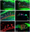
Locations of OC macrophages in the organ of Corti. (A, B) A typical location of OC macrophages, illustrated by Iba1 staining, in the region of Deiters’ and pillar cells in a cochlea examined 6 weeks post-treatment. Arrows indicate OC macrophages with irregular shapes. F-actin staining in (B) shows the footplates of Deiters’ cells and pillar cells. (C) Rounded macrophages (arrows) are present at the top portion of the Deiters’ cell region illustrated by IFIT3 immunostaining, a marker to label the central stalks of Deiters’ cells. Phalloidin staining reveals a loss of F-actin in the cuticular plates of OHCs. (D, E) Another common location of OC macrophages is at the junction of Deiters’ and Boettcher’s cells. Macrophages in this region exhibit an elongated morphology, with their long axis aligned with the cochlear spiral. (F) A schematic diagram illustrating the two primary locations of OC macrophages. The macrophages highlighted in orange are those located in the region of Deiters’ cells and pillar cells, whereas the macrophage highlighted in dark red is located at the junction between Deiters’ cells and Boettcher’s cells. IPC, inner pillar cell; OPC, outer pillar cell.
Figure 9
Figure 9
Differences in galectin-3 immunoreactivity between OC and BM Macrophages. (A, D) Galectin-3 immunostaining in cochleae examined 6 weeks post-treatment. The images show a basal section of the cochlea (approximately 70-80% from the apex). (A, B) show macrophages located at the junctural region between Deiters’ cells and Boettcher’s cells. (C, D) show macrophages within the region of Deiters’ cells and pillar cells. Arrows point to the macrophages with strong galectin-3 immunoreactivity. (E) and (F) Images show BM macrophages of the same regions. These macrophages show no detectable galectin-3 immunoreactivity. (G) Cross-sectional view of the sensory epithelium. The arrow indicates an OC macrophage with strong galectin-3 immunoreactivity, whereas the double-arrow identifies a galectin-3-negative BM macrophage. (H) Quantitative comparison of the mean gray value of galectin-3 fluorescence demonstrates significantly stronger immunoreactivity in OC macrophages than in BM macrophages. **** indicates p < 0.0001. OC, OC macrophages; BM, BM macrophages. n=16 macrophages from 6 cochleae.
Figure 10
Figure 10
Differences in CD68 immunoreactivity between OC and BM macrophages. (A, B) Typical images of CD68 immunoreactivity in BM macrophages located in the middle region of a normal control cochlea. Arrows indicate BM macrophages with weak CD68 immunoreactivity. (C, D) CD68 immunoreactivity in BM macrophages 6 weeks after cyclodextrin treatment. These macrophages exhibit varying levels of CD68 immunoreactivity in their cytoplasm. Some display levels comparable to those observed in control cochleae (double arrows), while others show stronger immunoreactivity (single arrow). (E, F) CD68 immunoreactivity in OC macrophages of a cyclodextrin-treated cochlea examined 6 weeks after treatment. Although variable among individual cells, all OC macrophages display stronger CD68 immunoreactivity (arrows), suggesting enhanced CD68 expression. (G) A cross-sectional view of the sensory epithelium from a cyclodextrin-treated cochlea shows the difference in CD68 immunoreactivity between OC and BM macrophages. The dotted ellipse highlights the region containing both OC and BM macrophages. Notably, CD68 immunoreactivity is stronger in OC macrophages (single arrow) compared to BM macrophages (double arrow). (H) Quantitative analysis of the mean gray level of CD68 immunoreactivity in cyclodextrin-treated cochleae. OC macrophages exhibit significantly higher CD68 immunoreactivity compared to BM macrophages. **** indicates p < 0.0001. OC, OC macrophages; BM, BM macrophages. n=12 macrophages from 3 cochleae.
Figure 11
Figure 11
Pathological changes in supporting cells during the chronic phase of cyclodextrin ototoxicity. (A, B) F-actin staining of the organ of Corti reveals sporadic loss of supporting cells in the middle region (A) and a more pronounced loss in the basal region (B) of a cochlea examined at 6 weeks after cyclodextrin treatment. Arrows indicate areas where supporting cells are absent. (C) Evidence of active degradation in supporting cells. The arrow points to an inner pillar cell exhibiting retained F-actin staining in the footplate but lacking a nucleus, suggestive of ongoing degeneration.
Figure 12
Figure 12
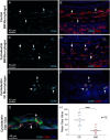
Galectin-3 expression in supporting cells under normal conditions and during the chronic phase of cyclodextrin ototoxicity. (A) Galectin-3 immunoreactivity in supporting cells under normal conditions. (B) The overlay of panel A with F-actin staining to illustrate the organ of Corti. Hensen’s cells exhibit strong galectin-3 immunoreactivity, whereas pillar cells and Deiters’ cells show little, if any, galectin-3 immunoreactivity. (C, D) Elevated galectin-3 immunoreactivity in supporting cells adjacent to an area of missing supporting cells. The arrow in (C) indicates a region with a missing pillar cell, as evidenced by the absence of F-actin staining. The dotted boxes in (D) highlight increased galectin-3 immunoreactivity in supporting cells surrounding the region of the missing pillar cell. As expected, these cells lack Iba1 immunoreactivity.
Figure 13
Figure 13
Macrophages in the Claudius cell region in cyclodextrin-treated cochleae. (A, B) Images show a macrophage in the Claudius cell region in a cochlea examined 6 weeks after cyclodextrin treatment. This cell displays Iba1 (A) and galectin-3 immunoreactivities (B). (C) Macrophages with strong galectin-3 immunoreactivity display irregular shapes (arrows). (D) A side view of the sensory epithelium shows a macrophage (arrow) located beneath Claudius cells. (E) Enlarged view of macrophages in the Claudius cell region. Macrophages display projections toward the organ of Corti (arrows). (F) A 3D view of the organ of Corti showing macrophages in the Claudius cell region. The macrophages exhibit radial processes (arrow) directed toward the organ of Corti. LW, lateral wall; OC, organ of Corti.
Figure 14
Figure 14
Identification of OC macrophages in aging cochleae. (A, B) CD45 immunolabeling of the basal region of a cochlea collected from a mouse at 19 months of age. (A) shows a confocal image containing only the organ of Corti layer, excluding the basilar membrane, to illustrate OC macrophages. The arrows point to OC macrophages. Panel (B) shows a confocal image of the basilar membrane layer, excluding the organ of Corti, to illustrate BM macrophages. Notice that OC macrophages appear smaller with irregular or elongated shapes and multiple processes, whereas BM macrophages exhibit a larger, flat morphology. (C, D) CD68 immunoreactivity in OC and BM macrophages in the aging cochlea examined at 19 months of age. OC macrophages display stronger CD68 immunoreactivity than BM macrophages. Arrows in images point to macrophages.

References

    1. Hirose K, Discolo CM, Keasler JR, Ransohoff R. Mononuclear phagocytes migrate into the murine cochlea after acoustic trauma. J Comp Neurol. (2005) 489:180–94. doi: 10.1002/cne.v489:2 - DOI - PubMed
    1. Lang H, Ebihara Y, Schmiedt RA, Minamiguchi H, Zhou D, Smythe N, et al. Contribution of bone marrow hematopoietic stem cells to adult mouse inner ear: mesenchymal cells and fibrocytes. J Comp Neurol. (2006) 496:187–201. doi: 10.1002/cne.v496:2 - DOI - PMC - PubMed
    1. Shi X. Resident macrophages in the cochlear blood-labyrinth barrier and their renewal via migration of bone-marrow-derived cells. Cell Tissue Res. (2010) 342:21–30. doi: 10.1007/s00441-010-1040-2 - DOI - PubMed
    1. Hu BH, Zhang C, Frye MD. Immune cells and non-immune cells with immune function in mammalian cochleae. Hear Res. (2018) 362:14–24. doi: 10.1016/j.heares.2017.12.009 - DOI - PMC - PubMed
    1. Liu W, Molnar M, Garnham C, Benav H, Rask-Andersen H. Macrophages in the human cochlea: saviors or predators-A study using super-resolution immunohistochemistry. Front Immunol. (2018) 9:223. doi: 10.3389/fimmu.2018.00223 - DOI - PMC - PubMed

LinkOut - more resources