. 2025 Jul;21(7):e70396.
doi: 10.1002/alz.70396.

ApoE3 Christchurch and tau interaction as a protective mechanism against Alzheimer's disease

Affiliations

ApoE3 Christchurch and tau interaction as a protective mechanism against Alzheimer's disease

Paula Perez-Corredor et al. Alzheimers Dement. 2025 Jul.

Abstract

Introduction: We described a protected case with familial Alzheimer's disease, homozygous for apolipoprotein E3 (APOE3) Christchurch variant (ApoE3Ch), exhibiting low tau protein levels despite genetic predisposition to the disease due to presenilin (PSEN)1-E280A. We reported the loss of interaction between ApoE3Ch and heparan sulfate proteoglycans (HSPGs) as a critical protective pathway. Here, we characterized differential interacting partners for both wild-type and Christchurch variants to identify additional protective mechanisms of ApoE3Ch.

Methods: We performed pull-down of mouse brain lysates using His-tag-ApoE3 recombinant proteins and determined interacting partners of ApoE3 via mass-spectrometry. We then performed in vitro and in vivo assays to validate the top interactors.

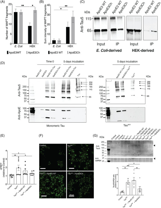
Results: We found enhanced binding of ApoE3Ch to tau and Dickkopf-1 (Dkk1, a WNT/β-catenin antagonist) that resulted in reduced tau aggregation in vitro. We demonstrated that ApoE3Ch interacts directly with Dkk1 and tau, reducing tau pathology. These findings supported the hypothesis of novel protective effects of direct ApoE3Ch interactions.

Highlights: ⁠Apolipoprotein E3 (ApoE3) Christchurch variant (ApoE3Ch) exhibits different protein interaction profiles compared to wild-type ApoE3, as revealed by proteomic analyses and pull-down experiments. The ApoE3Ch variant alters the protein's interaction with tau, thus affecting its aggregation in a tau biosensor cell assay and the retina of microtubule-associated protein tau (MAPT*P301S) transgenic mice. ⁠Gene ontology and pathway analyses indicate that ApoE3Ch interactors are associated with brain-related disorders and specific upstream regulators, including MAPT, a gene encoding for tau. ⁠Protein-protein interaction studies showed increased binding of ApoE3Ch to Dickkopf1 (Dkk1), a Wnt/β-catenin pathway antagonist, as compared to ApoE3WT, thus indicating that multiple protective mechanisms are regulated by the ApoE3Ch variant Our study uncovers a novel protective effect of the ApoE3Ch variant against tau pathology, thus proposing new insights into Alzheimer's disease mechanisms and potential therapeutic targets.

Keywords: Alzheimer's disease; ApoE3 Christchurch; Dkk1; apolipoprotein E; oligomerization; tau.


Conflict of interest statement

Dr. J. Arboleda‐Velasquez is listed as co‐inventor on a patent to leverage therapeutics based on the ApoE3 Christchurch findings filed by Mass General Brigham. Drs. Arboleda‐Velasquez and Kim are co‐founders of Epoch Biotech, a company focused on developing therapeutics inspired by the Christchurch variant. All other authors have no conflicts to disclose. Author disclosures are available in the Supporting Information.

Figures

FIGURE 1
FIGURE 1
Proteomic analyses of ApoE3 variants. (A) Experimental design of the ApoE co‐immunoprecipitation experiments. (B) Representative silver stain of the IP fractions using recombinant ApoE3 WT and ApoE3Ch proteins from E. coli (left blot) or HEK‐derived (right blot). (C). Number of interactor proteins identified by mass spectrometry (n = 3, independent pull‐down experiments). (D) Pie charts summarizing the number of unique and shared proteins binding to ApoE3 WT and ApoE3Ch. ApoE3, apolipoprotein E3; ApoE3Ch, apolipoprotein E3 Christchurch variant; HEK, human embryonic kidney; IP, immunoprecipitated; WT, wild‐type.
FIGURE 2
FIGURE 2
GO analyzes of interacting partners of ApoE3 WT and ApoE3Ch from different sources. (A) Pathway enrichment analysis based on the fold change of the proteins binding both ApoE3 WT and ApoE3Ch, E. coli‐derived (upper graph) and HEK‐derived (bottom graph). (B) BP, CC, and MF ontologies of ApoE3 E. coli (upper graph) and HEK‐derived ApoE3 binding partners (bottom). (C) Volcano plots showing log2 ratios of interactor protein binding (ApoE3Ch/ApoE3 WT). Significant interactors are shown in red. ApoE3, apolipoprotein E3; ApoE3Ch, apolipoprotein E3 Christchurch variant; BP, biological process; CC, cellular component; GO, gene ontology; HEK, human embryonic kidney; MF, molecular function; WT, wild‐type.
FIGURE 3
FIGURE 3
Ingenuity pathway analysis. (A) Comparative analysis of disease and biological function in ApoE3Ch versus ApoE3 WT interactors highlighting brain‐related disorders. (B) Comparison analysis of upstream regulators in ApoE3Ch versus ApoE3 WT interactors. (C) Pathway diagram with MAPT as the top upstream regulator showing indirect link to PSEN1, PSEN2, and APP, which are in turn linked to CTNNB1 and TP53. APP, amyloid precursor protein; ApoE3, apolipoprotein E3; ApoE3Ch, apolipoprotein E3 Christchurch variant; MAPT, microtubule‐associated protein tau; PSEN, presenilin; WT, wild‐type.
FIGURE 4
FIGURE 4
ApoE3 WT and ApoE3Ch differentially interacts with tau. (A, B) Number of MAPT fragments and sum intensity obtained via mass spectrometry analysis (*≤ 0.05, **≤ 0.01, Two‐way ANOVA, Fisher's LSD post‐hoc test, n = 3). (C) Co‐immunoprecipitation of human tau recombinant protein using His‐tag ApoE3 WT and ApoE3Ch proteins and detected via WB using anti‐tau antibody. (D) Representative Western blot of ApoE3 WT and ApoE3Ch after incubation with monomeric recombinant human tau or tau PFFs, arrows point the different tau aggregate bands. (E) Levels of FRET signal in tau RD P301S FRET Biosensor cells stimulated with tau PFF and treated with APOE3 variants (*≤ 0.05, **p ≤ 0.01, one‐way ANOVA, Fisher's LSD post‐hoc test, n = 6). (F) Scale bar: 100 µm. Representative maximum intensity projections of the live imaging acquisition of the tau aggregates in the tau RD P301S FRET Biosensor cells stimulated with tau PFFs and treated with the ApoE3 variants. (G) WB of tau aggregates in tau RD P301S FRET Biosensor cells treated with ApoE3 variants and ratio quantification between the total tau aggregates at ∼ 150 kDa of molecular weight and GAPDH, quantified bands are indicated by an arrowhead. (*p ≤ 0.05, **≤ 0.01, ***p ≤ 0.001, one‐way ANOVA, Fisher's LSD post‐hoc test, n = 6). ANOVA, analysis of variance; ApoE3, apolipoprotein E3; ApoE3Ch, apolipoprotein E3 Christchurch variant; GAPDH, glyceraldehyde‐3‐phosphate dehydrogenase; LSD, least significant difference; PFF, preformed protofibrils; WB, Western blotting; WT, wild‐type.
FIGURE 5
FIGURE 5
Effect of Christchurch variant on ApoE‐induced tau pathology in the mouse retina. (A) Representative immunofluorescence of subretinally injected MAPT P301S tau transgenic mice with recombinant ApoE3 and ApoE3Ch variants, as compared to vehicle, showing reduced levels of AT8‐positive cells in the presence of the Ch variant. (B) Relative AT8+ tau levels in dissected retina isolated from mice treated with either vehicle, ApoE3, or ApoE3Ch, showing that in the presence of ApoE3Ch, AT8+ cells are significantly reduced (**p < 0.005, One‐way ANOVA, n = 3‐4). (C) immunofluorescence staining of intravitreal injected 9 months old MAPT P301S tau mice with recombinant ApoE3 and ApoE3 Ch proteins, showing the reduced levels of ptau S396‐positive cells in the presence of the Ch variant. (D) Intravitreal injection of ApoE3 Ch in the retina significantly decreased phosphorylation of tau (* = p < 0.05, one‐way ANOVA, n = 2–5). Vehicle treated wild‐type mice were used as control for MAPT P301S mice (PS19). For both panels A and C, nuclei are stained with DAPI in blue, vascular formation is stained with Isolectin (IB4) in green and tau phosphorylation are stained with AT8 anti tau (A) or ptau S396 antibody (C) in red. Scale bar = 50 µm. ANOVA, analysis of variance; ApoE3, apolipoprotein E3; ApoE3Ch, apolipoprotein E3 Christchurch variant; DAPI, 4′,6‐diamidino‐2‐phenylindole.

References

    1. Alzheimer's Association . 2019 Alzheimer's disease facts and figures. Alzheimer's Association; 2019.
    1. Bekris LM, Yu CE, Bird TD, Tsuang DW. Genetics of Alzheimer disease. J Geriatr Psychiatry Neurol. 2010;23(4):213‐227. - PMC - PubMed
    1. Lalli MA, Cox HC, Arcila ML, et al. Origin of the PSEN1 E280A mutation causing early‐onset Alzheimer's disease. Alzheimers Dement. 2014;10(5):S277‐S283.e10. - PMC - PubMed
    1. Acosta‐Baena N, Sepulveda‐Falla D, Lopera‐Gomez CM, et al. Pre‐dementia clinical stages in presenilin 1 E280A familial early‐onset Alzheimer's disease: a retrospective cohort study. Lancet Neurol. 2011;10(3):213‐220. - PubMed
    1. Arboleda‐Velasquez JF, Lopera F, O'Hare M, et al. Resistance to autosomal dominant Alzheimer's disease in an APOE3 Christchurch homozygote: a case report. Nat Med. 2019;25(11):1680‐1683. - PMC - PubMed

Substances