. 2025 Aug 19;99(8):e0041425.
doi: 10.1128/jvi.00414-25. Epub 2025 Jul 10.

The HCMV tegument protein UL88 degrades MyD88 and reduces innate immune activation

Affiliations

The HCMV tegument protein UL88 degrades MyD88 and reduces innate immune activation

Rinki Kumar et al. J Virol. .

Abstract

The human cytomegalovirus (HCMV) encodes the tegument protein UL88, which supports virus spread by mediating the degradation of the innate immune signaling adapter protein, myeloid differentiation primary response 88 (MyD88). MyD88 transduces signals in multiple innate immune pathways, including acting downstream of pattern recognition receptors and IL-1 cytokine family members. MyD88 is rapidly and robustly upregulated following exposure to HCMV, irrespective of viral gene expression and, even after infection, primarily within uninfected cells in a culture. However, UL88 was required to downregulate cellular MyD88 protein levels as HCMV spread through a culture. The N-terminal 181 amino acids of UL88 were required to associate with and downregulate MyD88 protein. MyD88 expression significantly suppressed virus spread by triggering the production of a heat-labile soluble factor. This factor was produced between ~3 and 6 days after initial infection and did not increase the expression of well-characterized interferon-stimulated genes (ISGs). Indeed, increased MyD88 expression downregulated the expression of almost all ISGs examined. UL88 overexpression suppressed IL-1β-induced NF-κB activation within a cell. UL88 also suppressed virus-induced translocation of NF-κB to the nucleus of uninfected neighboring cells in an infected monolayer. Furthermore, UL88 overexpression was required for effective HCMV spread following transfer of the virus from monocytes to a fibroblast monolayer. These data indicate that UL88 is a novel antagonist of the immune response that acts to enhance the natural spread of HCMV by targeting MyD88 and provides vital insight into the innate immune responses that can control HCMV spread.IMPORTANCEThe significant role of many viral genes encoded by HCMV that are not essential for replication in cell culture is often overlooked. Our study reveals the importance of UL88 for regulating the innate immune response by showing evidence for interaction with and downregulation of MyD88 protein. The UL88-dependent regulation of MyD88 is physiologically relevant, as infection is enhanced in the absence of MyD88, and spread from myeloid cells to fibroblasts is blunted in the absence of UL88. These results highlight yet another important interaction between HCMV and the immune system.

Keywords: HCMV; MyD88; UL88; immune activation.


Conflict of interest statement

The authors declare no conflict of interest.

Figures
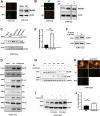
Fig 1
Fig 1
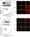
MyD88 levels during HCMV WT and UL88-STOP virus infection. (A) Protein lysates were harvested from fibroblasts either mock-infected or infected with TB40/E-mCh virus at an MOI of 0.05 at 3 dpi. Blots were probed for MyD88 and actin (loading control). Representative images of the infections show infected cells in red. (B) Protein lysates were harvested from fibroblasts either mock-infected or infected with untreated (UT) or UV-inactivated (UV) TB40/E-mCh or UL88-STOP-mCh virus at an MOI of 0.05 at 3 dpi. Blots were probed for MyD88 and actin (loading control). Representative images of the infections show infected cells in red. UT: untreated, UV: UV inactivated. (C) Protein lysates were harvested from fibroblasts infected with TB40/E WT, UL88-deficient (UL88-STOP and UL88-GalK), or UL88-Rev HCMV at an MOI of 3 at 96 hpi. Blots were probed for viral proteins UL88, IE1, and GAPDH as a loading control. (D) Protein lysates from fibroblasts, either mock-infected or infected with TB40/E WT or UL88-STOP HCMV at an MOI of 0.05, were harvested at 15 dpi. The lysates were analyzed by western blot (WB) and probed for MyD88, JAK1, NF-κB, IRF3, IRF1, EAA1, tubulin, and pp65 (viral protein). (E) The graph shows densitometry analysis from three independent experiments for each set at day 12 hpi. (F) Protein lysates were harvested from fibroblasts infected with either UL88-Rev or UL88-GalK virus at an MOI of 0.05 and harvested at d12 post-infection. Lysates were analyzed by western blot and probed for MyD88 and tubulin (loading control). (G) Protein lysates from fibroblasts, either mock-infected or infected with TB40/E WT or UL88-STOP TB40/E HCMV at an MOI of 0.05, were harvested every 3 days until 15 dpi. Blots were probed for MyD88, tubulin, and viral protein (pp65). (H) MRC-5 were either mock-infected or infected with untreated (UT) or UV-inactivated (UV) TB40/E-mCh or UL88-STOP-mCh virus at an MOI of 0.05. Representative images of the infections show infected cells in red at d12 post-infection. (I) Protein lysates from MRC-5 cells infected with TB40/E-mCh or UL88-STOP-mCh virus for 12 d (as pictured in H) were probed for MyD88 and actin (loading control). (J) RNA was harvested at 12 dpi from fibroblasts infected with TB40/E WT or UL88-STOP virus at an MOI of 0.05. MyD88 mRNA levels were analyzed by RT-qPCR and normalized to GAPDH. Fold changes are shown from three independent experiments. Results are shown as mean ± SD. *P < 0.05. Representative images of the infections show infected cells in red. UT: untreated, UV: UV inactivated.
Fig 2
Fig 2
Effect of downregulating MyD88 on HCMV infection. (A–C) Fibroblasts (MRC-5 or HDF) and ARPE-19 were transduced with lentivirus expressing UL88 or vector and subjected to puromycin selection. Cells were harvested for either WB or RNA analysis. (A) MRC-5 cells expressing either vector or UL88 were analyzed by western blot and probed for MyD88, UL88, and tubulin. The graph shows MyD88 protein levels quantified by densitometry analysis from four independent experiments. Results are shown as mean ± SD. *P < 0.05. (B) Cell lysates from HDFs expressing GFP vector, GFP-UL88 vector, or UL88 were analyzed by western blot and probed for MyD88 and tubulin. (C) Cell lysates from ARPE-19 cells expressing GFP vector or GFP-UL88 were analyzed by western blot and probed for GFP, UL88, MyD88, and p115 (loading control). (D) Cells expressing either GFP-vector alone or GFP-UL88 were stimulated with IL-1β for 6 hours. Cells were fixed in PFA and stained for p65-NF-κB (red), GFP (green), and DAPI (blue). Cells were imaged on the confocal. (E) At least 10 different fields were imaged for each sample. The number of nuclei and NF-κB-positive nuclei was counted. The percentage of p65-NF-κB-positive nuclei is graphed.
Fig 3
Fig 3
MyD88 is degraded by the N terminus of UL88 via the lysosomal pathway. (A) MRC-5 cells expressing either vector or UL88 were treated with DMSO, MG132 (10 µM), or Chloroquine (50 µM). Lysates were analyzed by western blotting. Blots were probed for UL88, MyD88, and p115 (loading control). Representative blots are shown. (B) Graph shows quantitation from three independent experiments as described in A. (C) 293 cells were co-transfected with HA-MyD88 and either GFP vector, GFP-UL88 full length, GFP-UL88 C, or GFP-UL88 N. Lysates were harvested 48 h post-transfection and analyzed by western blotting. Blots were probed for HA, GFP, and p115 (loading control). (D) 293 cells co-transfected with MyD88-flag and GFP-UL88 full length, GFP-UL88 C, or GFP-UL88 N. Lysates were harvested at 48 h post-transfection and subjected to co-immunoprecipitation using magnetic anti-flag beads. Samples were analyzed by western blot and probed for GFP, flag, and p115 as a loading control. Arrows point to GFP-UL88, GFP-UL88 N, and Flag-MyD88 in the IP blot.
Fig 4
Fig 4
Lentivirus-mediated overexpression of MyD88 inhibits virus spread. (A) Lentivirus-mediated downregulation of MyD88. MRC-5 cells were transduced with control lentivirus or lentivirus encoding shRNA targeting MyD88 (shMyD88-1 and shMyD88-2) and selected using puromycin. Knockdown efficiency was assessed by western blot analysis. Blots were probed for MyD88 and p115 as loading controls. (B) The transduced and selected MRC-5 cells were infected with TB40/E-mCh virus at an MOI of 0.05, and the spread of the virus was monitored by confocal imaging. Representative images from days 3, 9, and 15 are shown. (C) The spread of infection was quantified as the area of fluorescence using the NIS Element software. (D) MRC-5 cells were transduced with lentivirus from control or two different cassettes encoding the MyD88 gene (MyD88-1 and MyD88-2) and subjected to puromycin selection. Cell lysates were harvested and run on a Western blot to analyze MyD88 levels. Tubulin served as a loading control. (E) The transduced and selected MRC-5 cells were infected with TB40/E-mCh virus at an MOI of 0.05, and the spread of the virus was monitored every 3 days by confocal imaging up to day 21. Representative images from days 3, 9, and 15 are shown. (F) The spread of infection was quantified as the area of fluorescence using the NIS Element software.
Fig 5
Fig 5
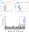
Effect of MyD88 overexpression on ISGs and spread of HCMV infection. (A, B) MRC-5 cells were transduced with lentivirus expressing a control or MyD88 gene and subjected to puromycin selection. Cells were either mock-infected or infected with TB40/E-mCh virus at an MOI of 0.05 and harvested at 7 dpi for RNA isolation, and samples were subjected to RT-qPCR analysis using the RT2 Profiler PCR array. (A) Volcano plot of the genes modulated in uninfected Myd88-transduced cells. The blue box represents genes that were downregulated, and the red box represents genes that were upregulated more than fourfold. (B) Volcano plot of the genes modulated in HCMV-infected MyD88-transduced cells. The blue box represents genes that were downregulated, and the red box represents genes that were upregulated more than fourfold. (C) The graph shows the representative ISGs that remained unchanged during HCMV infection in both conditions and ISGs that were significantly downregulated (red box) or upregulated by more than fourfold (blue box).
Fig 6
Fig 6
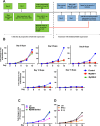
A MyD88-dependent heat-labile soluble factor reduces HCMV spread. (A) Schematic representation of the experiment methodology for this figure. (B) Supernatants from infected MRC-5 cells expressing either vector or MyD88 at an MOI of 0.05, with TB40/E-mCh, were collected every 3 days for 15 days and filtered through a 0.2 µm disc filter. The filtered supernatants from each day were then added to another monolayer of MRC-5 infected with TB40/E-mCh at an MOI of 0.05 every 3 days by removing the equivalent volume of the filtered supernatant from the well (therefore, 1:1 ratio with the media in the well). Cells were monitored for virus spread by imaging every 3 days. (C) Supernatants from infected MRC-5 cells expressing either vector or MyD88 were collected at 7 dpi and filtered through a 0.2 µm disc filter. They were then either untreated or denatured by boiling at 95°C before storage. The unboiled and boiled supernatants were added to another monolayer of MRC-5 infected with TB40/E-mCh at an MOI of 0.05 every 3 days by removing the equivalent volume of the filtered supernatant from the well (therefore a 1:1 ratio with the media in the well). The spread of the virus was monitored by imaging every 3 days. (D) A monolayer of MRC-5 infected with TB40/E-mCh at an MOI of 0.05 was treated with mock (UT), IFN-β (50 ng/mL), or IL1-β (100 ng/mL). The spread of the virus was monitored every 3 days by confocal imaging up to day 15. The spread of infection was quantified as the area of fluorescence using the NIS Element software. Results are shown as mean ± SD. *P < 0.05.
Fig 7
Fig 7
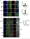
UL88 protein suppresses virus-induced NF-κB translocation. (A) MRC5 cells were plated on coverslips and infected with TB40/E-mCh, UL88-STOP-mCh, or UL88-Rev-mCh at an MOI of 0.05. Cells were fixed on day 7 and day 9 and subjected to IF analysis: DAPI (blue) and NF-κB (green), infection (red). NF-κB translocation into the nucleus was detected via staining, and cells with nuclear NF-κB signal were classified as “NF-κB-positive nuclei.” The percent of NF-κB-positive nuclei over total cells counted was calculated from at least 10 fields containing 100 cells each for each sample, as shown in the graph. (B) MRC5 cells expressing either GFP-vector or GFP-UL88 were plated on coverslips and infected with mCherry-tagged WT or UL88-STOP TB40/E virus at an MOI of 0.05. Cells were fixed on day 7 and day 9 and subjected to IF analysis: DAPI (blue), GFP (green), NF-κB (gray), and infection (red). NF-κB translocation into the nucleus was detected via staining, and cells with nuclear NF-κB signal were classified as “NF-κB-positive nuclei.” The percent of NF-κB-positive nuclei over total cells counted was calculated from at least 10 fields containing 100 cells each for each sample, as shown in the graph.
Fig 8
Fig 8
UL88 facilitates monocyte to fibroblast spread and downregulates HCMV-induced proinflammatory gene expression. (A) THP-1 cells were infected by spinoculation with an MOI of 20 with nonfluorescent TB40/E, UL88-STOP, or UL88-Rev, the latter two of which express GFP driven from the IE2 promoter or were left uninfected. GFP fluorescence was measured by flow cytometry at d5 post-infection. (B) Schematic representation of the experiment methodology for C and D. At day 3, 2.5 × 10⁵ THP-1, of which ~ 2.5 x 10⁴ were HCMV-infected, were transferred onto a monolayer of ~2.5 x 10⁵ HDFs, and virus spread was monitored every 3 days by confocal imaging up to day 15. (C) Representative images of TB40/E-GFP and UL88-STOP from day 3 and day 7 are shown. (D) The spread of infection was quantified as the area of fluorescence using the NIS Element software. The graph shows fold change in area of fluorescence for each virus. (E) MRC-5 cells were infected with TB40/E-mCh or UL88-STOP-mCh virus at an MOI of 0.05. Cells were harvested for RNA isolation at 7 dpi. Antiviral gene expression was profiled using a set of 84 ISGs by qPCR. Results are plotted as a volcano plot depicting the modulated genes compared between TB40/E WT HCMV and UL88-STOP HCMV infection. (F) ISGs known to be modulated in HCMV infection are shown with their relative fold change in mRNA levels (normalized to GAPDH) between TB40/E WT and UL88-STOP virus infection. The graph shows the genes that remained unchanged (green box), downregulated (red box), or upregulated (blue box) in TB40/E WT vs UL88-STOP HCMV-infected cells. Results are from biological triplicates.

References

    1. Adachi O, Kawai T, Takeda K, Matsumoto M, Tsutsui H, Sakagami M, Nakanishi K, Akira S. 1998. Targeted disruption of the MyD88 gene results in loss of IL-1- and IL-18-mediated function. Immunity 9:143–150. doi: 10.1016/s1074-7613(00)80596-8 - DOI - PubMed
    1. Deguine J, Barton GM. 2014. MyD88: a central player in innate immune signaling. F1000Prime Rep 6:97. doi: 10.12703/P6-97 - DOI - PMC - PubMed
    1. Griesenauer B, Paczesny S. 2017. The ST2/IL-33 axis in immune cells during inflammatory diseases. Front Immunol 8:475. doi: 10.3389/fimmu.2017.00475 - DOI - PMC - PubMed
    1. Li J, Lin S, Chen Q, Peng L, Zhai J, Liu Y, Yuan Z. 2010. Inhibition of hepatitis B virus replication by MyD88 involves accelerated degradation of pregenomic RNA and nuclear retention of pre-S/S RNAs. J Virol 84:6387–6399. doi: 10.1128/JVI.00236-10 - DOI - PMC - PubMed
    1. Murphy Schafer AR, Smith JL, Pryke KM, DeFilippis VR, Hirsch AJ. 2020. The E3 ubiquitin ligase SIAH1 targets MyD88 for proteasomal degradation during dengue virus infection. Front Microbiol 11:24. doi: 10.3389/fmicb.2020.00024 - DOI - PMC - PubMed

MeSH terms