. 2025 Jul 15;122(28):e2424666122.
doi: 10.1073/pnas.2424666122. Epub 2025 Jul 10.

A noncanonical cGAS-STING pathway drives cellular and organismal aging

Affiliations

A noncanonical cGAS-STING pathway drives cellular and organismal aging

Rafael Cancado de Faria et al. Proc Natl Acad Sci U S A. .

Abstract

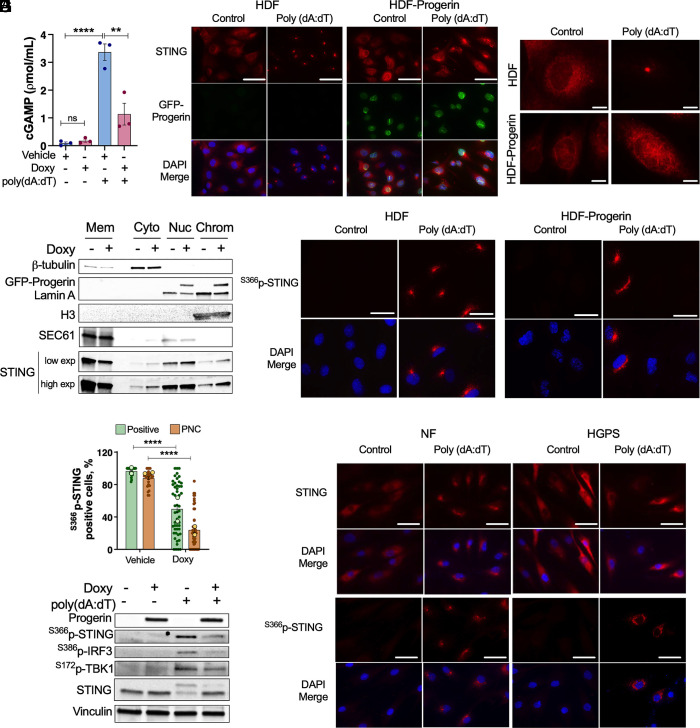
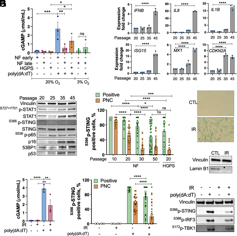
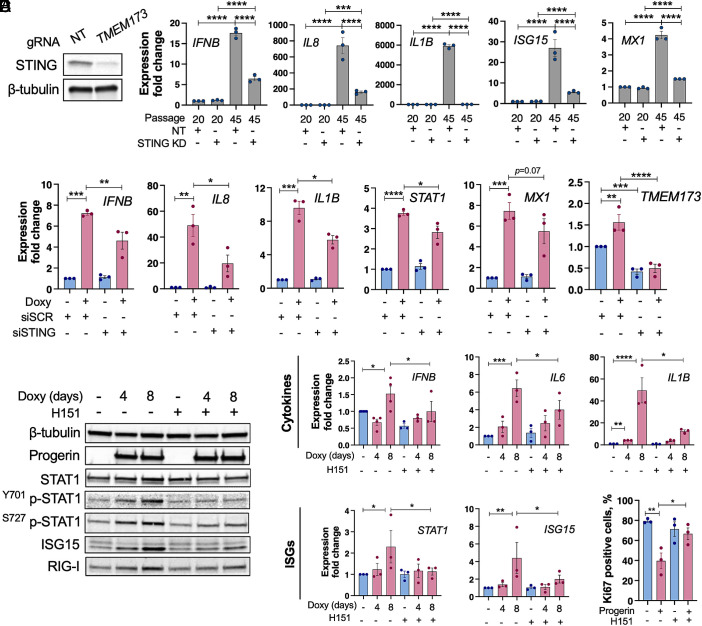
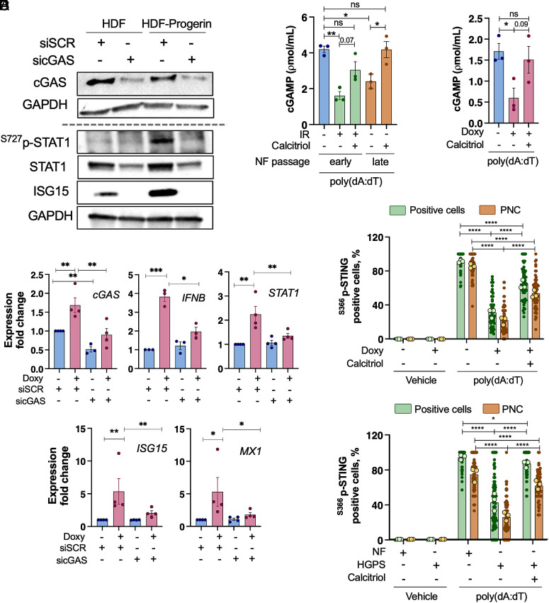
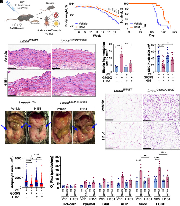
Accumulation of cytosolic DNA has emerged as a hallmark of aging, inducing sterile inflammation. Stimulator of interferon genes (STING) protein translates the sensing of cytosolic DNA by cyclic-GMP-AMP synthase (cGAS) into an inflammatory response. However, the molecular mechanisms whereby cytosolic DNA-induced cGAS-STING pathway leads to aging remain poorly understood. We show that STING does not follow the canonical pathway of activation in human fibroblasts passaged (aging) in culture, senescent fibroblasts, or progeria fibroblasts (from Hutchinson-Gilford progeria syndrome patients). Despite cytosolic DNA buildup, features of the canonical cGAS-STING pathway like increased cGAMP production, STING phosphorylation, and STING trafficking to perinuclear compartment are not observed in progeria/senescent/aging fibroblasts. Instead, STING localizes at endoplasmic reticulum, nuclear envelope, and chromatin. Despite the nonconventional STING behavior, aging/senescent/progeria cells activate inflammatory programs such as the senescence-associated secretory phenotype and the interferon response, in a cGAS and STING-dependent manner, revealing a noncanonical pathway in aging. Importantly, progeria/aging/senescent cells are hindered in their ability to activate the canonical cGAS-STING pathway with synthetic DNA, compared to young cells. This deficiency is rescued by activating vitamin D receptor signaling, unveiling mechanisms regulating the cGAS-STING pathway in aging. Significantly, in HGPS, inhibition of the noncanonical cGAS-STING pathway ameliorates cellular hallmarks of aging, reduces tissue degeneration, and extends the lifespan of progeria mice. Our study reveals that a new feature of aging is the progressively reduced ability to activate the canonical cGAS-STING pathway in response to cytosolic DNA, triggering instead a noncanonical pathway that drives senescence/aging phenotypes.

Keywords: aging; cGAS; cytosolic DNA; senescence-associated secretory phenotype; stimulator of interferon genes.


Conflict of interest statement

Competing interests statement:The authors declare no competing interest.

Figures

Fig. 1.
Fig. 1.
Progeria cells are impaired in the ability to activate the canonical cGAS–STING pathway. (A) HDF induced to express GFP-progerin (HDF-teton-GFP-progerin) with doxycycline (0.5 μg/mL) for 8 d (DMSO as vehicle). cGAMP synthesis measured by ELISA. cGAMP concentration was also assessed 4 h after transfection of 1 μg/mL poly(dA:dT). Data represent average ± SEM of three biological repeats. (B) IF with STING antibody (red) in the same cells as in (A). GFP-progerin is shown in green and DNA staining (DAPI) in blue. (Scale bar: 50 μM.) (C) Higher magnification images showing STING localization. (Scale bar: 10 μm.) (D) Subcellular fractionation to monitor STING presence at membranes, cytoplasm, nucleus, and chromatin fractions in HDF-teton-GFP-progerin cells. β-tubulin used as cytoplasm fraction control, SEC61 as ER marker, Lamin A as nuclear fraction control, and H3 as chromatin marker. Mem: membrane fraction; Cyto: cytoplasmic soluble fraction; Nuc: nuclear fraction including nuclear envelope; Chrom: chromatin fraction. (E) IF with S366p-STING antibody (red) and DAPI (blue) in HDF-teton-GFP-progerin. (Scale bar: 50 μm.) (F) Percentage of S366p-STING positive cells or cells with S366p-STING at the PNC. Mean of each of two independent experiments is shown (light circles), as well as mean ± SEM of all the image fields of both experiments (~25 image fields/experiment and >200 cells/experiment). Statistical significance via two-way ANOVA. (G) Immunoblot of HDF-teton-GFP-progerin transfected with poly(dA:dT) shows phosphorylation of STING, TBK1, and IRF3 in control HDF that is reduced upon progerin expression. Vinculin used as loading control. (H) IF showing STING and S366p-STING localization ± poly(dA:dT) stimulation in NF or HGPS fibroblasts. (Scale bar: 50 μm.) DAPI (blue) used for nuclear staining.
Fig. 2.
Fig. 2.
Canonical cGAS–STING pathway is hindered during passage of cells in culture and in DNA damage-induced senescent cells. (A) cGAMP production by ELISA in NF of early passage < 20 and late passage > 32, and HGPS patients (passage < 16). cGAMP also measured after transfection with 1 μg/mL poly(dA:dT) for 4 h, and in cells cultured under 20% or 3% oxygen. Data represent average ± SEM of three biological repeats. (B) Immunoblots of inflammation markers upregulated in NF during passage (STAT1, S727+Y701p-STAT1, STING, and S536p-p65), and other markers of senescence (p16, p53, and 53BP1). Not detected S366p-STING at any passage. Vinculin was loading control. (C) qRT-PCR of SASP transcripts IFNB, IL8, and IL1B; senescence marker CDKN2A; and ISGs (ISG15 and MX1). Graphs show expression fold change of triplicates from a representative experiment from at least two biological repeats. (D) Percentage of S366p-STING positive cells or cells with S366p-STING at PNC, calculated as in Fig. 1F. Mean of each of experiment (light circles) and average ± SEM of all the image fields of both experiments (>200 cells/experiment). Statistical significance by two-way ANOVA. (E) NF early passage (P20) irradiated (20 Gy) and cultured for 3 wk. β-galactosidase activity shows positivity in growth-arrested irradiated cells. (F) Lamin B1 immunoblot from the same cells as in (E). Vinculin was loading control. (G) cGAMP levels by ELISA. NF (P20) and senescent NF (P20+IR+3 wk) were transfected with poly(dA:dT) to activate the canonical cGAS–STING pathway. The graph shows mean ± SEM of three biological repeats. (H) Percentage of S366p-STING positive cells or cells with S366p-STING at the PNC, in same cells as in (E). Average of two biological repeats (n > 150 cells/experiment). Statistical analysis via two-way ANOVA. (I) Immunoblot of NF early passage control and senescent (3 wk after IR) that were transfected with poly(dA:dT); phosphorylation of STING, TBK1, and IRF3 was monitored. Vinculin was loading control.
Fig. 3.
Fig. 3.
STING is required for SASP and IFN response in aging/progeria cells. (A) Immunoblots show depletion of STING in NF using gRNA/CRISPR/Cas9. Graphs show transcript levels of inflammatory cytokines (IFNB, IL8, IL1B) and ISGs (ISG15, MX1) upon STING depletion in NF of early (P20) and late passage (P45). One representative experiment out of two independent biological repeats shown. Note how the increase in all inflammatory markers in late passage NF is reduced by STING depletion. (B) HDF-teton-GFP-progerin were transfected with siRNA targeting STING, and inflammatory cytokines (IFNB, IL8, IL1B) and ISGs (STAT1, MX1) monitored by qRT-PCR. Note how the increased expression of inflammatory genes is ameliorated by STING depletion. (C) HDF-teton-GFP-progerin induced to express progerin for 4 to 8 d were treated with vehicle (DMSO) or H151 (STING inhibitor) during the 4 to 8 d (0.5 μM H151). Representative blots from three independent experiments showing markers of sterile inflammation/IFN response: STAT1, p-STAT1, RIG-I, and ISG15. β-tubulin used as loading control. (D and E) qRT-PCR of inflammatory cytokines (IFNB, IL6, and IL1B) and ISGs (STAT1 and MX1) in HDF-teton-GFP-progerin expressing progerin for 4 to 8 d and treated with H151 or vehicle. Graph shows average ± SEM of three independent experiments. (F) Percentage of Ki67 positive cells (by IF) for proliferative capacity assessment in HDF-teton-GFP-progerin cells. The graph shows average ± SEM of three independent experiments.
Fig. 4.
Fig. 4.
cGAS and calcitriol regulate the canonical and noncanonical cGAS–STING pathways. (A) HDF-teton-GFP-progerin expressing progerin for 8 d (doxy) were transfected with siRNA targeting cGAS or siRNA control. Immunoblots show a decrease in cGAS levels. Cell lysis protocol was modified to detect cGAS (Materials and Methods). Immunoblots also show decreased STAT1, S727p-STAT1, and ISG15. (B) qRT-PCR in the same cells as in (A) to monitor cGAS depletion and expression of inflammatory markers (IFNB, STAT1, ISG15, and MX1). (C) cGAMP levels by ELISA in IR-induced senescent cells, and in early and late passage NF transfected with 1 μg/mL poly(dA:dT). Cells were incubated with calcitriol (100 nM) where indicated. Data represent average ± SEM of three biological repeats. (D) HDF-teton-GFP-progerin expressing progerin for 8 d (and control) were incubated with calcitriol (100 nM) or vehicle and transfected with poly(dA:dT). cGAMP was measured by ELISA. Data represent average ± SEM of three biological repeats. (E) Percentage of cells positive for S366p-STING and percentage of S366p-STING at the PNC in HDF and (F) NF and HGPS fibroblasts. The mean of each of three biological repeats is shown. In each experiment, ~25 image fields were analyzed (n > 200). The average ± SEM of all fields in three experiments is represented. Statistical significance determined by two-way ANOVA.
Fig. 5.
Fig. 5.
Pharmacological inhibition of STING improves health and lifespan of progeria mice. (A) Schematic of the treatment of LmnaG609G/G609G (G609G) and Lmna+/+ (WT; wild-type) with H151 used for lifespan studies or euthanized (according to Institutional Animal Care and Use Committee guidelines) at 90 d of age for tissue analysis. Female and male mice were treated, and aorta and WAT were analyzed. (B) Body weight of mixed male and female G609G mice treated with H151 (IP 50 μM/Kg, n = 13) or Vehicle (n = 16) and fed a standard chow diet. Body weight was monitored three times per week. (C) Kaplan–Meier survival curves of mixed male and female G609G mice fed chow diet and treated with H151 (n = 13) or vehicle (n = 16). Median survival of chow-fed G609G mice is 120.5 d for vehicle-treated mice and 152 d for H151-treated mice (26% improvement). (D) H&E staining of aortic arch from chow fed G609G mice treated with H151 or vehicle (90 d of age). (Scale bar: 50 μm.) (E) Quantification of elastin fragmentation frequency per 10 mm2 (n = 3 mouse aortas per group). (F) Quantification of SMC nuclei per 500 μm2 in six different areas of each independent mouse (n = 3 mouse aortas per group). (G) Pictures show WT and G609G mice treated with vehicle or H151 and fed a standard chow diet (90 d of age). Blue arrows highlight the presence of epididymal fat in each mouse. (H) Histology of epididymal WAT stained with H&E (n = 3 mouse eWAT per group). (Scale bar: 100 μm.) (I) Quantification of adipocyte size in μm2 of approximately 500 adipocytes per sample (n = 3 mice). (J) Mitochondrial respiration assessment in eWAT from G609G and WT mice treated with vehicle or H151 using Oroboros instrument. Graphs show average ± SEM of oxygen flux after additions of octanoyl-l-carnitine (Oct-carn); pyruvate and malate (Pyr/Mal); glutamate (Glut); adenosine diphosphate (ADP), succinate (Succ), and FCCP (n = 4-5).

Update of

Similar articles

References

    1. Ivanov A., et al. , Lysosome-mediated processing of chromatin in senescence. J. Cell Biol. 202, 129–143 (2013). - PMC - PubMed
    1. Dou Z., et al. , Cytoplasmic chromatin triggers inflammation in senescence and cancer. Nature 550, 402–406 (2017). - PMC - PubMed
    1. Harding S. M., et al. , Mitotic progression following DNA damage enables pattern recognition within micronuclei. Nature 548, 466–470 (2017). - PMC - PubMed
    1. Mackenzie K. J., et al. , cGAS surveillance of micronuclei links genome instability to innate immunity. Nature 548, 461–465 (2017). - PMC - PubMed
    1. Takahashi A., et al. , Downregulation of cytoplasmic DNases is implicated in cytoplasmic DNA accumulation and SASP in senescent cells. Nat. Commun. 9, 1249 (2018). - PMC - PubMed

LinkOut - more resources