. 2025 Oct 16;66(4):2402250.
doi: 10.1183/13993003.02250-2024. Print 2025 Oct.

Pharmacological inhibition of Epac1 protects against pulmonary fibrosis by blocking FoxO3a neddylation

Affiliations

Pharmacological inhibition of Epac1 protects against pulmonary fibrosis by blocking FoxO3a neddylation

Katherine Jankowski et al. Eur Respir J. .

Abstract

Background: Idiopathic pulmonary fibrosis (IPF) is marked by progressive lung scarring with no existing cure, emphasising the need for new therapeutic targets. Current evidence suggests that cyclic adenosine monophosphate (cAMP) mitigates lung fibroblast proliferation via the protein kinase A pathway, but the impact of exchange proteins directly activated by cAMP 1 (Epac1) on IPF remains unexplored.

Objective: To investigate the role of Epac1 in IPF progression.

Methods: We examined lung samples from IPF patients and controls, and from a bleomycin-induced mouse model of pulmonary fibrosis. The effects of Epac were analysed in knockout mice and through modulation using viral vectors. The Epac1-specific small compound inhibitor AM-001 was evaluated in vitro using lung fibroblasts from patients with IPF, in vivo in bleomycin mice and ex vivo in IPF precision-cut lung slices.

Results: Increased Epac1 expression was observed in lung tissues from IPF patients, fibrotic fibroblasts and bleomycin-challenged mice. Genetic or pharmacological inhibition of Epac1 with AM-001 decreased proliferation in normal and IPF fibroblasts, and reduced expression of profibrotic markers such as α-smooth muscle actin, transforming growth factor-β/SMAD family member 2/3, and interleukin-6/signal transducer and activator of transcription 3 pathways. Epac1-specific inhibition consistently protected against bleomycin-induced lung injury and fibrosis, suggesting significant therapeutic potential. Global gene expression profiling indicated a reduced profibrotic gene signature and neddylation pathway components in Epac1-deficient fibroblasts and human-derived lung cells. Mechanistically, the protective effects may involve inhibiting the neddylation pathway and preventing neural precursor cell expressed, developmentally downregulated 8 (NEDD8) activation, which in turn reduces the degradation of forkhead box protein O3 by NEDD8. Additionally, these effects may be enhanced while also limiting the proliferation of lung-infiltrating monocytes.

Conclusions: Our findings demonstrate that Epac1 regulates fibroblast activity in pulmonary fibrosis, and that targeting Epac1 with the pharmacological specific inhibitor AM-001 offers a promising therapeutic approach for treating IPF disease.


Conflict of interest statement

Conflict of interest: F. Lezoualc'h is a co-founder and scientific advisor of Revadix SAS. All the other authors declare no affiliations with or involvement in any organisation or entity with any financial interest in the subject matter or materials discussed in this manuscript.

Figures

None
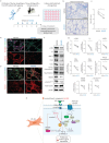
Mechanism of action and therapeutic impact on idiopathic pulmonary fibrosis (IPF) of exchange factor directly activated by cAMP 1 (Epac1) inhibition by AM-001. cAMP: cyclic adenosine monophosphate; FoxO3: forkhead box protein O3; IL-6: interleukin 6; NEDD8: neural precursor cell expressed, developmentally downregulated 8; P: phosphorylated; TGF-β: transforming growth factor β; STAT3: signal transducer and activator of transcription 3; Ub: ubiquinated.
FIGURE 1
FIGURE 1
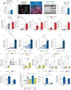
Exchange factor directly activated by cAMP 1 (Epac1) expression in idiopathic pulmonary fibrosis (IPF) and fibrosis-related genes and proliferation in lung fibroblasts (FBs). a) Epac1 mRNA levels were assessed in lung tissues from IPF patients (n=8) and healthy donors’ lungs (n=8) using quantitative reverse-transcriptase PCR (qRT-PCR). b) Representative immunofluorescence staining for Epac1 (red) and α-smooth muscle actin (α-SMA) (green) in lung tissues from healthy donors and IPF patients. Nuclei were counterstained with DAPI (blue). Scale bar: 100 μm. c) Representative immunoblot (left panel) and quantification (right panel) of Epac1 expression, normalised to GAPDH, in isolated lung FBs from IPF patients (n=3) and from normal human lung (NHL) (n=3). d) Proliferation was measured in media supplemented with low serum (0.1% S) or high serum (10% S) concentrations using a BrdU assay for 72 h in NHL-FBs overexpressing adenovirus (Ad).Epac1 and Ad.GFP (Ad.CT) (n=4). e) Transforming growth factor β1 (TGF-β1) mRNA expression in NHL-FBs infected with Ad.CT or Ad.Epac1 alone or co-treated with TGF-β1 (5 ng·mL−1) for 48 h (n=3). f) mRNA expression levels of profibrotic markers collagen type 1 ɑ1 (COL1A1), collagen type 3 ɑ1 (COL3A1) and connective tissue growth factor (CTGF) in NHL-FBs overexpressing either Ad.CT or Ad.Epac1 with or without TGF-β1 (5 ng·mL−1) for 48 h (n=3). g) mRNA expression levels of TGF-β1 and profibrotic markers (COL1A1, COL3A1, CTGF) in NHL-FBs co-treated with Sp-8-(4-chlorophenylthio)-2′-O-methyl-cAMP (8-CPT) (5 ng·mL−1), an Epac1 activator, and TGF-β1 (5 ng·mL−1) for 48 h (n=4). h) Epac1 mRNA (left panel) and protein expression (right panel) were measured by qRT-PCR and Western blot, respectively, in NHL-FBs overexpressing either a non-silencing short hairpin RNA (shNS) or a specific short hairpin RNA against Epac1 (shEpac1) (n=3). i–j) Analysis of TGF-β1, COL1A1, COL3A1 and CTGF mRNA expression in NHL-FBs overexpressing either shNS or shEpac1 was treated with vehicle or TGF-β1 for 24 h (n=3). k) Epac1 mRNA expression in NHL-FBs or IPF-FBs overexpressing shNS or shEpac1 (n=3). l) NHL-FB and IPF-FB proliferation were assessed using a BrdU assay under the specified experimental conditions (n=6). m, n) α-SMA and IL-6 mRNA expression levels in NHL-FBs and IPF-FBs overexpressing shNS or shEpac1 (n=3). The data are presented as mean±sem. *: p<0.05; **: p<0.01; ***: p<0.001; ****: p<0.0001.
FIGURE 2
FIGURE 2
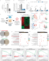
Pharmacological inhibition of exchange factor directly activated by cAMP 1 (Epac1) by AM-001. a) Epac1 activity using the bioluminescence resonance energy transfer (BRET) CAMYEL sensor (cAMP sensor using YFP-Epac-RLuc) in idiopathic pulmonary fibrosis (IPF) fibroblasts (FBs) stimulated with cAMP alone or in combination with AM-001 (n=4). b) Normal human lung (NHL) FB proliferation when treated with transforming growth factor β1 (TGF-β1) (5 ng·mL−1) alone or in combination with AM-001 (20 μM) in media supplemented with low serum (0.1% S) or high serum (5% S) concentrations, and cell proliferation was measured using a BrdU proliferation assay (n=4). c) mRNA expression levels of fibrosis markers TGF-β1, collagen type 1 ɑ1 (COL1A1), collagen type 3 ɑ1 (COL3A1) and connective tissue growth factor (CTGF) in NHL-FBs pretreated with AM-001 (20 μM) or DMSO (as vehicle control) for 24 h, followed by activation by TGF-β1 (5 ng·mL−1) for an additional 24 h (n=3). Quantification was performed using quantitative reverse-transcriptase PCR. d) Experimental design for RNA-sequencing (RNA-seq): NHL-FBs were assigned to three groups: 1) Control group: stimulated with TGF-β1 for 48 h and co-treated with vehicle; 2) AM-001 treatment group: stimulated with TGF-β1 for 48 h in the presence of the Epac1-selective inhibitor AM-001; and 3) Epac1 knockdown group: infected with a specific short hairpin RNA (shRNA) targeting Epac1 (shEpac1) for 72 h, followed by TGF-β1 stimulation for 48 h. e) Heatmap, principal component (PC) analysis and volcano plot displaying genes that were either upregulated or downregulated in response to shEpac1 (upper panel) or AM-001 (lower panel) compared to that in control cells (n=3 per group). f) Venn diagrams showing the overlap of genes commonly downregulated and upregulated by AM-001 and shEpac1 treatment. The intersecting areas highlight the genes affected by both treatments. g, h) Reactome and Kyoto Encyclopedia of Genes and Genomes (KEGG) pathway enrichment analysis for genes significantly differentially expressed in response to TGF-β1-stimulated NHL-FBs treated with AM-001 and in Epac1-depleted NHL-FBs+TGF-β1. i) An enrichment plot was generated through gene set enrichment analysis, focusing on Reactome pathways related to collagen formation, collagen biosynthesis and modifying enzymes, collagen chain trimerisation, and FOXO-mediated transcription in indicated conditions. The data are presented as mean±sem. ns: nonsignificant; padj: adjusted p-value; var.: variance. *: p<0.05; **: p<0.01; ***: p<0.001; ****: p<0.0001.
FIGURE 3
FIGURE 3
Effects of pharmacological inhibition of exchange factor directly activated by cAMP 1 (Epac1) in idiopathic pulmonary fibrosis (IPF) fibroblasts (FBs) on the signal transducer and activator of transcription 3 (STAT3), phosphoinositide 3-kinase (PI3K)/protein kinase B (AKT) and SMAD2/3 pathways. a) Epac1 mRNA levels in normal human lung (NHL) FBs and IPF-FBs treated for 48 h with either transforming growth factor β1 (TGF-β1) (5 ng·mL−1) or SB431542 (10 µM), a potent and selective inhibitor of the TGF-β type I receptor (n=3). b) Proliferation was assessed by BrdU labelling in both NHL-FBs and IPF-FBs after treatment with AM-001 (20 μM) in the presence of low serum (0.1% S) or high serum (10% S) medium concentrations for 72 h (n=6). c–e) mRNA expression levels of TGF-β1 (c, n=3), α-smooth muscle actin (α-SMA) (d, n=6) and interleukin 6 (IL-6) (e, n=6) by quantitative reverse-transcriptase PCR in nonactivated NHL-FBs and IPF-FBs treated with either vehicle or AM-001 (20 μM) for 48 h. f) Representative immunoblot analysis of phosphorylated STAT3 (p-STAT3) and total STAT3 levels in both NHL-FBs and IPF-FBs in the presence or absence of AM-001 (20 μM). The left panel shows a representative immunoblot, and the right panel shows the p-STAT3/total STAT3 ratio quantification (n=3). g) Representative immunoblot analysis of phosphorylated SMAD2/3 (p-SMAD2/3), total SMAD2/3, phosphorylated AKT (p-AKT), total AKT and GAPDH (as loading controls) in NHL-FBs and IPF-FBs treated with vehicle or AM-001 (20 μM) (n=3–4). The left panel shows representative immunoblots and the right panels shows the quantification of the SMAD signalling pathway (p-SMAD2/3/total SMAD1) and the PI3K/AKT signalling pathway (p-AKT/total AKT). The data are presented as mean±sem. ns: nonsignificant. *: p<0.05; **: p<0.01; ***: p<0.001.
FIGURE 4
FIGURE 4
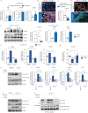
Molecular responses to AM-001 and neddylation inhibition in lung fibroblasts (FBs). a) The transcript levels of neural precursor cell expressed, developmentally downregulated 8 (NEDD8)-activating enzyme E1 (NAE1), ubiquitin conjugating enzyme E2 M (UBE2M) and NEDD8 in lung tissue samples from healthy donors (n=7) and idiopathic pulmonary fibrosis (IPF) patients (n=7) were assessed by quantitative reverse-transcriptase PCR (qRT-PCR). b) Co-immunostaining of NEDD8 (red) and α-smooth muscle actin (α-SMA) (green) in lung tissue sections from both healthy donors and IPF patients. Nuclei were counterstained with DAPI (blue). Scale bars: 100 μm. c) Forkhead box protein O3 (FoxO3a) mRNA levels (n=8). d) Representative images of co-immunostaining of FoxO3a (red) with α-SMA (green) in lung tissue sections from healthy donors and IPF patients. Nuclei were counterstained with DAPI (blue). Scale bars: 100 μm. e) Representative immunoblot analysis of FoxO3a, UBE2M and NEDD8 in normal human lung (NHL) FBs and IPF-FBs (left panel). Right panels show the ratio quantification using GAPDH as the loading control (n=3). f) Transcript levels of NAE1, NEDD8 and UBE2M in NHL-FBs and IPF-FBs after 48 h of treatment with either vehicle or AM-001 (20 μM) for 48 h (n=3). g) FoxO3a mRNA expression in NHL-FBs and IPF-FBs after 48 h of treatment with either vehicle or AM-001 (20 μM) for 48 h (n=3). h) Representative immunoblot analysis showing the levels of phosphorylated FoxO3a at Thr32 (p-FoxO3aThr32), total FoxO3a and NEDD8 under the indicated conditions. GAPDH was used as a loading control. i) mRNA expression levels of fibrosis markers transforming growth factor β1 (TGF-β1), collagen type 1 ɑ1 (COL1A1), collagen type 3 ɑ1 (COL3A1) and connective tissue growth factor (CTGF) were measured by qRT-PCR in NHL-FBs and IPF-FBs treated with MLN4924 (5 µM) alone, a small molecule inhibitor of NAE, or in combination with AM-001 (20 μM) for 48 h (n=3). j) Representative immunoblot analysis of p-FoxO3aThr32, total FoxO3a and NEDD8 protein levels in NHL-FBs or IPF-FBs treated with vehicle, MLN4924 alone or in combination with AM-001 for 48 h. k) Immunoprecipitation of haemagglutinin (HA)-tagged NEDD8 (HA-NEDD8) followed by immunoblot analysis of NEDD8 and total FoxO3a in IPF-FBs transfected with HA-NEDD8, in IPF-FBs pretreated with proteasome inhibitor MG132 (10 µM) to inhibit protein degradation, and under the indicated conditions. Data are presented as mean±sem. 8-CPT: Sp-8-(4-chlorophenylthio)-2′-O-methyl-cAMP; IB: immunoblot; IgG: immunoglobulin G; ns: nonsignificant. *: p<0.05; **: p<0.01; ***: p<0.001; ****: p<0.0001.
FIGURE 5
FIGURE 5
Exchange factor directly activated by cAMP 1 (Epac1) deficiency prevents bleomycin (BLM)-induced lung fibrosis. a) Schematic of the experimental design for the induction of BLM-induced lung fibrosis. Wild-type (WT) littermates and Epac1 knockout (KO) mice (Epac1−/−) were subjected to a single intratracheal (IT) aerosolised delivery of BLM at a dose of 4 U·kg−1, while the sham control group received saline as a vehicle. The animals were sacrificed 28 days after BLM administration. b) Epac1 and Epac2 mRNA levels, and Epac1 protein levels in WT and Epac1 KO mice were assessed by quantitative reverse-transcriptase PCR (qRT-PCR) and Western blotting (n=5). c) Representative images of Masson's trichrome staining to visualise fibrotic lesion changes of lung sections from WT and Epac1 KO mice challenged with BLM (left panel). Scale bar: 100 μm. The right panel shows the quantification of the fibrotic area, expressed as the fold change relative to the saline-treated WT group (n=6–8). d) The Ashcroft scoring system was used to assess the severity of lung fibrosis in Epac1 KO relative to WT mice challenged with saline (n=6–8). e, f) mRNA expression levels of profibrotic markers transforming growth factor β1 (TGF-β1), collagen type 1 ɑ1 (COL1A1), collagen type 3 ɑ1 (COL3A1) and connective tissue growth factor (CTGF) were measured by qRT-PCR in lung tissues from WT and Epac1 KO mice treated with saline (vehicle) or BLM (n=5–7). g) Neural precursor cell expressed, developmentally downregulated 8 (NEDD8) mRNA expression in lung tissue was measured by qRT-PCR (n=5–6). h) Co-immunostaining for NEDD8 (red) and α-SMA (green) in lung sections from WT and Epac1 KO mice challenged with BLM. Nuclei were counterstained with DAPI (blue). Scale bar: 100 μm. i) Forkhead box protein O3 (FoxO3a) mRNA expression by qRT-PCR in lung tissue from WT and Epac1 KO mice challenged with vehicle or BLM (n=5–7). j) Co-immunostaining for FoxO3a (red) and α-SMA (green) in lung sections from WT and Epac1 KO mice challenged with BLM. Nuclei were counterstained with DAPI (blue). Scale bars: 100 μm. k, l) Representative immunoblot analysis (k) and quantification (l) of Epac1, NEDD8, phophorylated FoxO3a (p-FoxO3a), total FoxO3a, phosphorylated SMAD2/3 (p-SMAD2/3) and total SMAD2/3 protein expression in saline-treated WT and Epac1 KO mice or challenged with BLM (n=3). β-actin was used as a loading control. The data are presented as mean±sem. ns: nonsignificant. *: p<0.05; **: p<0.01; ***: p<0.001; ****: p<0.0001.
FIGURE 6
FIGURE 6
Pharmacological inhibition of exchange factor directly activated by cAMP 1 (Epac1) by AM-001 attenuates bleomycin (BLM)-induced pulmonary fibrosis (PF). a) Schematic outlining the experimental design. C57BL/6 mice received a single intratracheal (IT) delivery of BLM (4 U·kg−1) for 28 days (∼4 weeks). AM-001 was then administered via intraperitoneal (IP) injection every other day at a dose of 10 mg·kg−1 for an additional 2 weeks. b) Representative images of Masson's trichrome staining (left panel) of lung sections from control saline-treated group and BLM-challenged mice treated with either vehicle or AM-001. The right panel shows the quantification of the fibrotic area, expressed as the fold change relative to the saline control group (n=5–7). c) Lung hydroxyproline content, an index of collagen deposition, was measured in saline-treated control mice and BLM-challenged mice administered either vehicle or AM-001 (n=5–7). d) mRNA expression levels of profibrotic markers transforming growth factor β1 (TGF-β1), collagen type 1 ɑ1 (COL1A1), collagen type 3 ɑ1 (COL3A1) and connective tissue growth factor (CTGF) in lung tissues were measured by quantitative reverse-transcriptase PCR (qRT-PCR) in lung tissue from the control saline-treated group and BLM-challenged mice treated with either vehicle or AM-001 (n=3–7). e) The transcript levels of neural precursor cell expressed, developmentally downregulated 8 (NEDD8)-activating enzyme E1 (NAE1), ubiquitin conjugating enzyme E2 M (UBE2M) and NEDD8 in lung tissues from both the control saline-treated group and BLM-challenged mice treated with either vehicle or AM-001 were analysed by qRT-PCR (n=3–7). f) Co-immunostaining of NEDD8 (red), forkhead box protein O3 (FoxO3a) (red) and α-smooth muscle actin (α-SMA) (green) in lung sections from the control saline-treated group and BLM-challenged mice treated with either vehicle or AM-001. Nuclei were stained with DAPI (blue). Scale bars: 100 μm. g) Representative immunoblot analysis and h) quantification of Epac1, phosphorylated FoxO3a (p-FoxO3a), total FoxO3a, NEDD8, phosphorylated SMAD2/3 (p-SMAD2/3) and total SMAD2/3 in lung homogenates from the indicated group of mice (n=3). The data are presented as mean±sem. ns: nonsignificant. *: p<0.05; **: p<0.01; ***: p<0.001; ****: p<0.0001.
FIGURE 7
FIGURE 7
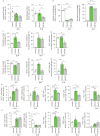
Phenotypical and functional characterisation of immune responses induced by AM-001. C57BL/6 mice received either a single intratracheal delivery of bleomycin (BLM) of 4 U·kg−1 (vehicle) or saline (controls). After 21 days, BLM-treated mice received AM-001 via intraperitoneal injections every alternate day at a dose of 10 mg·kg−1 for an additional 2 weeks (AM-001). The gating strategy used to characterise immune cell subsets is provided in supplementary figure S14. a) Percentage of neutrophils (Ly6G+CD3CD19) and macrophages (CD11c+F4/80+Ly6GCD3CD19) within CD45+ alive cells (n=4–5). b) Percentage of alveolar (SiglecF+) and recruited (SiglecF) macrophages within the total population (n=4–5). c) Percentage of M1 (Ly6C+, top) and M2 (Ly6C, bottom) subsets within recruited macrophages, along with their expression of major histocompatibility complex II (MHC-II) and Ki67 within each subset (n=4–5). d) Percentage of CD4+ T-cells (CD4+CD3+CD19) within CD45+ alive cells and CD69+ resident CD4+ T-cells, and expression of Ki67, CD25 and programmed cell death protein 1 (PD-1) within the CD4+ T-cell subset (n=4–5). e) Percentage of CD8+ T-cells (CD8+CD3+CD19) within CD45+ alive cells and CD69+ resident CD8+ T-cells, and expression of Ki67, CD25 and PD-1 within the CD8+ T-cell subset (n=4–5). Data are presented as mean±sem. *: p<0.05; **: p<0.01; ***: p<0.001; ****: p<0.0001.
FIGURE 8
FIGURE 8
Pharmacological inhibition of exchange factor directly activated by cAMP 1 (Epac1) by AM-001 attenuates fibrosis in a patient-derived ex vivo precision-cut lung slices (PCLS) model of human lung fibrosis. a) Idiopathic pulmonary fibrosis (IPF) human-derived PCLS preparation and treatment with AM-001 every other day with 20 μM AM-001 or vehicle for a total of 10 days. Lung sections from three different patients were used. Detailed information regarding the clinical information of the patients included in this study is presented in supplementary table S3. b) Representative images of collagen fibres measured with Masson Trichrome staining and c) quantified in IPF-PCLS treated with AM-001 or vehicle as a control (n=3). Scale bar: 500 μm. d, e) Representative images of co-immunostaining of neural precursor cell expressed, developmentally downregulated 8 (NEDD8) (upper panel, red) and forkhead box protein O3 (FoxO3a) (lower panel, red) with α-smooth muscle actin (α-SMA) in IPF-PCLS treated with vehicle or AM-001. Nuclei were counterstained with DAPI (blue). f, g) Representative immunoblot (left panels) and quantification (right panels) of α-SMA, fibronectin (Fn), matrix metallopeptidase 2 (MMP-2), collagen 1 (COL1), collagen 3 (COL3), transforming growth factor β1 (TGF-β1), NEDD8, FoxO3a phosphorylation at Thr32 (p-FoxO3aThr32 and total FoxO3a protein level in PCLS derived from three IPF patients, treated with either AM-001 or vehicle (Veh) (n=3). h) Pathway schematic of AM-001 effects on Epac1 and the downstream molecular pathway. Data are presented as mean±sem. AKT: protein kinase B; FB: fibroblast; GDP: guanosine diphosphate; GTP: guanosine triphosphate; STAT3: signal transducer and activator of transcription 3; Ub: ubiquitination; *: p<0.05.

Update of

Comment in

References

    1. Selman M, Lopez-Otin C, Pardo A. Age-driven developmental drift in the pathogenesis of idiopathic pulmonary fibrosis. Eur Respir J 2016; 48: 538–552. doi: 10.1183/13993003.00398-2016 - DOI - PubMed
    1. Raghu G, Amatto VC, Behr J, et al. Comorbidities in idiopathic pulmonary fibrosis patients: a systematic literature review. Eur Respir J 2015; 46: 1113–1130. doi: 10.1183/13993003.02316-2014 - DOI - PubMed
    1. Travis WD, Costabel U, Hansell DM, et al. An official American Thoracic Society/European Respiratory Society statement: update of the international multidisciplinary classification of the idiopathic interstitial pneumonias. Am J Respir Crit Care Med 2013; 188: 733–748. doi: 10.1164/rccm.201308-1483ST - DOI - PMC - PubMed
    1. Meiners S, Eickelberg O, Konigshoff M. Hallmarks of the ageing lung. Eur Respir J 2015; 45: 807–827. doi: 10.1183/09031936.00186914 - DOI - PubMed
    1. Thannickal VJ, Murthy M, Balch WE, et al. Blue Journal conference. Aging and susceptibility to lung disease. Am J Respir Crit Care Med 2015; 191: 261–269. doi: 10.1164/rccm.201410-1876PP - DOI - PMC - PubMed

MeSH terms

LinkOut - more resources