. 2025 Jul 10;8(9):e202503343.
doi: 10.26508/lsa.202503343. Print 2025 Sep.

TLR-induced STK25 activation promotes IRF5-mediated inflammation

Affiliations

TLR-induced STK25 activation promotes IRF5-mediated inflammation

Matthew R Rice et al. Life Sci Alliance. .

Abstract

The transcription factor interferon regulatory factor 5 (IRF5) functions as an important mediator of the inflammatory response downstream of MyD88-dependent TLRs. Whereas dysregulation of IRF5 activity has been implicated in the development of numerous autoimmune diseases including systemic lupus erythematosus, the factors that modulate TLR-induced IRF5 post-translational modifications are poorly understood. The focus of this study was to identify novel kinases in TLR-MyD88-IRF5 signaling. We performed a kinome-wide siRNA screen in human THP-1 monocytic cells and identified serine/threonine protein kinase 25 (STK25) as a positive regulator of pro-inflammatory cytokine production via phosphorylation of IRF5 at Thr265, leading to IRF5 transcriptional activation. We further found that STK25 undergoes autophosphorylation in response to multiple TLR triggers. Findings were validated in Stk25-deficient primary immune cells revealing a significant attenuation in R848-induced IRF5 nuclear translocation and pro-inflammatory cytokine production. Finally, we detected increased levels of STK25 autophosphorylation in immune cells from systemic lupus erythematosus donors compared with healthy controls. These findings implicate STK25 as a new regulator of TLR7/8 signaling through the modulation of IRF5 activation.


Conflict of interest statement

The authors declare that they have no conflict of interest.

Figures
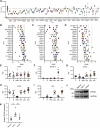
Figure 1.
Figure 1.. Identification of kinases involved in the regulation of R848-induced inflammation.
(A) A comprehensive siRNA screen was conducted in THP-1 cells to identify kinases that modulate R848-mediated pro-inflammatory cytokine release. The 20 proteins that exhibited the most significant reduction in R848-induced TNF-α production following siRNA knockdown are displayed in ranked order from left to right. Previously characterized regulators of TLR signaling are underlined in red. Targets underlined in blue have never been associated with TLR7/8-induced pro-inflammatory cytokine production. The black dotted line represents the mean of the R848-stimulated control group (N = 63 biological replicates). Each letter represents an independent siRNA construct (N = 3 technical replicates). (A, B, C, D) The color scheme used in (A) is conserved in (B, C, D). (B) The strictly standardized mean difference (SSMD) scores for each siRNA construct used in the identification of key regulators of R848-induced TNF-α production in the THP-1 screen are shown in descending order of significance. An SSMD score of −1 for a particular siRNA molecule was considered a positive hit and targets with two or more hits were selected for validation in follow-up studies with human primary cells. The black dotted line represents an SSMD score of 0, and the red dotted line represents an SSMD score of −1. (C, D) Human primary monocytes (C) and monocyte-derived dendritic cells (D) were stimulated with R848 for 24 h following siRNA knockdown of targets identified in the primary screen. SSMD scores for each siRNA construct were determined using the degree of siRNA-mediated inhibition of R848-induced TNF-α and IL-6 production from cells isolated from two independent donors. (E, F, G, H, I) Relative mRNA expression of STK16 (E), MAP3K19 (F), SPHK2 (G), STK25 (H), and IRF5 (I) in sorted peripheral blood leukocytes from healthy control donors (N = 11–26). Normalized transcripts per million values were obtained from GSE149050. T, T cells; B, B cells; PMN, neutrophils; cDC, conventional dendritic cells; pDC, plasmacytoid dendritic cells; cMo, classical monocytes. (J) Immunoblot analysis of STK25 protein expression in untreated human immortalized cell lines. Blots were probed with antibodies against STK25 and GAPDH. (K) Densitometric analysis of STK25 protein levels after normalization to the expression of GAPDH (N = 3–4 biological replicates). *P < 0.05, U, unstimulated, S, R848-stimulated, ns, not significant. Data represent mean ± SEM.
Figure S1.
Figure S1.. Identification of STK25-mediated IRF5 phosphorylation sites by mass spectrometry.
(A) Phosphorylation of a biotinylated C-terminal construct of IRF5 (residues 222–467, 3 μM) by kinases involved in TLR signaling in an in vitro scintillation proximity assay (N = 4 biological replicates). (B) IRF5 peptide coverage from three independent replicates. Residues in red were detected and residues in blue were determined to be phosphorylated. (C) Conservation of Thr183 across multiple human isoforms of IRF5. (D) Conservation of Thr183 in IRF5 protein sequences from multiple species. (E) Spectral data for the phosphorylation of IRF5 at Tyr313. Data represent mean ± SEM.
Figure 2.
Figure 2.. STK25 phosphorylates IRF5 at Thr265 in vitro.
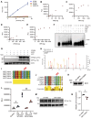
(A) Dose-dependent phosphorylation of a biotinylated C-terminal construct of IRF5 (residues 222–467, 3 μM) by candidate kinases in an in vitro scintillation proximity assay (N = 4 biological replicates). (B, C, D, E) Phosphorylation of full-length IRF5 by IKKβ (B, C) or STK25 (D, E) in an in vitro luminescent kinase assay. (B) Dose-dependent phosphorylation of IRF5 (500 ng) by IKKβ. (C) Dose-dependent phosphorylation of IRF5 by a fixed amount of IKKβ (100 ng). (D) Dose-dependent phosphorylation of IRF5 (500 ng) by STK25. (E) Dose-dependent phosphorylation of IRF5 by a fixed amount of STK25 (350 ng). (F) Phos-tag immunoblot analysis of the kinetics of STK25- and IKKβ-mediated phosphorylation of full-length IRF5 in vitro. Blots were probed with an antibody against total IRF5. p-IRF5, phosphorylated IRF5. (G) Immunoblot analysis of IRF5 phosphorylation at threonine residues following incubation with STK25 in an in vitro kinase assay for 1 h at RT. Blots were probed with antibodies against total p-Thr and IRF5. Representative of three independent experiments. p-Thr, phosphorylated threonine. (H) Mass spectrometry-based identification of Thr265 as an STK25-dependent IRF5 phosphorylation site. In vitro kinase reactions with IRF5 and STK25 were incubated for 1 h at RT, subjected to SDS–PAGE, and the gel was stained with Coomassie blue. Protein bands were excised, destained, and digested with trypsin. Representative of three independent experiments. (I) Conservation of Thr265 across multiple human isoforms of IRF5. (J) Conservation of Thr265 in IRF5 protein sequences from multiple species. (K) WT-IRF5 and IRF5-T265A were generated via an in vitro cell-free protein expression system and evaluated by immunoblot analysis. Blots were probed with an antibody against total IRF5. (L) Phosphorylation of WT-IRF5 and IRF5-T265A by STK25 in an in vitro luminescent kinase assay (N = 3 biological replicates). (M) Immunoblot analysis of WT-IRF5 and IRF5-T265A phosphorylation at threonine residues following incubation with STK25 in an in vitro kinase assay for 1 h at RT. Blots were probed with antibodies against total p-Thr and IRF5. (N) Densitometric analysis of WT-IRF5 and IRF5-T265A phosphorylation at threonine residues after normalization to the expression of total IRF5 (N = 3 biological replicates). *P < 0.05, **P < 0.01. Data represent mean ± SEM.
Figure 3.
Figure 3.. STK25-mediated phosphorylation of IRF5 at Thr265 induces transcriptional activation in HEK293T cells.
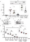
(A) HEK293T cells were co-transfected either alone or in combination with IRF5-FLAG, IRF5-T265A-FLAG, or STK25 in addition to an interferon-stimulated response element (ISRE) firefly luciferase (ISRE-Luc) reporter and a cytomegalovirus (CMV) Renilla luciferase (CMV-RL) internal control. The firefly luciferase signal was normalized to the Renilla luciferase signal to determine the relative light units (RLU). For each experiment, the relative ratio for each sample was normalized to a sample with IRF5-FLAG, ISRE-Luc, and CMV-RL (N = 12–13 transfections from three independent experiments). (B) Immunoblot analysis of HEK293T cells co-transfected with IRF5-FLAG or IRF5-T265A-FLAG. Blots were probed with antibodies against IRF5 and β-actin. (C) Immunoblot analysis of STK25-deficient HEK293T cells generated via CRISPR-Cas9-mediated gene editing. Blots were probed with antibodies against STK25 and β-actin. (A, D) WT and STK25-deficient (KO) HEK293T cells were co-transfected as in (A) to evaluate normalized ISRE-Luc reporter activity (N = 5 biological replicates per genotype). *P < 0.05, **P < 0.01, ***P < 0.001, ****P < 0.0001, ns, not significant. Data represent mean ± SEM.
Figure 4.
Figure 4.. STK25 responds to TLR7/8 activation in THP-1 cells and regulates TLR-induced pro-inflammatory cytokine production in murine primary immune cells.
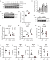
(A) Immunoblot analysis of STK25 autophosphorylation at Thr174 in THP-1 cells stimulated with R848 for 0.5, 1, 2, 4, 6, or 24 h. Blots were probed with antibodies against p-STK25 (T174), STK25, and GAPDH. (B) Densitometric analysis of p-STK25 (T174) protein levels after normalization to total STK25 protein levels and the expression of GAPDH (N = 2 independent experiments). (C) Immunoblot analysis of STK25 expression in THP-1 cells stimulated with R848 for 24 h. (D) Densitometric analysis of STK25 protein levels after normalization to the expression of β-actin (N = 4 biological replicates). UT, untreated. (E) Immunoblot analysis of STK25 protein expression in Stk25+/+ (WT) and Stk25−/− (KO) splenocytes. Blots were probed with antibodies against STK25 and GAPDH. (F, G) Imaging flow cytometry analysis of IRF5 nuclear translocation in peripheral blood CD11b+ monocytes (F) and B220+ B cells (G) from WT and KO mice following stimulation with R848 or CpG-B for 2 h (N = 3 biological replicates per genotype). (H, I) Flow cytometry analysis of the frequencies of IL-6+CD11b+ (H) and TNF-α+CD11b+ (I) splenocytes from WT and KO mice following stimulation with R848 for 18 h (N = 5–8 biological replicates per genotype). (J, K, L) Quantification of IL-6 production in culture supernatants of WT and KO splenocytes stimulated with R848 (J), CpG-B (K), or LPS (L) for 24 h (N = 4–5 biological replicates per genotype). *P < 0.05, **P < 0.01, ***P < 0.001. ns, not significant. Data represent mean ± SEM.
Figure S2.
Figure S2.. STK25 undergoes autophosphorylation in response to multiple TLR ligands, with minimal influence on cell viability in murine splenocytes.
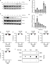
(A) Immunoblot analysis of STK25 autophosphorylation at Thr174 in THP-1 cells stimulated with LPS for 0.5, 1, 2, 4, 6, or 24 h. Blots were probed with antibodies against p-STK25 (T174), STK25, and GAPDH. (B) Densitometric analysis of p-STK25 (T174) protein levels after normalization to total STK25 protein levels and the expression of GAPDH (N = 2 independent experiments). (C) Immunoblot analysis of STK25 autophosphorylation at Thr174 in Ramos B cells stimulated with CpG-B for 0.5, 1, 2, 4, 6, or 24 h. Blots were probed with antibodies against p-STK25 (T174), STK25, and GAPDH. (D) Densitometric analysis of p-STK25 (T174) protein levels after normalization to total STK25 protein levels and the expression of GAPDH (N = 2 independent experiments). (E, F, G, H, I) Flow cytometry analysis of WT and KO splenocyte viability for uncultured (N = 8–13 biological replicates per genotype) (E), untreated (N = 4–5 biological replicates per genotype) (F), R848-stimulated (N = 6 biological replicates per genotype) (G), CpG-B-stimulated (N = 6 biological replicates per genotype) (H), and LPS-stimulated (N = 6 biological replicates per genotype) (I) cells used for 24 h cytokine release assays. (J) Immunoblot analysis of blot overlay assays conducted with full-length IRF5 (100 ng) and varying amounts (50–200 ng) of full-length IKKβ, Lyn, or STK25. Blots were probed with an antibody against IRF5.
Figure 5.
Figure 5.. Basal autophosphorylation of STK25 at Thr174 is increased in PBMCs from patients with systemic lupus erythematosus.
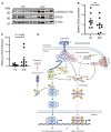
(A) Immunoblot analysis of STK25 autophosphorylation at Thr174 in PBMCs from healthy donors and patients with systemic lupus erythematosus. Blots were probed with antibodies against p-STK25 (T174), STK25, and β-actin. (B) Densitometric analysis of total STK25 protein levels after normalization to the expression of β-actin (N = 8–9 samples per condition). (C) Densitometric analysis of p-STK25 (T174) protein levels after normalization to total STK25 protein levels and the expression of β-actin (N = 8–9 samples per condition). Data represent mean ± SEM. (D) Proposed model for STK25 as an IRF5 kinase downstream of TLR7/8/9 signaling (pathway highlighted by the red dashed arrows). The activation of TLR7/8 by ssRNA or TLR9 by CpG-B DNA induces the autophosphorylation of STK25 at Thr174 and stimulates the formation of the Myddosome, a signaling complex comprised of MyD88, IRAK1/4, TRAF6, and IRF5. The activation of IRAK1 by IRAK4 leads to the recruitment of TRAF6. The ubiquitination (colored circles) of TRAF6 provides a binding site for TAB2/3, thereby facilitating the activation of TAK1. NEMO, a component of the IKK complex, interacts with ubiquitinated TRAF6 to promote the activation of IKKβ by TAK1. IKKβ-mediated phosphorylation of IκBα induces its ubiquitination and degradation by the 26S proteasome. The inhibition of IκBα permits the activation and nuclear translocation of the RelA and p50 subunits of NF-κB. In the nucleus, NF-κB regulates the expression of pro-inflammatory cytokines. In the alternate arm of the pathway, IRF5 undergoes TRAF6-catalyzed ubiquitination in addition to IKKβ- and/or STK25-dependent phosphorylation at Ser446 and Thr265, respectively. The activation of IRF5 also involves an interaction with the adaptor protein, TASL, which binds to SLC15A4. Whereas IKKβ is required for TASL-mediated activation of IRF5, it is unknown if STK25 modulates the TASL-dependent pathway. Altogether, these modifications induce IRF5 dimerization and nuclear translocation. In the nucleus, transcriptionally active IRF5 modulates the expression of pro-inflammatory cytokines and type I interferons. Created with BioRender.com.

References

    1. Kawasaki T, Kawai T (2014) Toll-like receptor signaling pathways. Front Immunol 5: 461. 10.3389/fimmu.2014.00461 - DOI - PMC - PubMed
    1. Schoenemeyer A, Barnes BJ, Mancl ME, Latz E, Goutagny N, Pitha PM, Fitzgerald KA, Golenbock DT (2005) The interferon regulatory factor, IRF5, is a central mediator of toll-like receptor 7 signaling. J Biol Chem 280: 17005–17012. 10.1074/jbc.M412584200 - DOI - PubMed
    1. Takaoka A, Yanai H, Kondo S, Duncan G, Negishi H, Mizutani T, Kano SI, Honda K, Ohba Y, Mak TW, et al. (2005) Integral role of IRF-5 in the gene induction programme activated by Toll-like receptors. Nature 434: 243–249. 10.1038/nature03308 - DOI - PubMed
    1. Balkhi MY, Fitzgerald KA, Pitha PM (2008) Functional regulation of MyD88-activated interferon regulatory factor 5 by K63-linked polyubiquitination. Mol Cell Biol 28: 7296–7308. 10.1128/MCB.00662-08 - DOI - PMC - PubMed
    1. Ren J, Chen X, Chen ZJ (2014) IKKβ is an IRF5 kinase that instigates inflammation. Proc Natl Acad Sci U S A 111: 17438–17443. 10.1073/pnas.1418516111 - DOI - PMC - PubMed

MeSH terms

LinkOut - more resources