Review
. 2025 Jul 11;10(1):216.
doi: 10.1038/s41392-025-02264-1.

Pattern recognition receptors: function, regulation and therapeutic potential

Affiliations
Review

Pattern recognition receptors: function, regulation and therapeutic potential

Ruochan Chen et al. Signal Transduct Target Ther. .

Abstract

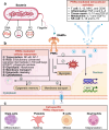
Pattern recognition receptors (PRRs) are sensors in the immune system, detecting pathogen-associated molecular patterns (PAMPs) and damage-associated molecular patterns (DAMPs). They serve as essential links between the innate and adaptive immune responses, initiating defense mechanisms against pathogens and maintaining immune homeostasis. This review examines the classification, structure, and signaling cascades of key PRR families, including toll-like receptors (TLRs), C-type lectin receptors (CLRs), nucleotide-binding oligomerization domain-like receptors (NLRs), AIM2-like receptors (ALRs), and others. It explores the dual roles of PRRs in immune defense and regulation, particularly through inhibitory PRRs (iPRRs), which prevent immune overactivation. The review also investigates the ligand recognition mechanisms and signaling pathways, highlighting the involvement of PRRs in disease progression and immune modulation. Notable signaling pathways, including NF-κB, MAPK, cGAS-STING, and MYD88-mediated and non-MYD88-mediated cascades, are discussed in the context of immune responses. Mechanisms that fine-tune PRR-mediated responses include transcriptional and fpost-transcriptional regulation, protein degradation, subcellular localization, and the recruitment of amplifiers and inhibitors, along with metabolic and microbial factors. These regulatory strategies ensure immune signaling remains adaptable and precise, preventing excessive inflammation. The review also explores the therapeutic potential of targeting PRRs in treating infectious, inflammatory, autoimmune, and malignant diseases, underscoring their importance in advancing immunological research and precision medicine.


Conflict of interest statement

Competing interests: The authors declare no competing interests.

Figures

Fig. 1
Fig. 1
Key discoveries in PRRs. a The proposal of the Infectious-Non-Self (INS) model. In 1989, Charles A. Janeway introduced a revolutionary hypothesis within the self-non-self model of immunity. He proposed that the innate immune system possesses a unique capacity to detect microbial infections (named PAMPs) via receptors, which are primarily expressed on APCs and are named PRRs. b The proposal of the Danger/injury model. Building on the foundation established by self-non-self models, Polly Matzinger introduced and elaborated the danger/injury model in immunology in 1994. This revolutionary proposal represented a significant shift in immunological thought. It posited that PRRs can be activated not only by foreign antigens but also by endogenous cellular alarm signals (named DAMPs) released by distressed or injured cells. This insight profoundly reshaped our view of the immune system’s responsiveness to cellular distress. c The Discovery of TLR4. In 1997, Jules Hoffmann found that mutated “Toll” receptors in fruit flies showed increased vulnerability to fungal infections. This discovery laid the foundation for understanding the role of Toll-like receptors in the innate immune response. In 1998, Alexander Poltorak and his colleagues identified mutations in the Tlr4 gene while investigating the defects in lipopolysaccharide (LPS) signaling in C3H/HeJ and C57BL/10ScCr mouse strains. This discovery provided definitive evidence for the pivotal role of TLR4 in LPS recognition and unveiled its significance in the innate immune response of mammals. d The Discovery of other TLRs. Subsequently, other TLRs including TLR3, TLR5, and TLR 9 have been discovered in succession during 2000-2001. e The introduction of the inflammasome. The term “inflammasome” was introduced by Jurg Tschopp and his colleagues in 2002. The first inflammasome to be identified was NACHT, LRR, and PYD domains-containing protein 1 (NLRP1) in 2002, and NLRP3 quickly followed this in 2004. f The discovery of cGAS. The cGAS pathway, discovered in 2013 by Professor Zhijian Chen, is capable of recognizing pathogenic double-stranded DNA molecules and serves as one of the pivotal signals in initiating antiviral innate immune defense. This finding obtained the Lasker Award for Basic Medical Research in September 2024. This figure is created by BioRender (https://app.biorender.com)
Fig. 2
Fig. 2
Structure of different PRRS in the cell membrane and the extracellular. a The structure of TLRs. TLRs featured an ectodomain adorned with LRRs that mediate signal recognition, transmembrane regions, and the cytoplasmic TIR domains that activate downstream signaling pathways. Upon recognizing PAMPs or DAMPs, TLRs interact with MYD88 or TRIF and trigger downstream signaling cascades, ultimately leading to the release of inflammatory cytokines, IFN-Is, chemokines, and antimicrobial peptides. b The structure of RLRs. RLRs family encompasses three members: LGP2, MDA-5, and RIG-I. Structurally, RLRs consist of two N-terminal CARDs, a central DDX/ATPase domain, and a C-terminal regulatory domain. In RIG-I, the C-terminus comprises the RD and the CTD, while MDA-5 lacks the RD, thus being devoid of self-inhibitory capabilities. In contrast to other RLR family members, LGP2 lacks the CARD domain, precluding its ability to recruit homologous molecules for signal transduction. However, researches showed that LGP2 could regulates RIG-I and MDA-5 signaling. c The structure of CLRs. CLRs including CLEC4E (MINCLE), CLEC7A (Dectin-1), Dectin-2, CLEC8A, CLEC9A (DNGR1), CLEC12A and MRs. Dectin-1 features an extracellular CTLD and an intracellular tail linked to an ITAM, while Dectin-2 lacks the ITAM sequence and does not possess signal transduction capabilities. MRs’ extracellular segment comprises two parts: the proximal membrane end with eight consecutive CTLDs, responsible for ligand endocytosis and transport, and the distal membrane end with a cysteine-rich lectin domain that recognizes sulfated carbohydrate conjugates. d The structure of Extracellular soluble PRMs. Extracellular soluble PRMs include diverse molecular families, primarily pentraxins, collectins, and ficolins. Pentraxins are categorized into two families: short and long molecules. These molecules are predominantly synthesized in the liver in response to inflammatory signals and interleukins. The long pentraxin family, represented by PTX3, uniquely features an extended N-terminal domain. Collectins and ficolins are both oligomers composed of basic subunits, each consisting of three polypeptide chains with four domains: an N-terminal cysteine-rich domain, a collagen-like sequence, an α-helical neck region, and a functional pattern recognition domain at the C-terminus. The functional domain differs between collectins and ficolins; collectins feature a globular CRD, while ficolins possess a fibrinogen-like domain. This figure is created by BioRender (https://app.biorender.com)
Fig. 3
Fig. 3
Structure of different PRRS in the cytoplasm and nucleus. a The structure of cGAS. Upon cytosolic DNA detection, cGAS synthesizes 2'3’-cGAMP from ATP and GTP. The produced 2'3’-cGAMP acts as a second messenger, binding to and activating the ER-resident protein STING. This interaction leads to the dimerization and autophosphorylation of STING, which then recruits and activates TBK1. Subsequently, TBK1 phosphorylates the transcription factor IRF3, promoting its dimerization, nuclear translocation, and activation of ISGs. This pathway is crucial for the induction of type I interferon responses against intracellular pathogens. b The functional domains of HNRNPA2/B1. The N-terminal of HNRNPA2/B1 proteins contains two RNA recognition motifs (RRM1 and RRM2), and the C-terminal is a glycine-rich low-complexity region (LC) containing an RGG box, an M9 nuclear localization signal (NLS), and a core prion-like domain (PrLD), reference from. c The structure of NLRs. NLRs are composed of three main domains: one is the central nucleotide-binding domain, also known as the NACHT domain (synthesized by the abbreviations of the following four kinds of NLR members: NAIP, CIITA, HETE, and TEP1; LRRs at the C-terminus, which are used to identify ligands; and the N-terminal effector domain, which is the protein interaction domain, such as CARD or PYD. Based on their distinct N-terminal effector domains, the NLRs can be categorized into five subfamilies. NLRA, NLRB, NLRC, NLRP, and NLRX1. d The structure of CLRs, exemplified by AIM. Structurally, AIM2 consists of a C-terminal HIN-200 domain and an N-terminal PYD. Acting as a cytosolic DNA receptor, AIM2 directly interacts with dsDNA through its HIN-200 domain. The PYD of AIM2 engages with the corresponding PYD of the inflammasome adapter protein ASC. Subsequently, the CARD of ASC connects with the CARD of procaspase-1, leading to the formation of the AIM2 inflammasome. This figure is created by BioRender (https://app.biorender.com)
Fig. 4
Fig. 4
The signaling pathways mediated by iPRRs and their regulation. iPRRs predominantly transmit inhibitory signals through motifs of ITIMs or ITSM located in their cytoplasmic tails. Upon receptor ligation, these motifs undergo phosphorylation, triggering the recruitment of inhibitory effectors containing SH2 domains, such as SHP-1, SHP-2, SHIP, and Csk. The known group of iPRRs currently includes Siglec-10, CD300a/f, Siglecs 2,3,5–11, CEACAM1, LILRB1 (CD85j), LILRB3 (CD85a), TIGIT, PVR, LAIR-1, and SIRL-1. In scenarios where tolerating damage is advantageous for the host, DAMPs or PAMPs can trigger iPRRs to dampen the immune response. Conversely, when damage cannot be tolerated, DAMPs or PAMPs signal via activating PRRs to initiate a robust immune response. The relative abundance of PRRs and iPRRs, along with their corresponding ligands, dictates the intensity of the ensuing immune reaction. This figure is created by BioRender (https://app.biorender.com)
Fig. 5
Fig. 5
PAMPs and DAMPs play critical roles in immune response and inflammatory response. Various DAMPs or PAMPs (a) are recognized by PRRs present on various immune or non-immune cells (b). This recognition initiates a cascade of downstream signaling pathways, ultimately culminating in the release of a diverse spectrum of cytokines, chemokines, adhesion molecules, and other inflammatory mediators. In unison, these processes orchestrate the initiation, activation, and amplification of immune response (c) and inflammatory response (d), thereby facilitating a coordinated response to tissue injury or stress. This figure is created by BioRender (https://app.biorender.com)
Fig. 6
Fig. 6
Double-edged sword effect of PRR in vivo. a The double effects of PRRs in antimicrobial responses. The role of PRRs in immunity to infection is complex. The functions of PRRs can be detrimental; for example, they can alter the expression of other key PRRs, inhibit antigen presentation, facilitate viral infection of myeloid cells, and promote pathological inflammation, including Th2 responses in the context of allergies. In addition, PRRs play a vital role in immune defense by recognizing PAMPs on the cell surface, inducing intracellular signaling cascades that mediate pathogen uptake and killing, production of inflammatory mediators, and the induction of several other protective cellular responses, such as the production of ROS and NET formation. Also, PRRs can promote antigen presentation and shape the development of adaptive immunity, including Th1, Th17, and CTL responses to provide protection. b The double effects of PRRs in recognition of self-components. PRRs serve a critical function in the immune response by enhancing the processing and presentation of antigens derived from necrotic cells, which is pivotal for the evolution of adaptive immune responses, notably those directed against neoplastic entities. Inflammation and adaptive immunity induced by self-recognition of PRRs can lead to additional beneficial outcomes, such as wound healing or tissue repair. Activation signals from PRRs and other receptors are balanced by inhibitory signals from inhibitory PRRs to prevent detrimental responses. However, dysregulated, excessive, or persistent activation signals due to dysregulation of inhibitory signaling and accumulation of undigested self-ligands can lead to immunopathology, including autoimmune and/or autoinflammatory diseases. This figure is adapted from Reis et al. with some modifications. This figure is created by BioRender (https://app.biorender.com)
Fig. 7
Fig. 7
Cellular responses induced by PRRs in microbial infection. a PRRs-mediated extracellular activities. Cell-extrinsic responses triggered by PRRs signaling encompass a range of activities that impact the immediate (or systemic) milieu surrounding the cell responsible for the detection of PAMPs. b PRRs-mediated cellular responses. Cell-intrinsic responses induced by PRR signaling occur within the cell that detected a PAMP and contribute to the activities indicated in (a). c Cell-specific PRRs responses. Cell-type-specific responses induced by PRR signaling occur uniquely within the indicated cell type and are mediated by TLR–PAMP interactions. This figure is created by BioRender (https://app.biorender.com)
Fig. 8
Fig. 8
Distinct signaling pathways in the recognition of PAMPs and DAMPs by PRRs. a Inflammasomes complex. Recognition of PAMPs or DAMPs by PRRs stimulates the NF-κB-mediated upregulation of inactive pro-IL-1β and multiple receptor proteins, such as NLRP1, NLRP3, NLRC4, NLRC12, as well as the proteins AIM2 and pyrin. The downstream signaling involves the engagement of a receptor protein activator, which drives the assembly of the inflammasome, proximity-induced autoproteolysis of caspase-1, and the subsequent cleavage of IL-1β, IL-18, and GSDMD, ultimately leading to the induction of inflammation and pyroptosis. b MYD88-dependent and MYD88-independent pathways. TLRs are universally expressed across diverse immune system cells, with TLRs 1, 2, 4, 5, and 6 positioned prominently on the cell surface, while TLRs 3, 7, 8, and 9 are intimately associated with endosomal membranes. Upon recognition of various DAMPs or PAMPs by these TLRs, they initiate a complex signaling cascade, leveraging adaptor proteins MYD88 (MYD88-dependent pathways) and TRIF (MYD88-independent pathways) as crucial intermediates. These adaptors, in turn, activate downstream MAPKs and IKK, which collaborate to facilitate the production of inflammatory cytokines by activating transcription factors like AP-1 and NF-κB, respectively. c The mitogen-activated protein kinase (MAPK) signaling. Typical MAPKs activated by various extracellular stimuli are characterized by a three-tiered signaling cascade, starting with MAP3KK, followed by MAP2K, and culminating in the activation of MAPK. In mammalian cells, three major MAPK families have been identified: ERK1/2, JNK1/2/3, and the p38 (α, β, γ, δ) isoforms signaling cascades. The transcription factors regulated by MAPK signaling include ELK1, ATF-2, AP1, STAT1, c-Fos, MEF-2, c-Myc, CREB, C/EBPα, and c-Jun. The MAPK pathway precisely influences cell proliferation, stress responses, inflammation, differentiation, functional synchronization, transformation, and apoptosis. d The NF-κB signaling. Upon the recognition of PAMPs or DAMPs by PRRs, a critical signaling cascade is initiated. This leads to the activation of the IκB Kinase (IKK) complex, which is composed of IKKα, IKKβ, and NEMO (also known as IKKγ). The IKK complex then phosphorylates IκB (inhibitor of κB), a protein that keeps the NF-κB complex (made up of p50 and p65 subunits) inactive in the cytoplasm. Following phosphorylation, IκB is marked for ubiquitination (Ub) and is subsequently degraded, allowing the NF-κB complex to be released. The liberated NF-κB complex translocates to the nucleus, where it triggers the transcription of genes involved in immune responses. This signaling pathway is essential for a swift reaction to infections and inflammatory stimuli, and it also plays a pivotal role in governing cell survival and proliferation. e The cGAS–STING–IFN pathway. In the cytoplasm, the presence of free cytosolic DNA or endosome-encapsulated DNA triggers the activation of the cGAS enzyme. cGAS synthesizes cGAMP, which serves as a second messenger that binds to and activates STING. This activation initiates a signaling cascade that involves the transcription factors IRF3 and IRF7, leading to the production of Type I IFNs. Concurrently, STING also activates the NF-κB pathway, which promotes the expression of inflammatory cytokines such as IL-6 and TNF. The coordination of these signaling events within the nucleus results in a robust DNA-driven immune response, which is essential for the defense against intracellular pathogens and the regulation of immune homeostasis. f The Ca2+ signaling. Extracellular Ca2+ signaling mediated by ion channels in the cell membrane can modulate the activation of these PRR subfamilies. This modulation favors the promotion of IRF3/7 activation, initiating IFN-associated innate immune responses and enhancing NF-κB-related inflammatory reactions. Furthermore, recent evidence has revealed that TLR7 augments the cAMP-PKA pathway, ultimately leading to increased expression of SERCA and RYR2 in the sarcoplasmic reticulum, thereby disrupting Ca2+ signaling within cardiomyocytes. This figure is created by BioRender (https://app.biorender.com)
Fig. 9
Fig. 9
Transcriptional regulation in PRRs-mediated inflammatory response. a Activation and repression mediated by transcription factors. For example, the SUMOylation of the WRKY33 TF enhances its interaction with MAPK3 and MAPK6 of PRRs’ downstream signaling pathway. b Regulation of enhancers and promoters in PRRs. For example, NF-κB possesses the ability to activate poised enhancers and promoters among TLR4. c Epigenetic modifications in PRRs, including three pivotal epigenetic events—DNA methylation, histone acetylation, and noncoding RNA. For example, Patients with COVD-19 showed heightened methylation of the TLR4 and TNF-α. This figure is created by BioRender (https://app.biorender.com)
Fig. 10
Fig. 10
Noncoding RNA regulation in PRRs. A variety of noncoding RNA families, such as miRNAs, lncRNAs, tRNA fragments, and circRNAs, have been recognized as pivotal regulators in biological processes like transcription, splicing, and translation. These regulatory roles can significantly influence PRRs-mediated immune responses, adapted from Carpenter et al. with some modifications
Fig. 11
Fig. 11
Translation regulations in PRRs. a Regulation of translation initiation in PRRs. For example, phosphorylation, hyperphosphorylation and dephosphorylation eukaryotic translation initiation factor induces regulation of translation, affecting most cellular PRR mRNAs’ expression, acting through the TICAM1or mTOR- mediated pathway. b Gene-specific regulation in PRRs. For example, TIA1, an ARE-binding protein, can repress the translation of TNF and other cytokine mRNAs following TLR stimulation by impeding their association with polyribosomes. c Regulation of translation elongation and termination in PRRs. For example, MAP3K8 and CLEC4E inhibit the translational machinery by dephosphorylating eukaryotic translation elongation factors, thereby suppressing PRRs-dependent cytokine production. This figure is created by BioRender (https://app.biorender.com)
Fig. 12
Fig. 12
Different mechanistic models for cross-regulation of PRR signaling pathways. In principle, the cooperation of distinct PRR and non-PRRs signals cross-regulates to generate effector responses through three types of mechanisms: synergy, enhancement, and suppression. a Synergy. Different receptor-mediated signals in a responding cell efficiently generate an amplified inflammatory response. This phenomenon can occur between PRRs of the same class, different PRR types, distinct downstream signaling pathways of PRRs, and even between PRRs and non-PRRs. For example, NODs and TLRs synergistically induce the production of cytokines and antimicrobial peptides. b Enhancement. One receptor-mediated effector response (E1) is augmented by another receptor-induced effector mechanism (E2). This “enhancement” effect can be implemented through a direct mechanism (M1) or a positive feedback mechanism (M2). For example, TLR2 as well as TLR4 can upregulate NLRC5 expression and promote the assembly of the PANoptosome. c Suppression. A one receptor-mediated effector response (E1) is inhibited by a distinct receptor-induced effector mechanism (E2) to introduce amplified inflammatory response. This “suppressive” effect can be achieved either through a direct mechanism (M1) or via a negative feedback loop (M2). For example, C-type lectin domain family 2 member D (CLEC2D) has been shown to form dimers with TLR2, leading to inhibiting the activation of the transcription factor IRF5 and subsequent IL-12 production. This figure is created by BioRender (https://app.biorender.com)
Fig. 13
Fig. 13
PRR regulation in carcinogenesis. The functional pleiotropy of PRRs in carcinogenesis is likely governed by cell type, the nature of the upstream activating ligand, the composition of multisubunit receptor complexes, downstream signaling pathways, disease stage, and tissue type. PRR-induced pro-inflammatory cytokines play dual roles in tumor immunity, including antitumor immunity to inhibit tumor growth and immune evasion to promote tumor growth. For example, NLRP6 regulates the secretion of IL-18 and antimicrobial peptides in enterocytes, as well as mucin production in goblet cells, which contributes to its ability to prevent the invasion of pathobionts that initiate colitis-associated colorectal cancer, thereby inhibiting tumor growth. The functional cytokine introduce by cGAS–STING pathway activates the production of anti-inflammatory cytokines, which contribute to an immunosuppressive TME. This figure is created by BioRender (https://app.biorender.com)
Fig. 14
Fig. 14
The role of PRR in ageing. a Stimulation of TLR5 by a flagellin-fused protein has been shown to effectively extend the lifespan and enhance the health span of mice of both genders. This effect is mediated by an increased surface expression of TLR5 and a subsequent elevation in IL-22 secretion. Additionally, aging diminishes TLR activation by agonists, leading to decreased production of inflammatory cytokines. b Hematopoietic stem cells (HSCs) lacking IRF8 do not respond to CpG, an agonist of TLR9 in mice. In contrast, continuous CpG stimulation not only increases the number of wild-type HSCs but also reduces their ability to form bone marrow colonies in ageing mice. c Extracellular HMGB1 stimulates the release of inflammatory cytokines through the TLR2/4 and NF-κB signaling pathways, thereby enhancing the production of the senescence-associated secretory phenotype (SASP). Blocking TLR2 and TLR4 can alleviate cellular aging and mitigate age-related diseases. d Inhibiting IFN-I response mediated by cGAS activation mitigates aging-associated cognitive decline and dysfunction in neurodegenerative diseases (NDDs) models. This figure is created by BioRender (https://app.biorender.com)

References

    1. Metschnikoff, E. Lecture on phagocytosis and immunity. Br. Med. J.1, 213–217 (1891). - PMC - PubMed
    1. Paul, E. Croonian lecture—on immunity with special reference to cell life. Proc. R. Soc. Lond.66, 424–448 (1900).
    1. Paul, W. E. Bridging innate and adaptive immunity. Cell147, 1212–1215 (2011). - PubMed
    1. Jenkins, M. K. & Schwartz, R. H. Pillars article: antigen presentation by chemically modified splenocytes induces antigen-specific T cell unresponsiveness in vitro and in vivo. J. Exp. Med. 165, 302–319 (1987). - PubMed
    1. Land, W. et al. The beneficial effect of human recombinant superoxide dismutase on acute and chronic rejection events in recipients of cadaveric renal transplants. Transplantation57, 211–217 (1994). - PubMed

MeSH terms

LinkOut - more resources