. 2025 Jul 10;15(1):24812.
doi: 10.1038/s41598-025-10196-7.

Exploring the anti-inflammatory mechanism of geniposide in rheumatoid arthritis via network pharmacology and experimental validation

Affiliations

Exploring the anti-inflammatory mechanism of geniposide in rheumatoid arthritis via network pharmacology and experimental validation

Meng Huang et al. Sci Rep. .

Abstract

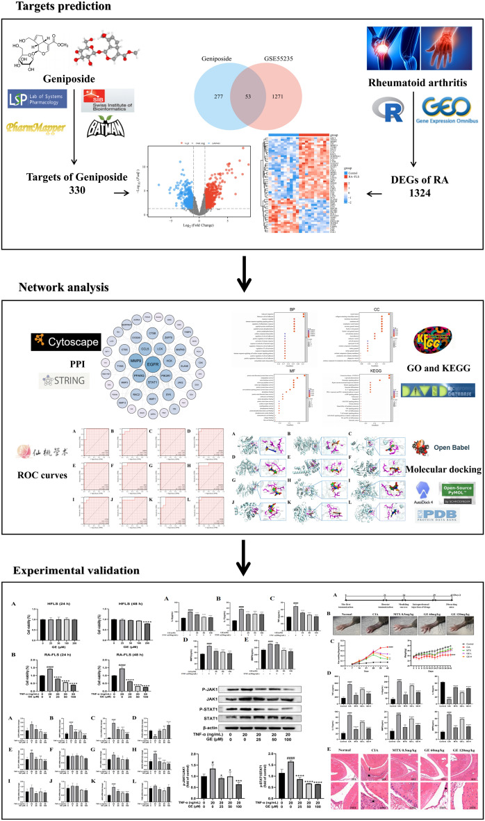
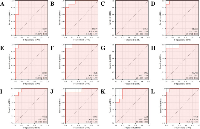
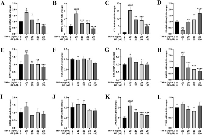
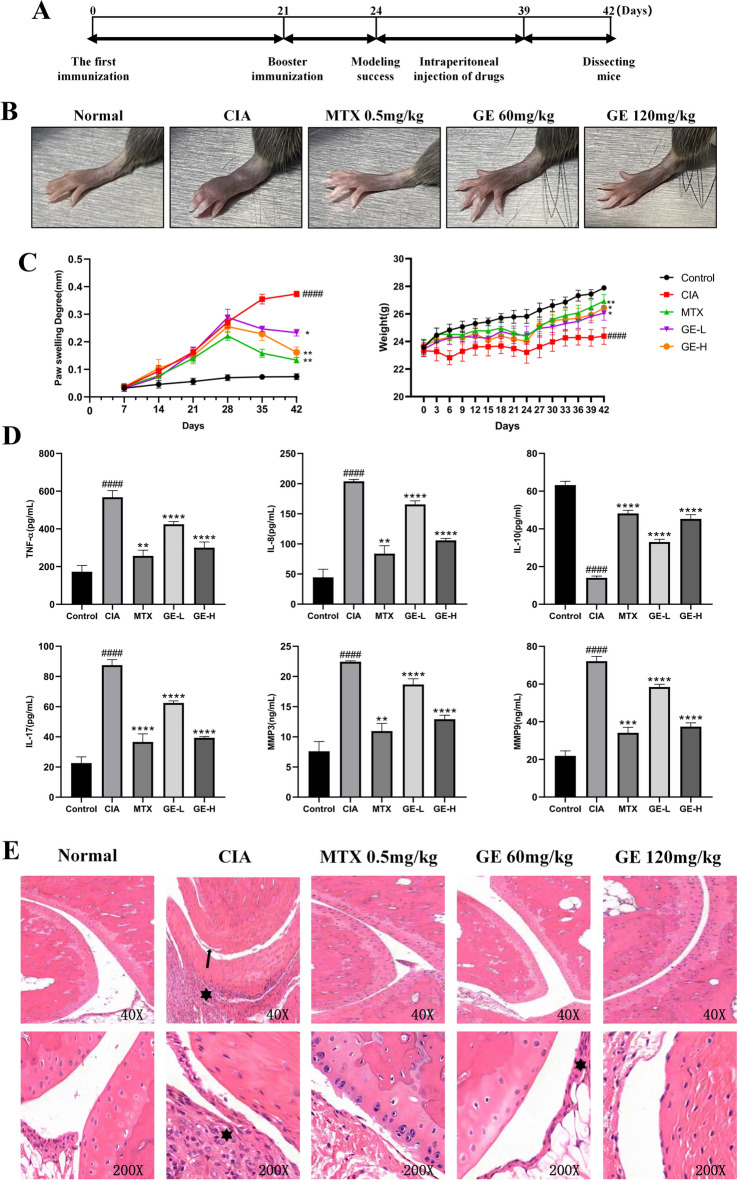
Rheumatoid arthritis (RA) is a chronic autoimmune disease that affects millions worldwide, characterized by joint pain, swelling, and functional impairment. Current treatments like Non-steroidal anti-inflammatory drugs (NSAIDs), corticosteroids, and biologics, have limitations including side effects and resistance problems, which create a need for new therapeutic strategies. This study aims to explore the potential therapeutic role and mechanisms of Geniposide (GE), a natural compound extracted from Gardenia jasminoides, in RA treatment. Through network pharmacology methods and using target prediction databases including TCMSP, SwissTargetPrediction, Pharmmapper, and Batman, 330 potential targets of GE were identified. In RA, 1324 differentially expressed genes (DEGs) were identified from the GSE55235 dataset. By intersecting the datasets, 53 shared targets were identified, which were further analyzed using Gene Ontology (GO) and Kyoto Encyclopedia of Genes and Genomes (KEGG) enrichment, with pathways like IL-17 and JAK-STAT being significantly highlighted. Additionally, protein-protein interaction (PPI) network analysis identified 12 key targets (EGFR, MMP-9, CCL5, PPARG, STAT1, HCK, SYK, MAPK8, CTSB, RAC2, JAK2, TYMS) with high degree values. Furthermore, molecular docking studies confirmed strong binding affinities between GE and the identified targets. Experimental validation demonstrated that GE inhibited RA-FLS cell proliferation in a dose-dependent manner by using MTT assays and reduced the level of pro-inflammatory cytokines (IL-17, IL-8, TNF-α, MMP-3, MMP-9) as measured by ELISA. RT-qPCR and Western blot analyses further confirmed that GE modulated the mRNA expression of key targets and inhibited the phosphorylation of JAK1 and STAT1 proteins, respectively. Finally, we verified the anti-inflammatory effect of GE on CIA mice through in vivo experiments. These findings suggest that GE has anti-RA effects by targeting several key molecules and pathways. This provides a theoretical basis for developing GE as a novel therapeutic for RA.


Conflict of interest statement

Declarations. Competing interests: The authors declare no competing interests.

Figures

Fig. 1
Fig. 1
Flow chart of the analysis process in the study.
Fig. 2
Fig. 2
Intersecting targets of GE and RA.. (A) 2D structure of drug GE. (B) Volcano plot representing differentially expressed genes in the datasets GSE55235: grey indicates genes with no difference, red represents upregulated genes, and blue denotes downregulated genes. (C) Intersection targets of disease and drug. (D) Heatmap showcasing of differentially expressed genes. The x-axis represents samples from the two datasets, while the y-axis displays the differentially expressed genes. Red indicates high expression and blue signifies low expression.
Fig. 3
Fig. 3
Screening key targets of GE against RA. (A) The PPI network of 53 common targets is drawn by cytoscape. The darker the color and the larger the shape, the higher the Degree value. (B) Hub genes were screened from the PPI network using the Betweenness (BC), Closeness (CC), Degree (DC), and Network (NC) methods. (C) Subnetwork of the PPI network of 12 hub targets.
Fig. 4
Fig. 4
Functional analysis of differentially expressed genes. (A-C) The GO enrichment analysis results showed that leukocyte migration, leukocyte cell–cell adhesion was significantly enriched in RA. (D) KEGG results showed that IL-17 and JAK-STAT signaling pathways were significantly enriched in RA.
Fig. 5
Fig. 5
ROC curves of the 12 key genes in the datasets GSE55235 for RA diagnosis.
Fig. 6.
Fig. 6.
3D mode diagram of molecular docking. (A) EGFR and GE. (B) MMP-9 and GE. (C) CCL5 and GE. (D) PPARG and GE. (E) STAT1 and GE. (F) HCK and GE. (G) SYK and GE. (H) MAPK8 and GE. (I) CTSB and GE. (J) RAC2 and GE. (K) JAK2 and GE. (L) TYMS and GE.
Fig. 7
Fig. 7
Effect of different concentrations of GE on cell viability in RA-FLS and HFLS cells for 24 h and 48 h. (A) GE added HFLS produced cytotoxicity at 200 μM for 24 h and 48 h. (B) GE inhibited the proliferation of RA-FLS cells in a dose-dependent manner. Compared with the blank control group, ####P < 0.0001. Compared with the TNF-α stimulus group, ****P < 0.0001.
Fig. 8
Fig. 8
Effect of different concentrations of GE on inflammatory cytokine release in RA-FLS cells. After 24 h of GE treatment, the expression level of inflammatory factors in the supernatant of RA-FLS cells was determined by ELISA assay. (A) IL-8. (B) IL-17. (C) TNF-α. (D) MMP-3. (E) MMP-9. Compared with the blank control group, ###P < 0.001. Compared with the TNF-α stimulus group, **P < 0.01, ****P < 0.0001.
Fig. 9
Fig. 9
GE inhibits the mRNA expression of key targets in TNF-α-induced RA-FLS. (A-L) The effect of GE on the mRNA levels of core genes in RA-FLS stimulated by TNF-α. Compared with the blank control group, #P < 0.05, ##P < 0.01, ###P < 0.001, ####P < 0.0001. Compared with the TNF-α stimulus group, *P < 0.05, **P < 0.01, ***P < 0.001, ****P < 0.0001.
Fig. 10
Fig. 10
GE inhibits the activation of JAK/STAT pathway in TNF-α-induced RA-FLS. FLS for p-JAK1 and p-STAT1 were starved in 10% FBS medium, pretreated with GE and 20 ng/ml TNF-α for 48 h before collection. The phosphorylation of JAK1 and STAT1 in FLS were measured by western blot. Compared with the blank control group, #P < 0.05, ##P < 0.01, ###P < 0.001, ####P < 0.0001. Compared with the TNF-α stimulus group, *P < 0.05, **P < 0.01, ***P < 0.001, ****P < 0.0001.
Fig. 11
Fig. 11
GE attenuated the severity of arthritis in CIA mice. (A) In vivo experiment flow chart. (B) Observation of foot paw swelling in CIA model mice induced by GE. (C) Effects of GE on body weight and hind paw swelling of CIA model mice. (D) ELISA was used to detect the effect of GE on the inflammation of CIA model mice. (E) HE staining results of CIA mice joints after GE treatment. Black arrows indicate cartilage destruction, and asterisks indicate inflammatory cell infiltration. Compared with Control group, ####P < 0.0001. Compared with CIA group, *P < 0.05, **P < 0.01, ***P < 0.001, ****P < 0.0001.

References

    1. McInnes, I. B. & Schett, G. The pathogenesis of rheumatoid arthritis. N. Engl. J. Med.365(23), 2205–2219 (2011). - PubMed
    1. Fan, W., Zhou, Z. Y., Huang, X. F., Bao, C. D. & Du, F. Deoxycytidine kinase promotes the migration and invasion of fibroblast-like synoviocytes from rheumatoid arthritis patients. Int. J. Clin. Exp. Pathol.6(12), 2733–2744 (2013). - PMC - PubMed
    1. Downton, P., Dickson, S. H., Ray, D. W., Bechtold, D. A. & Gibbs, J. E. Fibroblast-like synoviocytes orchestrate daily rhythmic inflammation in arthritis. Open Biol.14(7), 240089 (2024). - PMC - PubMed
    1. Tarasiuk-Stanislawek, K. et al. Idiopathic remitting seronegative symmetrical synovitis with pitting edema syndrome mimicking symptoms of polymyalgia rheumatica: A case report. J. Med. Case Rep.16(1), 334 (2022). - PMC - PubMed
    1. Lindegaard, H. et al. Low field dedicated magnetic resonance imaging in untreated rheumatoid arthritis of recent onset. Ann Rheum Dis.60(8), 770–776 (2001). - PMC - PubMed

MeSH terms