. 2025 Jul 10;15(1):24913.
doi: 10.1038/s41598-025-09743-z.

DSP and DPP are dispensable for initiation of dentin and enamel mineralization but critical for circumpulpal dentin mineralization

Affiliations

DSP and DPP are dispensable for initiation of dentin and enamel mineralization but critical for circumpulpal dentin mineralization

Tian Liang et al. Sci Rep. .

Abstract

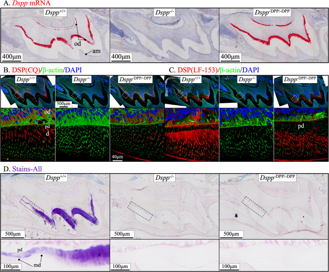
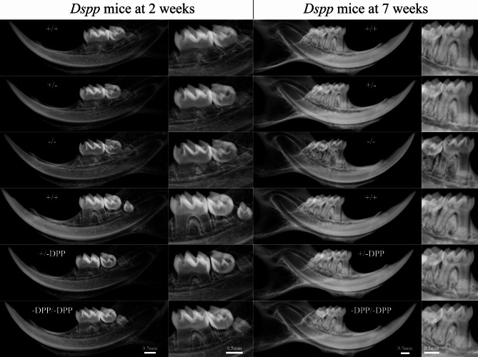
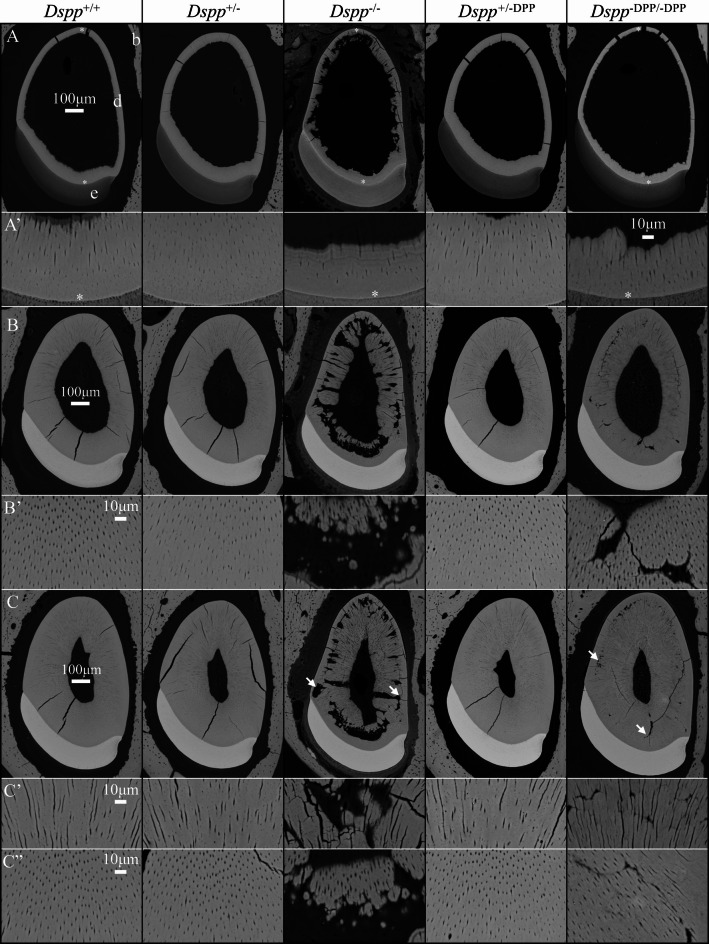
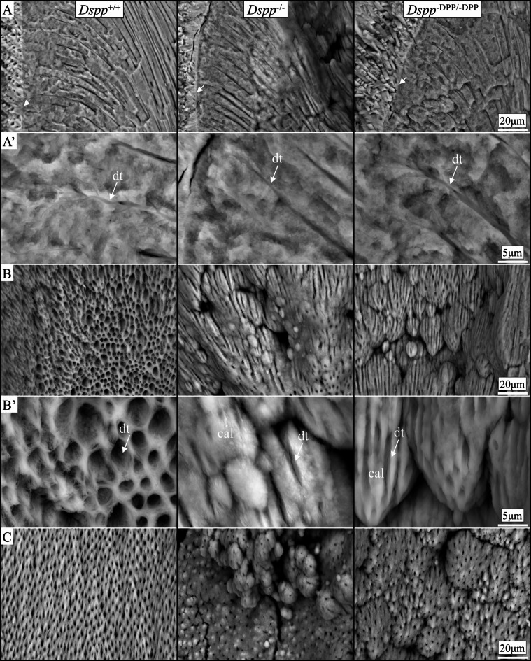
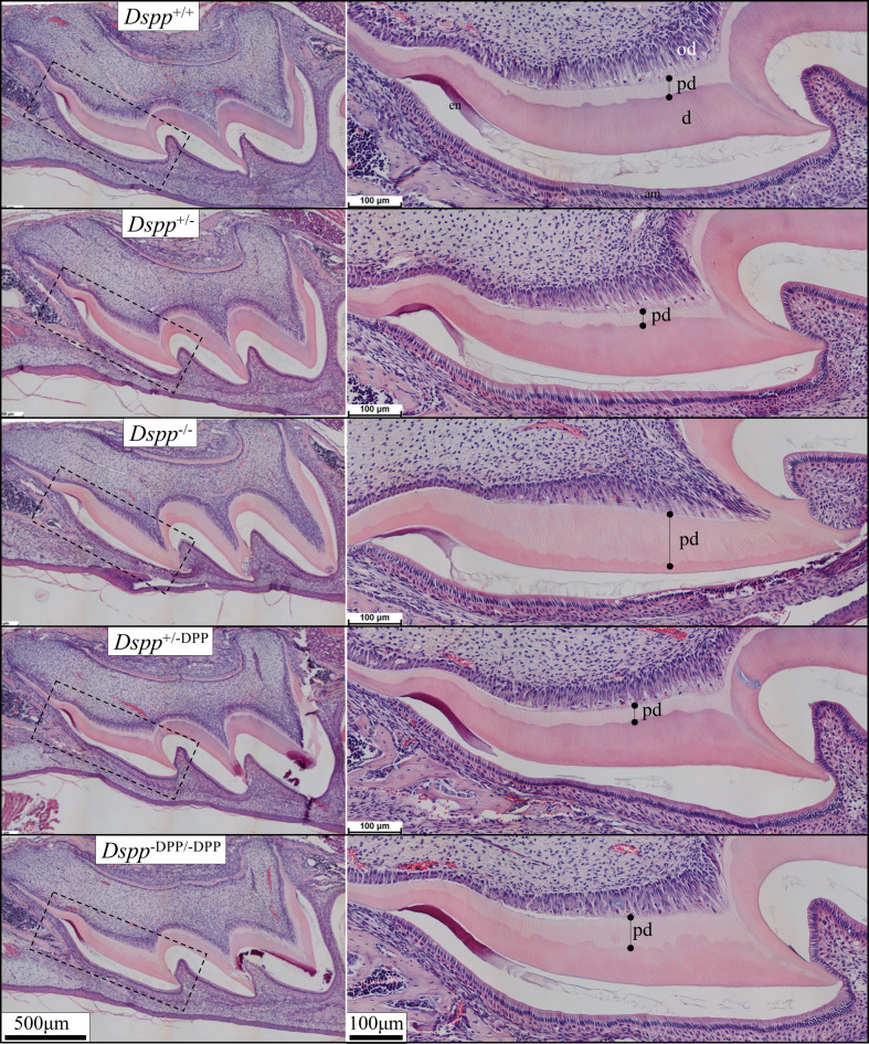
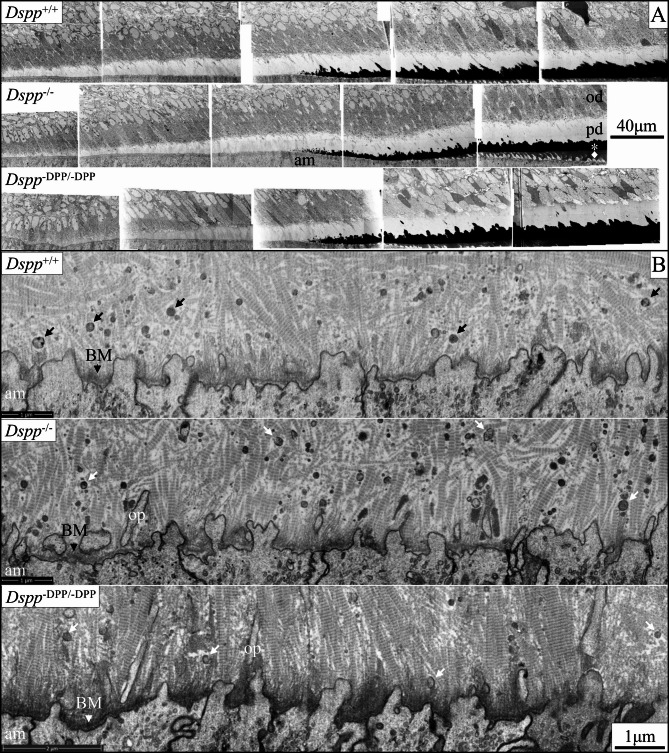
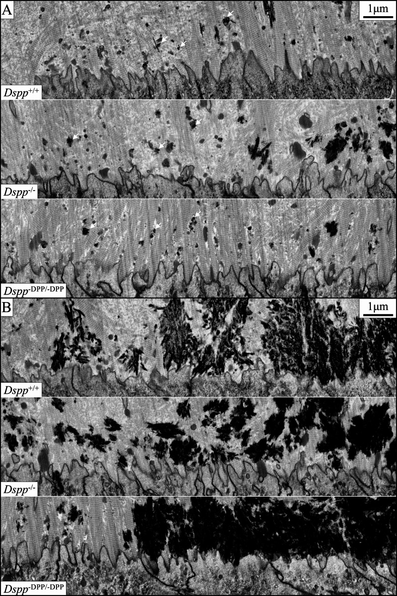
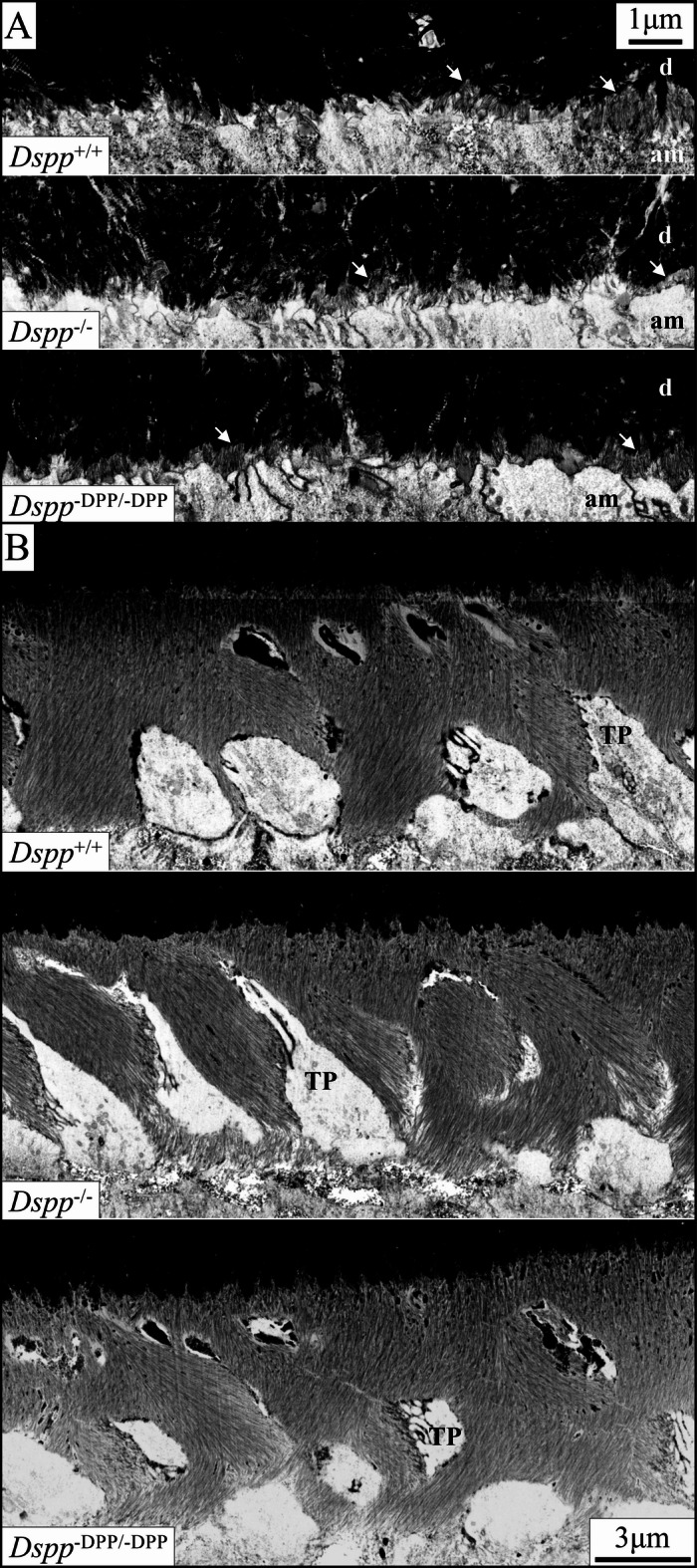
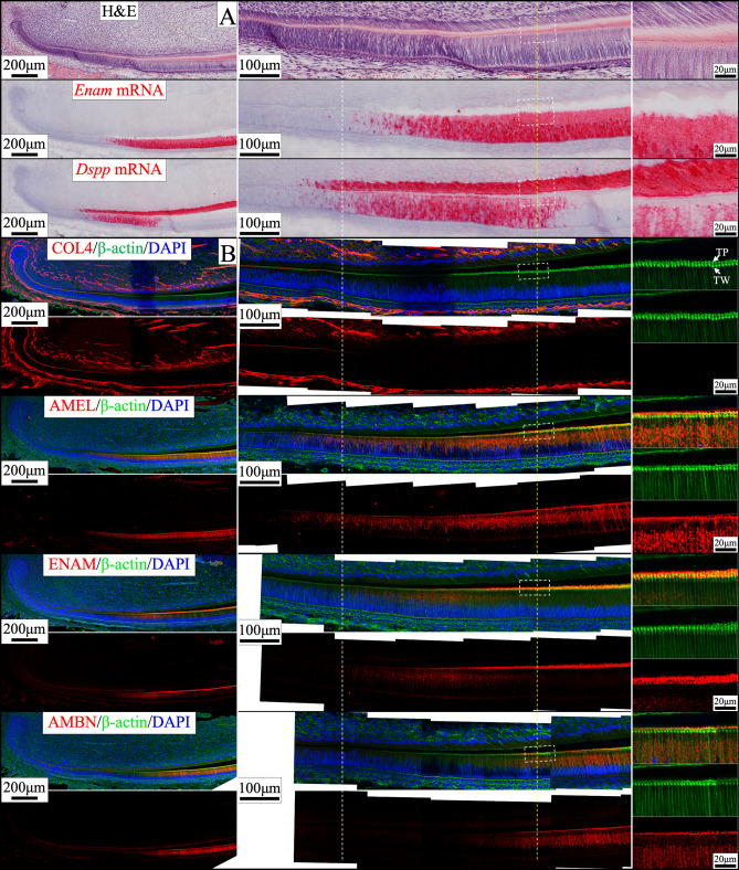
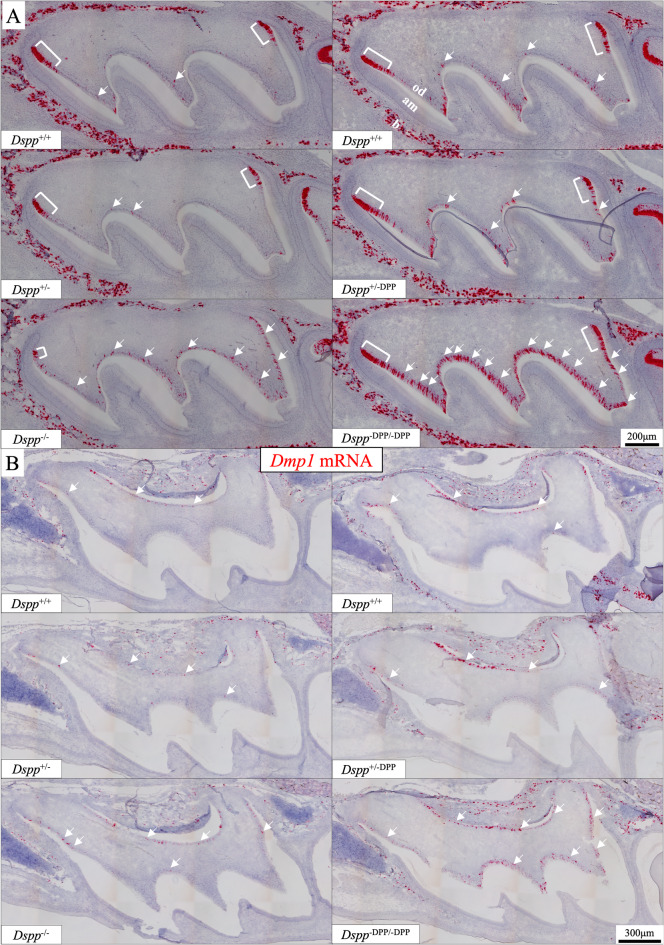
Dentin sialophosprotein (DSPP) is cleaved into the N-terminal dentin sialoprotein (DSP) proteoglycan and the C-terminal dentin phosphoprotein (DPP; the most acidic proteins in humans). To define the functions of DSP and DPP, we generated a Dspp-DPP mouse model using CRISPR/Cas9 technology and compared tooth mineralization in Dspp-DPP and Dspp-/- mice in the C57BL/6 background. In both mice, the initiation of dentin mineralization was associated with matrix vesicles. Odontoblasts appeared normal with odontoblastic processes. In Dspp-/- mice, limited mineralized dentin was observed. In Dspp-DPP/-DPP mice, dentin globules (calcospherites) with varied mineral density and unmineralized interglobular dentin were observed throughout the circumpulpal dentin. The area of predentin was smaller compared to Dspp-/- mice, but larger than wild-type mice. In Dspp-/- and Dspp-DPP/-DPP mice, enamel formation was comparable to wild-type. In both mice, Dmp1 expression in differentiating and differentiated odontoblasts was altered. We propose a model for dentin mineralization in which DSP, enriched in the peritubular dentin, propagates mineralization within the hypomineralized calcospherites in the intertubular dentin while DPP is essential for the maturation of calcospherite mineralization in the circumpulpal dentin. We conclude that DSP and DPP are dispensable for the initiation of dentin and enamel mineralization, but critical for circumpulpal dentin mineralization.

Keywords: Biomineralization; Dentin; Dentin Sialophosphoprotein; Dentin mineralization; Dentin phosphoprotein; Dentin sialoprotein.


Conflict of interest statement

Declarations. Competing interests: The authors declare no competing interests. Conflict of interest: The authors declare that the research was conducted without any commercial or financial relationships that could be construed as a conflict of interest.

Figures

Fig. 1
Fig. 1
Validation of the Dspp−/− and Dspp−DPP/−DPP mice and localization of DSP and DPP in Day 14 maxillary 1st molars. A. RNAscope in situ hybridization stains Dspp transcripts red in Dspp+/+ (wild-type) and Dspp−DPP/−DPP odontoblasts, but not in Dspp−/− odontoblasts. Refer to Fig S1-S3 for a complete set of RNAscope data on Dspp expression. B&C. Immunohistochemistry data show DSP signals (red) in Dspp+/+ and Dspp−DPP/−DPP odontoblasts and dentin matrix. A rabbit polyclonal antibody from Dr. Chunlin Qin (CQ) is used in B and another rabbit polyclonal antibody generated by Dr. Larry Fisher (LF-153) is used in C. Green and blue signals show β-actin and nuclei (DAPI), respectively. B shows moderate intracellular signals in the Golgi area and signals in the dentinal tubules, but not in the unmineralized predentin. C shows mild intracellular signals and intense extracellular signals concentrating in the peritubular dentin and diffusing to the intertubular dentin, but not in the predentin. The dentin mineralization front is relatively smooth in the Dspp+/+ dentin but tortuous in the Dspp−DPP/−DPP dentin. A globular pattern of DSP signals is obvious in the Dspp−DPP/−DPP mice, resembling dentin calcospherites. Signals in the dentin matrix are stronger in the newly mineralized circumpulpal dentin. Refer to Fig S4-S5 for the complete set. D. Stains-All staining shows the presence of DPP (purple blue) in Dspp+/+ dentin matrix, but not in Dspp−/− and Dspp−DPP/−DPP dentin matrix. In Dspp+/+ dentin matrix, staining is more intense in odontoblast processes and intertubular dentin. Staining in the dentin matrix is stronger in the newly mineralized circumpulpal dentin. A globular dentin mineralization front is observed. The predentin and the mantle dentin is not stained. Refer to Fig S6-S7 for the complete set of data. Key: od, odontoblasts; am, ameloblasts; d, mineralized dentin; pd, predentin; md, mantle dentin.
Fig. 2
Fig. 2
Radiography of mandibular incisors and molars from Dspp mutant mice. Dspp−/− mandibular incisors are more radiolucent, and the molar dentin is obviously thinner than the wild-type. Refer to Figs. S14-S16 for the comparisons among additional groups.
Fig. 3
Fig. 3
Limited dentin mineralization in Dspp−/− mice and uneven dentin mineralization in Dspp−DPP/−DPP mice. These panels compare backscattered scanning electron microscopy (bSEM) images of 1 mm incremental cross-sections of 7-week-old Dspp mandibular incisors. Panels A, B, C are from Levels 3, 6, and 8 (3 mm, 6 mm, and 8 mm from the apical loop), respectively. A’, B’, C’, and C” are magnified views of dentin mineralization patterns in these mice. Refer to Figs. S17-S29 for complete sets of images from multiple mice. Enamel formation is comparable in all mice. Dspp+/− and Dspp+/−DPP dentin mineralization is comparable to wild-type (Dspp+/+) dentin mineralization. The mantle dentin formation (*) in Dspp−/− and Dspp−DPP/−DPP mice is relatively normal (A and A’). The Dspp−/− mineralized dentin is thinner and hypomineralized. Islands of mineralized dentin and figure-like unmineralized processes extending from dental pulp to the outer part of the circumpulpal dentin (arrows in C) are observed. Dentinal tubules are present. The Dspp−DPP/−DPP mineralized dentin is hypomineralized and unevenly mineralized. Dentin globules (calcospherites) with varying mineral densities and fissure-like unmineralized dentin (arrows in C) are observed. Dentinal tubules are present. Key: b, alveolar bone; d, dentin; e, enamel.
Fig. 4
Fig. 4
Dentinal tubules penetrate the calcospherites in Dspp−/− and Dspp−DPP/−DPP mice. Shown are bSEM surface scanning images of 7-week-old mutant Dspp manually fractured mandibular 1st molars and incisors. Refer to Figs. S35-S38 for complete sets of images from multiple mice. A. Cross sections of dentin in molars. Dentinal tubules are evident, extending from dental pulp to the dentinoenamel junction (DEJ, arrows). A’. higher magnification lateral views of molar dentinal tubules (dt) near the DEJ. B. Pulp surface views of molar coronal dentin. In Dspp+/+ mice, the dentin mineralization front is relatively uniform with the pulpal openings of dentinal tubules. In Dspp−/− and Dspp−DPP/−DPP mice, dentin globules (calcospherites, cal) are obvious, with dentinal tubules passing between them. B’. Magnified views of B. C. Pulpal surface views of incisor dentin. The incisor findings are like those of the molars.
Fig. 5
Fig. 5
Unmineralized predentin is thicker in Dspp−/− and Dspp−DPP/−DPP mice. H&E-stained micrographs of 14-day-old Dspp mutant maxillary first molars. Boxed areas in the panels on the left are shown in higher magnification on the right. Predentin (pd) and dentin (d) are comparable in the wild-type (Dspp+/+) and the Dspp+/− and Dspp+/−DPP heterozygous mice. The thickness of unmineralized dentin (predentin) is increased in Dspp−DPP/−DPP mice and increased further in Dspp−/− mice, with corresponding reductions in the thickness of the mineralized dentin (d), so that the Dspp−/− mineralized dentin is extremely thin and most of the dentin matrix remains unmineralized. Refer to Figs. S39-S41 for complete sets of images from multiple mice. Key: od, odontoblasts.
Fig. 6
Fig. 6
Ultrastructurally, the onset of dentin mineralization in Dspp−/− and Dspp−DPP/−DPP mice is comparable to the wild-type. Focused ion beam-backscattered scanning electron microscopy (FIB-bSEM) of 7-week-old Dspp+/+, Dspp−/−, and Dspp−DPP/−DPP mandibular incisors. Refer to Figs. S42-S64 for complete sets of FIB-bSEM images. (A) Consecutive montages (~ 100 μm in width) of 5000x TLD images showing (on the left) odontoblasts (od) secreting predentin (pd), dentin mineral (*) deposition (center) in predentin near ameloblasts (am), followed by early enamel formation (◆) on the surface of coalesced dentin mineral (right). The initiation of dentin mineralization is comparable in Dspp+/+, Dspp−/−, and Dspp−DPP/−DPP mice. (B) 20000x images showing the partially degraded basement membrane (BM) and ameloblasts extending finger-like processes into the predentin. Note the oriented, banded collagen fibers and abundant unmineralized matrix vesicles (arrows) and darker mineralized matrix vesicles. Key: op, odontoblastic process.
Fig. 7
Fig. 7
The coalescing of dentin mineralization islands in the Dspp−/− and Dspp−DPP/−DPP mice. (A) Dentin mineral (arrow) is first observed in matrix vesicles near the distal membrane of ameloblasts. The matrix vesicles burst and seed mineral nuclei into the collagen fiber-rich predentin. (B) Once dentin mineral foci form and grow in the dentin matrix, they coalesce into a continuous layer of mineralized, collagen-rich, dentin. TLD detector, 20000x.
Fig. 8
Fig. 8
Enamel mineral ribbon initiation and Tomes’ process formation in the Dspp−/− and Dspp−DPP/−DPP mice are comparable to those of the wild-type. (A) Enamel mineral ribbons (arrows) initiate on a continuous layer of mineralized dentin (d) in close proximity to the irregular distal surface membrane of secretory ameloblasts (am). (B) Following the continuous elongation of the initial enamel mineral ribbons, the ameloblast distal membrane forms a Tomes’ processes that is divided into a protruding and tilted distal process that extends rod enamel ribbons and a proximal region covering the cell junctions that extends interrod enamel ribbons. TLD detector, 20000x.
Fig. 9
Fig. 9
The transient expression of Dspp in ameloblasts starts around the time when presecretory basement membrane is degraded and ends around the time when Tomes’ processes start to form. (A) H&E staining (top) and RNAscope in situ hybridization signal (red) for Enam and Dspp and (B) immunohistochemistry were performed on consecutive longitudinal sections of a 10-day-old wild-type mandibular incisors. Presecretory basement membrane (labeled by type IV collagen or COL4) and enamel matrix proteins (AMEL, ENAM, and AMBN), β-actin (showing cytoskeleton, particularly Tomes’ processes), and DAPI (showing nuclei) are labelled in red, green, and blue, respectively. The white line marks the time when the presecretory basement membrane is degraded, and the yellow line delineates the time when Tomes’ processes start to form. Boxed areas on the yellow line are magnified on the right. Key: TP, Tomes’ processes; TW, terminal web.
Fig. 10
Fig. 10
Comparison of odontoblast expression of Dmp1 in Dspp+, Dspp, and Dspp−DPP mice. RNAscope in situ hybridization of Dmp1 mRNA transcripts in Dspp mutant mice at Day 3 (panel A) and Day 14 (panel B) maxillary 1st molars. Comparisons should be made within an individual column. Each column displays 3 molars (wild-type, heterozygous, homozygous) collected from a single litter and processed together. Positive Dmp1 mRNA signal is red. Refer to Fig S65-S66 for a complete set of RNAscope data on Dmp1 expression. (A) Dmp1 mRNA is detected in bone cells (b) and odontoblasts (od), but not in ameloblasts (am). In Dspp+/+ mice (wild-type, WT, top panels), differentiating odontoblasts near the cervical loops express high levels of Dmp1 (brackets). Dmp1 expression is sharply reduced and detected occasionally in differentiated odontoblasts (arrows). In Dspp+/− and Dspp+/−DPP odontoblasts (middle panels), Dmp1 expression resembles the WT. In Dspp−/− odontoblasts (bottom left panel), differentiating odontoblasts express Dmp1 at a high level for a much shorter period near the cervical loops (bracket), while differentiated odontoblasts express Dmp1 in a sporadic pattern that was slightly higher than that of WT odontoblasts (arrows). In Dspp−DPP/−DPP odontoblasts (bottom right panel), differentiating odontoblasts express high level of Dmp1 (bracket), similar to those in WT odontoblasts. In differentiated odontoblasts, Dmp1 expression is detected in more than half of the odontoblasts (arrows). (B) Dmp1 expression is slightly upregulated in Dspp−/− odontoblasts (bottom left panel), and further elevated in Dspp−DPP/−DPP odontoblasts (bottom right panel).

Similar articles

References

    1. Weiner, S., Addadi, L. & Biomineralization At the cutting edge. Science298, 375–376. 10.1126/science.1078093 (2002). - PubMed
    1. Knoll, A. H. Biomineralization and evolutionary history. Rev. Mineral. Geochem.54, 329–356 (2003).
    1. Simmer, J. P. et al. The modified shields classification and 12 families with defined DSPP mutations. Genes (Basel). 13. 10.3390/genes13050858 (2022). - PMC - PubMed
    1. Simmer, J. P. et al. A genetic model for the secretory stage of dental enamel formation. J. Struct. Biol.213, 107805. 10.1016/j.jsb.2021.107805 (2021). - PMC - PubMed
    1. Smith, C. E. Cellular and chemical events during enamel maturation. Crit. Reviews Oral Biology Medicine: Official Publication Am. Association Oral Biologists. 9, 128–161. 10.1177/10454411980090020101 (1998). - PubMed

LinkOut - more resources