. 2025 Sep;133(5):633-647.
doi: 10.1038/s41416-025-03080-8. Epub 2025 Jul 10.

Cancer-associated fibroblasts are associated with neo-adjuvant treatment response in oesophageal adenocarcinoma

Collaborators, Affiliations

Cancer-associated fibroblasts are associated with neo-adjuvant treatment response in oesophageal adenocarcinoma

Robert C Walker et al. Br J Cancer. 2025 Sep.

Abstract

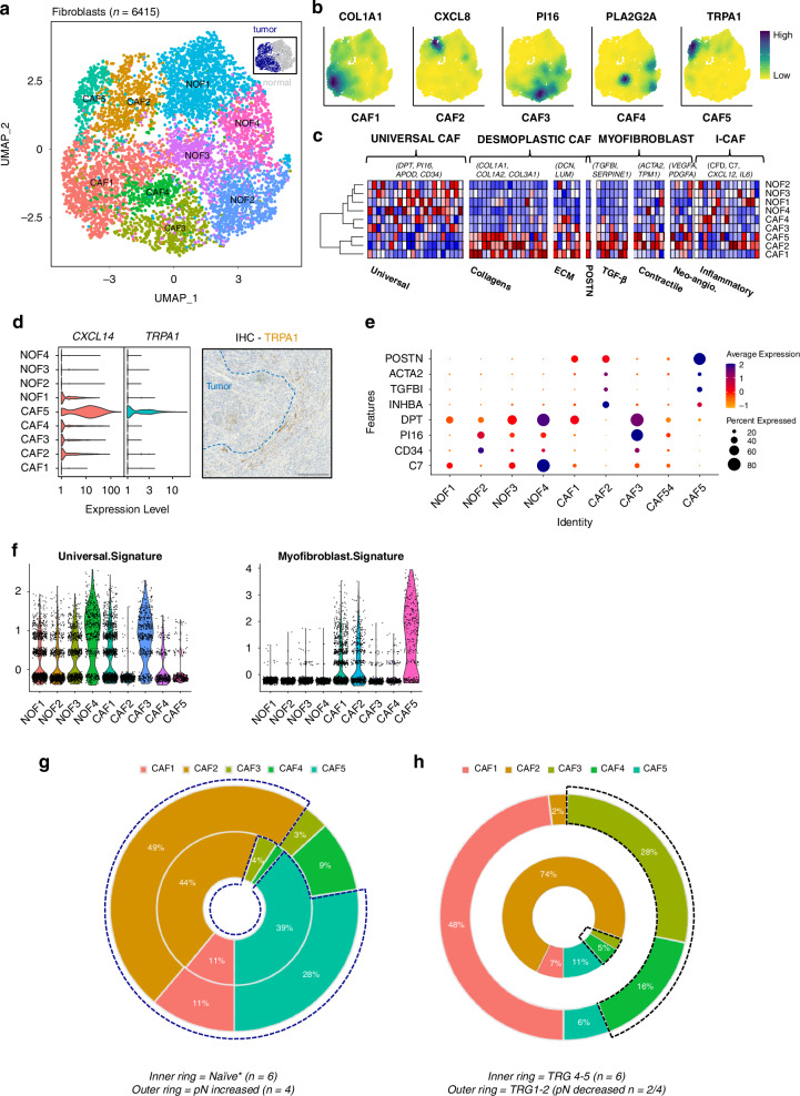
Background: Neoadjuvant treatment (NAT) in oesophageal adenocarcinoma (EAC) is characterised by differential responses between patients and treatment modalities. The components of the tumour microenvironment (TME) that contribute to this are unknown. We explored this, focusing on cancer-associated fibroblasts (CAF) an abundant TME component.

Methods: We performed histopathologic, single-cell RNA sequencing and transcriptomic analysis on 26 patients, stratified by pathological response to NAT, and validated a prognostic model in genomic consortia cohorts. Patient-derived cells were used to model CAF phenotypes in vitro.

Results: We observed changes in the TME in response to the NAT received. Specific changes in fibroblasts correlated with treatment response and altered gene expression associated with NAT type. Three myofibroblastic phenotypes dominate the TME, two of which persist in non-responders and could only be partially re-capitulated in vitro using co-culture with cancer cells or TGF-β. A two-gene NAT fibrotic signature was an independent prognostic indicator in chemo/chemoradiotherapy treated patients (HR = 2.47, p = 0.029).

Conclusions: This study provides a compendium of cell phenotypes in EAC across the current NAT treatment pathway that provides insights into CAF biology and cancer progression. MyoCAFs represent an axis to repurpose agents to enhance current therapies and immunotherapy.


Conflict of interest statement

Competing interests: The authors declare no competing interests.

Figures

Fig. 1
Fig. 1. Treatment response and the EAC TME.
a UMAP of cell lineages. Inset: UMAP of patient ID and a Pie chart of cell type proportions. b UMAP of tumour microenvironment components and cell source (T = tumour (red dots), N= normal (blue dots)) stratified by treatment status. Black arrows indicate the cancer-associated fibroblast cell clusters. c Feature Plots of canonical cell lineage marker expression in individual cells (each cell is a dot) where a gold colour indicates a specific transcript is not detected/expressed, and an increasing grey-black scale indicates increases in scaled expression levels. d scProportionTest plots of observed log2 fold differences between tumour (T) and normal (N) samples. FDR < 0.05 are indicated by the vertical dotted lines. e scProportionTest plots of observed log2 fold differences across different Mandard grades of treatment response. FDR < 0.05 are indicated by the vertical dotted lines, red arrows highlight cell types of interest. Sample sizes as indicated below each plot.
Fig. 2
Fig. 2. Cancer-associated fibroblast phenotypes in EAC.
a UMAP of fibroblast populations, inset UMAP identifies sample origin. b UMAPs of the CAF populations with selected lineage markers. c Heatmaps of canonical marker gene expression and selected cluster markers displaying universal, desmoplastic, myofibroblast and inflammatory CAF phenotypes. d Violin plot of CXCL14 and TRPA1 RNA expression and a representative IHC image of TRPA1 staining at the tumour/stroma boundary. e Dot plot of marker gene expression for fibroblast clusters. f Violin plots of the universal fibroblast signature and the myofibroblast signature across the NOF and CAF clusters. Fibroblast changes following treatment. g Neoadjuvant treatment naïve patients. The outer ring are the four cases with an increased in their pathological node status following surgery. The blue dashed line surrounds the myoCAF populations. h Chemo/CRT treated patients, stratified by different TRG statuses as indicated. The black dashed line surrounds the CAF3 and four populations.
Fig. 3
Fig. 3. Candidate genes associated with neoadjuvant response in EAC CAFs.
a Violin plots of gene expression in individual CAF cells. The five CAF clusters are labelled accordingly and stratified by treatment response. b Differential expression of candidate genes (from panel A, excluding CAF3 & 4 marker genes) comparing CAFs exposed to neoadjuvant treatment (as indicated in the chart legend) versus CAFs from surgically resected tumours without neoadjuvant therapy. The bars represent −log10(p-values) and the red dotted line is the p-value cut-off at 0.05.
Fig. 4
Fig. 4. Cell communication analysis of OAC tumours following neoadjuvant therapy.
a Top panel—Comparative Cell-Cell Interaction network in responders (P-value filter set at 0.2), where node size (Page rank odds-ratio) and edge thickness (% of interactions) represents importance of the cell communication between cells. The arrows indicate the signal direction and colour the activation status (Brown = Up and Blue = Down). The white asterisks indicate the top ranked cell types for NAT response in either patient group. Bottom panel - Bubble plots of cancer associated fibroblast interaction network changes (expressed as proportions) of cancer (x-axis), immune (y-axis) and stromal compartments (bubble radius) associated with NAT response. The asterisks indicate statistically significant differences in proportions between response groups (Two by Two Chi-squared test, two tailed Fisher’s Exact P-values; * ≤0.05, ***≤0.001). b As (a) for non-responders. Line plot of the distribution of ranked ligands for the indicated receiver cell type in the sender agonistic (all cell lineages) and sender-focused (CAF1-5) NicheNet analysis in non-responders to chemotherapy (c) and chemo-radiotherapy (d). Ligands in red are not in the top ranked ligands for all CAF-senders. APC antigen presentation cell. e, f A scaled DotPlot of target gene candidates across cell types stratified by chemotherapy response type. Colour scale of average expression and black circles denote the percentage of expressing cells. g, h A scaled DotPlot of the target gene candidates across cell types stratified by chemo-radiotherapy response type. Colour scale of average expression and black circles denote the percentage of expressing cells. Gene symbol colours: Green = Ligand; Blue = Receptor; Purple = target gene.
Fig. 5
Fig. 5. In vitro validation of gelsolin expression and myofibroblast programs in ex vivo patient-derived CAFs and EAC tumour tissue sections.
a GSN expression in the universal and myofibroblast populations across head and neck, oesophageal squamous, breast, gastric, pancreatic and colorectal cancer (*p < 0.0001). b HFFF2 fibroblasts treated with TGF-β for 72 h show CAF differentiation compared to vehicle (c) Relative expression of mRNA of myofibroblast genes ACTA2 and POSTN following CAF differentiation by TaqMan assay at 6 h, 24 h, 48 h and 72 h compared to vehicle (n = 3 replicate experiments). d Relative expression of mRNA of ZEB1 and ZEB2 (EMT regulation), ATF1 and GSN (cytoskeleton factors) following CAF differentiation by TaqMan assay at 6 h, 24 h, 48 h, and 72 h compared to vehicle (n = 3 replicate experiments). e Protein expression of GSN, αSMA and GAPDH a in TGF-β treated HFFF2 cells and vehicle quantified by Western Blotting and densitometry in biological triplicates (unpaired two-tailed t-test). f Volcano Plot of top ten over and under expressed genes and selected differentially expressed GOIs in patient-derived ex-vivo CAFs co-cultured with EAC cancer cell line, MFD1. Data from ref. [4]. *GOIs/Genes of interest include GSN; myoCAF markers: POSTN, ACTA2; EMT marker: ZEB1, VIM; CAF1 markers: COL1A1, COL3A1; CAF2 markers: CXCL8, IGFBP3; CAF5 markers: CXCL14, TRPA1, F3; Predicted transcription factor regulators: MYLK, STAT1. Adjusted P value cutoff set to an alpha of 0.05 (1.3 in –log10 space) and LogFC cutoff set to ±0.5. g Clustered heatmap of patient derived CAF mRNA expression heterogeneity by qPCR. Z-scored expression values as indicated by the colour scale. Underlined CAF samples were used for experiments shown in (c). h Fold change in mRNA expression in selected patient derived CAFs at 72 h following TGF-beta treatment. Median line shown on boxes. i Representative low power images of IHC expression patterns in EAC in the same tissue area for GSN, POSTN, α-SMA, CD3 and EpCAM from adjacent sections, and the corresponding H&E stain. H&E images are annotated with white dashed lines to label major tumour regions (T), stroma (S), and smooth muscle (M), where present. Three representative patients are shown. Example peritumoral fibroblast areas are marked with arrowheads. Scale bars—1000 μm.
Fig. 6
Fig. 6. Prognostic value of the 2-gene expression model in EAC genomic consortium cohorts.
Kaplan-Meier survival curves (Black line: 1st tertile scores (“Low group”). Red line: 3rd tertile scores (“High group”) and Cox Proportional Hazards tables for our 2-gene expression signature in the OAC cases from The Cancer Genome Atlas project (a) and OCCAMs consortium (b). Overall survival follow-up to 60 months from diagnosis. Only cases with complete data for all covariates were included in the multivariate analysis (c) Heatmap of the scRNA-seq derived cell signature scores from GSVA analysis of OCCAMs cases with whole genome expression data. Euclidean distance and average linkage were used to clustered samples. The curly brackets indicate cases with either high or low 2-gene signature scores from (a).

References

    1. Kim J, Bowlby R, Mungall AJ, Robertson AG, Odze RD, Cherniack AD, et al. Integrated genomic characterization of oesophageal carcinoma. Nature. 2017;541:169–74. - PMC - PubMed
    1. Pectasides E, Stachler MD, Derks S, Liu Y, Maron S, Islam M, et al. Genomic heterogeneity as a barrier to precision medicine in gastroesophageal adenocarcinoma. Cancer Discov. 2018;8:37–48. - PMC - PubMed
    1. Hoeppner J, Brunner T, Lordick F, Schmoor C, Kulemann B, Neumann UP, et al. Prospective randomized multicenter phase III trial comparing perioperative chemotherapy (FLOT protocol) to neoadjuvant chemoradiation (CROSS protocol) in patients with adenocarcinoma of the esophagus (ESOPEC trial). J Clin Oncol. 2024;42:LBA1–LBA. - PMC - PubMed
    1. Kelly RJ, Ajani JA, Kuzdzal J, Zander T, Van Cutsem E, Piessen G, et al. Adjuvant nivolumab in resected esophageal or gastroesophageal junction cancer. N Engl J Med. 2021;384:1191–203. - PubMed
    1. Janjigian YY, Van Cutsem E, Muro K, Wainberg Z, Al-Batran SE, Hyung WJ, et al. MATTERHORN: phase III study of durvalumab plus FLOT chemotherapy in resectable gastric/gastroesophageal junction cancer. Future Oncol. 2022;18:2465–73. - PubMed

MeSH terms

Supplementary concepts

LinkOut - more resources