. 2025 Jul 10;8(1):425.
doi: 10.1038/s41746-025-01831-8.

Deep learning on routine full-breast mammograms enhances lymph node metastasis prediction in early breast cancer

Affiliations

Deep learning on routine full-breast mammograms enhances lymph node metastasis prediction in early breast cancer

Daqu Zhang et al. NPJ Digit Med. .

Abstract

With the shift toward de-escalating surgery in breast cancer, prediction models incorporating imaging can reassess the need for surgical axillary staging. This study employed advancements in deep learning to comprehensively evaluate routine mammograms for preoperative lymph node metastasis prediction. Mammograms and clinicopathological data from 1265 cN0 T1-T2 breast cancer patients (primary surgery, no neoadjuvant therapy) were retrospectively collected from three Swedish institutions. Compared to models using only clinical variables, incorporating full-breast mammograms with preoperative clinical variables improved the ROC AUC from 0.690 to 0.774 (improvement: 0.001-0.154) in the independent test set. The combined model showed good calibration and, at sensitivity ≥90%, achieved a significantly better net benefit, and a sentinel lymph node biopsy reduction rate of 41.7% (13.0-62.6%). Our findings suggest that routine mammograms, particularly full-breast images, can enhance preoperative nodal status prediction. They may substitute key predictors such as pathological tumor size and multifocality, aiding patient stratification before surgery.


Conflict of interest statement

Competing interests: S.Z. and M.D. have received speaker’s fees and travel support from Siemens Healthcare AG and are listed as patent holders for US Patent PCT/EP2014/057372. The authors otherwise declare no conflicts of interest or financial interest.

Figures

Fig. 1
Fig. 1. Patient selection process.
The study cohort construction and patient exclusion criteria are presented. cN0 clinical node-negative, T invasive tumor grade.
Fig. 2
Fig. 2. Deep learning (DL) workflow.
The study employed a three-step DL approach. a SSL was conducted on unlabeled mammograms to pretrain the backbone feature extractor by maximizing the consistency between augmentations of the same patch. Contiguous patches were tiled from high-resolution mammograms to enable the extraction of subtle variations in local breast tissue. b SL was performed on mammograms of all views with five cancer outcomes to establish a neck module—a global feature extractor, on top of the backbone. For the neck module, a residual block and a vision Transformer were separately evaluated on the ROI or full-breast mammograms. Tumor ROIs were annotated by an automatic detecting tool. The five cancer outcomes investigated included LNM, number of LNMs, LVI, Tsize, and multifocality. c The extracted mammogram features and 11 preoperative clinical variables were concatenated to collaboratively train the final LNM classifier. Mammogram preprocessing details are provided in Supplementary Section 4.
Fig. 3
Fig. 3. Comparisons of ROC curves for LNM prediction between models using various modalities.
ROC curves of the development set (left column for site 1 and middle column for site 2) and the independent test set (right column, site 2) are presented. The number of patients, N, and positive rate for each set are presented in the subtitles. The areas under the curves are presented in the figure legends. LNM lymph node metastasis, PreopClinic preoperative clinicopathology, fullMammo full-breast mammogram, ROIMammo mammogram of region of interest, Tsize tumor size, Multifoc multifocality, CV cross-validation, ROC receiver operating characteristic.
Fig. 4
Fig. 4. Evaluation of three state-of-the-art self-supervised learning (SSL) methods.
SSL on unlabeled mammograms enhanced the representations for predicting cancer outcomes. The mean and standard deviation (error bars) were calculated across 1000 bootstrap samples. LNM lymph node metastasis, ROC AUC area under the receiver operating characteristics curve.
Fig. 5
Fig. 5. Comparisons of Transformer and ResBlock for mammogram modeling.
Transformer outperformed ResBlock in full-breast and ROI-based mammogram modeling for predicting cancer outcomes. The mean and standard deviation (error bars) were calculated across 1000 bootstrap samples. ROI region of interest, LNM lymph node metastasis, ROC receiver operating characteristics, AUC area under the curve.
Fig. 6
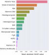
Fig. 6. Feature importance analysis.
Mammogram features played an important role in predicting lymph node metastasis. Feature importance estimated by the mean absolute SHAP value of the combined model of preoperative predictors and full-breast mammogram features is presented.
Fig. 7
Fig. 7. Visualization of activation maps.
Grad-CAM was used to interpret the mammogram patterns. The Transformer localized tumors more accurately for tumor size prediction and assigned higher importance to tumor and peritumor regions for predicting lymph node metastasis (LNM) and lymphovascular invasion (LVI) compared to the ResBlock model. Both selected patients A and B were LNM positive and LVI positive showing mediolateral views with visible lesions to demonstrate the activation maps of the Transformer and the ResBlock model (columns 1–2 for patient A and columns 3–4 for patient B). Model predictions were denoted as p. The expectations of LNM and LVI predictions for the Transformer model were 0.239 and 0.158, respectively, and 0.240 and 0.178 for the ResBlock model.

References

    1. Ferlay, J. et al. Cancer statistics for the year 2020: an overview. Int. J. Cancer149, 778–789 (2021). - PubMed
    1. Dillekås, H., Rogers, M. S. & Straume, O. Are 90% of deaths from cancer caused by metastases? Cancer Med.8, 5574–5576 (2019). - PMC - PubMed
    1. Carter, C. L., Allen, C. & Henson, D. E. Relation of tumor size, lymph node status, and survival in 24,740 breast cancer cases. Cancer63, 181–187 (1989). - PubMed
    1. Soerjomataram, I., Louwman, M. W., Ribot, J. G., Roukema, J. A. & Coebergh, J. W. W. An overview of prognostic factors for long-term survivors of breast cancer. Breast Cancer Res. Treat.107, 309–330 (2008). - PMC - PubMed
    1. Caudle, A. S., Cupp, J. A. & Kuerer, H. M. Management of axillary disease. Surg. Oncol. Clin.23, 473–486 (2014). - PubMed

LinkOut - more resources