. 2025 Nov;30(11):5072-5083.
doi: 10.1038/s41380-025-03090-1. Epub 2025 Jul 10.

Maternal stressors disrupt mouse placental proteome and fetal brain development in a sex-specific fashion through inflammation and oxidative stress

Affiliations

Maternal stressors disrupt mouse placental proteome and fetal brain development in a sex-specific fashion through inflammation and oxidative stress

Chiara Musillo et al. Mol Psychiatry. 2025 Nov.

Abstract

Adverse maternal conditions during pregnancy result in an increased risk for neuropsychiatric disorders in the offspring, although the underlying mechanisms are poorly understood. We have recently shown that two distinct insults, prenatal stress (PNS) or maternal high-fat diet (mHFD), increase inflammation and oxidative stress in the brain of adolescent female mice. Here, we sought to investigate the early mechanisms underlying such effects, focusing on the placenta and fetal brain, as well as the protective effects of the antioxidant N-acetyl-cysteine (NAC), in C57Bl6/N mice. We used a multi-disciplinary approach combining proteomic, metabolomic, lipidomic and histological analysis to characterize the structural and functional changes of the placenta; moreover, a targeted gene expression analysis was carried out in the brains of male and female fetuses to evaluate oxidative stress and inflammatory-related changes. Our data highlight comparable, but sex-specific, responses to the two maternal stressors, which target placenta and fetal brain, and are buffered by NAC administration. Placental function was specifically disrupted in males, with signaling pathways of cardio-metabolic risk emerging in this sex. By contrast, fetal brain was affected in females, with an increased expression of genes related to inflammation and oxidative stress. In conclusion, we provide evidence for an early origin of sex-dependent embedding of prenatal adverse experiences in different organs which might explain differential susceptibility to later disease trajectories.


Conflict of interest statement

Competing interests: The authors declare no competing interests.

Figures

Fig. 1
Fig. 1. Maternal body weight and markers of oxidative stress.
A PNS reduced the dams’ body weight specifically at G14 and G17.5 (interaction stress x time: F1,8 = 4.156, p = 0.0001; Tukey’s post hoc comparisons: ** p < 0.01 PNS vs CTRL). NAC administration reduced body weight from the 5th week and throughout the entire pregnancy (interaction treatment x time F1,8 = 23.01, p < 0.0001; Tukey’s post hoc comparisons: $$ p < 0.01 NAC vs Vehicle). B PNS increased 3-Nitrotyrosine levels while NAC prevented this effect (interaction stress x treatment: F1,28 = 4.553, * p = 0.0418). C Overall, HFD increased maternal body weight (main effect of diet: F1,31 = 23.579, ## p < 0.0001), while NAC administration prevented it (interaction diet x treatment x time: F13,403 = 2.014, p = 0.0186, Tukey’s post hoc comparisons: ** p < 0.01 HFD-NAC vs HFD-Vehicle). D mHFD increased 3-Nitrotyrosine levels while NAC prevented this effect (interaction stress x treatment: F1,26 = 4.330, * p = 0.0474). Two-way ANOVA (with repeated measures for body weight). Number of subjects: 6–12 within each experimental group. Data are mean ± SEM.
Fig. 2
Fig. 2. Thickness of the placental layers.
A PNS reduced the thickness of the labyrinth in both sexes (main effect of stress: F1,42 = 6.382, # p = 0.0154). Overall NAC reduced the junctional zone (main effect of stress: F1,42 = 11.37, $$ p = 0.0016), with female placentas being more affected (interaction sex x treatment: F1,42 = 3.061, p = 0.0875, Šídák’s post hoc comparisons: * p < 0.05 CTRL-NAC vs CTRL-Veh). B mHFD increased the thickness of the junctional zone (main effect of diet: F1,40 = 4.121, # p = 0.049), while NAC buffered this effect (main effect of treatment: F1,40 = 16.83, $$ p = 0.0002). The preventive effect of NAC is more evident in male placentas (interaction diet x treatment x sex: F1,40 = 4.307, p = 0.0444; Šídák’s post hoc comparisons: * p < 0.05 HFD-NAC vs HFD-Vehicle). Three-way ANOVA. Number of subjects: 4–8 within each experimental group. Data are mean ± SEM.
Fig. 3
Fig. 3. Number of DEPs in placenta samples.
A Number of up-regulated, down-regulated and total DEPs in each experimental group compared to their control condition for each sex. B, C Upset plots showing the intersection of DEPs in the PNS B and HFD C conditions. The color legends indicate DEP overlaps in males (blue), females (dark pink), in stress conditions (PNS/HFD) in males (cyan) or females (magenta), in mice administered with NAC (aquamarine) and in both males and females in the same treatment condition (orange).
Fig. 4
Fig. 4. Functional analysis of DEPs.
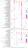
Dot plots show the KEGG pathways significantly enriched in the PNS (A) and HFD (B) conditions. Dot size is proportional to the number of featured DEPs; shades of color from red to blue indicate the level of significance.
Fig. 5
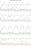
Fig. 5. Neuroinflammatory and homeostatic microglial markers in male and female fetal brains at E18.
A Cytokine mRNA expression in fetal brains following prenatal exposure to psychophysical stress. PNS increased levels of Il-1β in female fetal brains compared to males (interaction stress x treatment x sex: F1,38 = 3.61, p = 0.0651; Šídák’s post hoc comparisons: * p < 0.05 males PNS-Veh vs females PNS-Veh). PNS also upregulated Il-6 in females (interaction stress x sex: F1,39 = 5.54, p = 0.0237; Šídák’s post hoc comparisons: ** p < 0.01 PNS-Veh vs CTRL-Veh), NAC prevented this latter effect (interaction stress x treatment: F1,39 = 8.34, p = 0.0063; Šídák’s post hoc comparisons: * p < 0.05 PNS-NAC vs PNS-Veh). Male fetal brains showed higher levels of Tnf-α (main effect of sex: F1,38 = 4.34, £ p = 0.0439). Expression levels of Tgf-β and Igf-1 were not affected by either PNS nor NAC (interaction stress x treatment x sex Tgf-β: F1,39 = 0.0398, p = 0.8428; Igf-1: F1,37 = 0.21, p = 0.6495). B Cytokine mRNA expression in mHFD fetal brains. mHFD increased Il-1β and Igf-1 expression levels in both sexes (main effect of diet Il-1β ː F1,38 = 7.03, # p = 0.0116; Igf-1: F1,39 = 6.01, # p = 0.0188) as well as Tgf-β in females (interaction diet x sex: F1,39 = 8.09, p = 0.0070; Šídák’s post hoc comparisons: * p < 0.05 HFD-Veh vs CD-Veh). mHFD showed a trend to increase Tnf-α levels (interaction diet x sex: F1,38 = 4.45, p = 0.0415). Prenatal NAC overall reduce Il-6 levels (main effect of treatment: F1,38 = 5.43, p = 0.0252). C Markers of homeostatic and immune/redox-related functions of microglia in PNS fetal brains. Neither PNS nor NAC affected Cd 68, Trem 2 and Tmem 119 (Cd 68: F1,40 = 0.184, p = 0.6702; Trem 2: F1,40 = 0.460, p = 0.5014; Tmem 119: F1,36 = 6.25, pp = 0.0171). NAC treatment reduced iNos/Arg-1 mRNA ratio in PNS males (interaction stress x treatment x sex: F1,35 = 7.55, p = 0.0094, Šídák’s post hoc comparison: * p < 0.05 males PNS-NAC vs PNS-Veh). A significant interaction between stress x sex x treatment was found in Ucp2 levels (interaction stress x treatment x sex: F1,40 = 5.14, p = 0.0288). D Markers of homeostatic and immune/redox functions of microglia in mHFD fetal brains. mHFD had sex-dependent effects increasing levels of Tmem 119 and Cd 68 specifically in females (interaction diet x sex Cd 68: F1, 40 = 7.15, p = 0.0108; Šídák’s post hoc comparisons: * p < 0.05 females HFD-Veh vs CD-Veh; ** p < 0.01 males HFD-Veh vs females HFD-Veh; Tmem 119: F1,38 = 5.13, p = 0.0293; Šídák’s post hoc comparisons: * p < 0.05 females HFD-Veh vs CD-Veh; * p < 0.05 males HFD-Veh vs females HFD-Veh). Prenatal NAC overall reduced Cd 68 levels (main effect of treatment: F1,40 = 4.79, $ p = 0.0345). No changes were observed for Trem 2 levels (interaction diet x sex x treatment: F1,40 = 0.46, p = 0.5014). The iNos/Arg-1 ratio was found increased in mHFD fetal brains in both sexes (main effect of diet: F1,38 = 10.3, ## p = 0.0027). Generally, control males showed higher Ucp-2 mRNA levels when compared to females (interaction diet x treatment x sex F1,34 = 6.37, p = 0.0165; Šídák’s post hoc comparisons: * p < 0.05 males CD-Veh vs females CD-Veh). In particular, mHFD reduced Ucp-2 in males (** p < 0.01 HFD-Veh vs CD-Veh); likewise NAC downregulated it specifically in control males (** p < 0.01 CD-NAC vs CD-Veh). Three-way ANOVA. Number of subjects: 5–6 within each experimental group. Data are mean ± SEM.
Fig. 6
Fig. 6. Main sex-dependent effects following PNS or mHFD.
Both stressors increased systemic oxidative stress markers in the pregnant mother, an effect moderated by NAC. During prenatal life, greater sex-dependent effects characterised placental physiology in males and brain gene expression in females. Arrows pointing up indicate increasing - while arrows pointing down decreasing - effects on the outcomes. Rescuing effects of the prenatal administration of NAC are marked by a blue tick.

References

    1. Cirulli F, Musillo C, Berry A. Maternal obesity as a risk factor for brain development and mental health in the offspring. Neuroscience. 2020;447:122–35. - PubMed
    1. Davis J, Mire E. Maternal obesity and developmental programming of neuropsychiatric disorders: an inflammatory hypothesis. Brain Neurosci Adv. 2021;5:239821282110034. - PMC - PubMed
    1. Khambadkone SG, Cordner ZA, Tamashiro KLK. Maternal stressors and the developmental origins of neuropsychiatric risk. Front Neuroendocrinol. 2020;57:100834 10.1016/j.yfrne.2020.100834. - PMC - PubMed
    1. Sanchez CE, Barry C, Sabhlok A, Russell K, Majors A, Kollins SH, et al. Maternal pre-pregnancy obesity and child neurodevelopmental outcomes: a meta-analysis. Obes Rev. 2018;19:464–84. - PMC - PubMed
    1. Cirulli F, De Simone R, Musillo C, Ajmone-Cat MA, Berry A. Inflammatory signatures of maternal obesity as risk factors for neurodevelopmental disorders: role of maternal microbiota and nutritional intervention strategies. Nutrients. 2022;14:3150 10.3390/nu14153150. - PMC - PubMed

MeSH terms

LinkOut - more resources