

Validation of dementia-free status in the iranian brain imaging database using ADNI data: a transition point from middle age to older adulthood
- PMID: 40640729
- PMCID: PMC12243199
- DOI: 10.1186/s12883-025-04293-3
Validation of dementia-free status in the iranian brain imaging database using ADNI data: a transition point from middle age to older adulthood
Abstract
Objective: To validate the Iranian Brain Imaging Database (IBID) for dementia-free status in middle-aged to older adults (over 55 years old) by comparing dementia probability metrics with Alzheimer's disease (AD) patients from the Alzheimer's Disease Neuroimaging Initiative (ADNI).
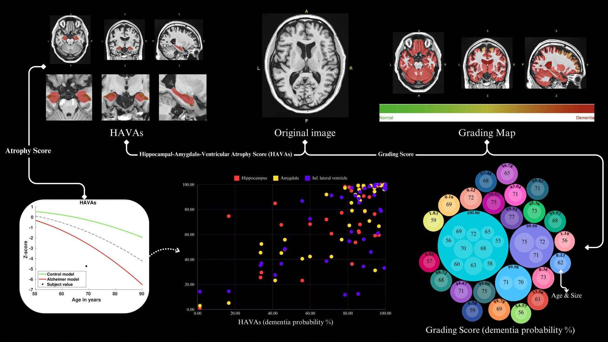
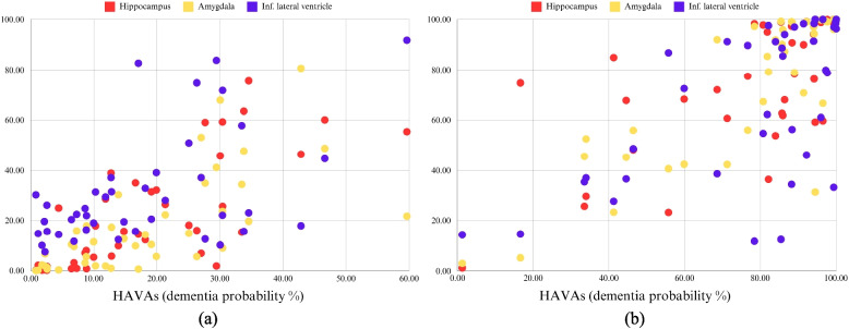
Methods: This cross-sectional study included 41 cognitively normal participants from IBID (age > 55 years; median (IQR) = 64 (62, 68) years; females = 51.2%) and 41 age/sex-matched AD patients from ADNI (age > 55 years; median (IQR) = 69 (62, 71) years; females = 53.7%). High-resolution 3D T1-weighted MPRAGE MRI data were harmonized across Siemens 3.0 Tesla scanners, preprocessed via the AssemblyNet-AD pipeline (volBrain), and analyzed for grading scores and hippocampal-amygdalo-ventricular atrophy scores (HAVAs), along with hippocampus, amygdala, and inferior lateral ventricle atrophy scores. Non-parametric Mann-Whitney U tests were used to compare group differences, and quantile regression (25th, 50th, and 75th quantiles) model atrophy severity-dependent associations.
Results: IBID participants exhibited minimal dementia probability (median grading score = 0.20% vs. ADNI = 99.93%; U = 84.0, p < 0.0001) and markedly lower atrophy across all regions (HAVAs: 14.76 vs. 86.41; U = 75.0, p < 0.0001), with non-overlapping interquartile ranges. Quantile regression revealed stable hippocampal contributions (q₀.₅ coefficient = 0.311, p < 0.001) and increasing group disparities at higher atrophy severities (q₀.₇₅: IBID vs. ADNI coefficient = - 11.82, p < 0.001).
Conclusion: We provided validated dementia-free IBID neuroimaging data to distinguish normal aging from dementia-AD status.
Keywords: Aging; Alzheimer's disease; Dementia-free classification; MRI; Neuroimaging; Structural atrophy.
© 2025. The Author(s).
Conflict of interest statement
Declarations. Ethics approval and consent to participate: The Ethics Committee of the National Institute for Medical Research Development (NIMAD) approved this study (Ethical Code: IR.NIMAD.REC.1396.319) in accordance with the Declaration of Helsinki. All individuals and/or their legal representatives gave written informed consent before entering the study. Consent for publication: Not applicable. Competing interests: The authors declare no competing interests.
Figures

Similar articles
-
Predicting cognitive decline: Deep-learning reveals subtle brain changes in pre-MCI stage.J Prev Alzheimers Dis. 2025 May;12(5):100079. doi: 10.1016/j.tjpad.2025.100079. Epub 2025 Feb 6. J Prev Alzheimers Dis. 2025. PMID: 39920001 Free PMC article.
-
¹⁸F-FDG PET for the early diagnosis of Alzheimer's disease dementia and other dementias in people with mild cognitive impairment (MCI).Cochrane Database Syst Rev. 2015 Jan 28;1(1):CD010632. doi: 10.1002/14651858.CD010632.pub2. Cochrane Database Syst Rev. 2015. PMID: 25629415 Free PMC article.
-
Baseline habitual dietary nitrate intake and Alzheimer's Disease related neuroimaging biomarkers in the Australian Imaging, Biomarkers and Lifestyle study of ageing.J Prev Alzheimers Dis. 2025 Jun;12(6):100161. doi: 10.1016/j.tjpad.2025.100161. Epub 2025 Apr 11. J Prev Alzheimers Dis. 2025. PMID: 40221237
-
Medial temporal lobe atrophy in Down syndrome along the Alzheimer's disease continuum.Brain. 2025 Jul 7;148(7):2509-2521. doi: 10.1093/brain/awaf133. Brain. 2025. PMID: 40243675
-
18F PET with florbetapir for the early diagnosis of Alzheimer's disease dementia and other dementias in people with mild cognitive impairment (MCI).Cochrane Database Syst Rev. 2017 Nov 22;11(11):CD012216. doi: 10.1002/14651858.CD012216.pub2. Cochrane Database Syst Rev. 2017. PMID: 29164603 Free PMC article.
References
Publication types
MeSH terms
LinkOut - more resources
Full Text Sources
Medical
