. 2025 Jul 10;23(1):772.
doi: 10.1186/s12967-025-06808-5.

Exosomes derived from hypoxia-preconditioned M2 macrophages alleviate degeneration in knee osteoarthritis through the miR‑124‑3p/STAT3 axis

Affiliations

Exosomes derived from hypoxia-preconditioned M2 macrophages alleviate degeneration in knee osteoarthritis through the miR‑124‑3p/STAT3 axis

Hao Li et al. J Transl Med. .

Abstract

Background: M2 macrophages-derived exosomes (M2φ-Exos) have been demonstrated to effectively alleviate osteoarthritis (OA) in animal models. Hypoxic preconditioning is commonly used to enhance the biological effects of stem cell-derived exosomes, but its impact on M2φ-Exos remains unclear. This study aims to investigate whether hypoxic preconditioning could enhance the biological effects of M2φ-Exos in OA treatment and to explore the underlying molecular mechanisms, with the goal of providing new insights for the development of safe and effective OA therapeutic strategies.

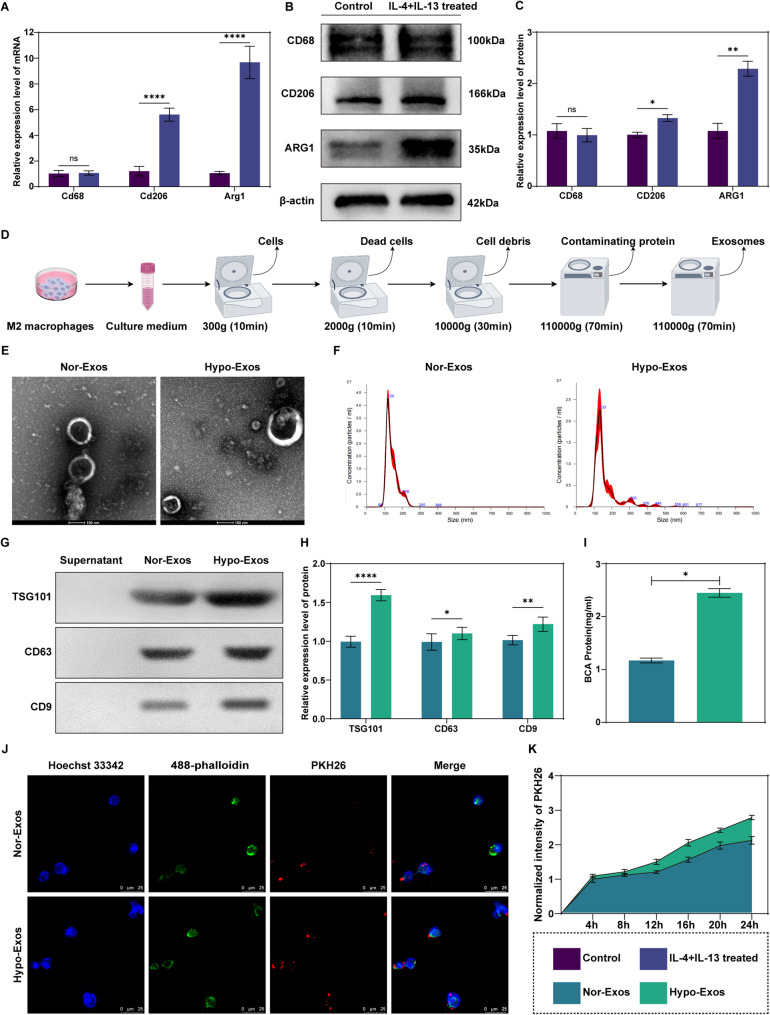
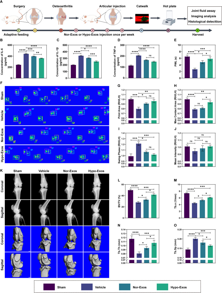
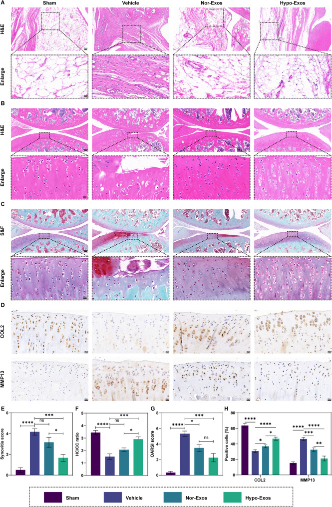
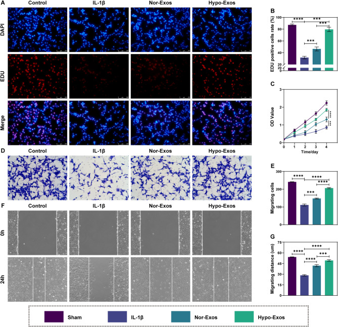
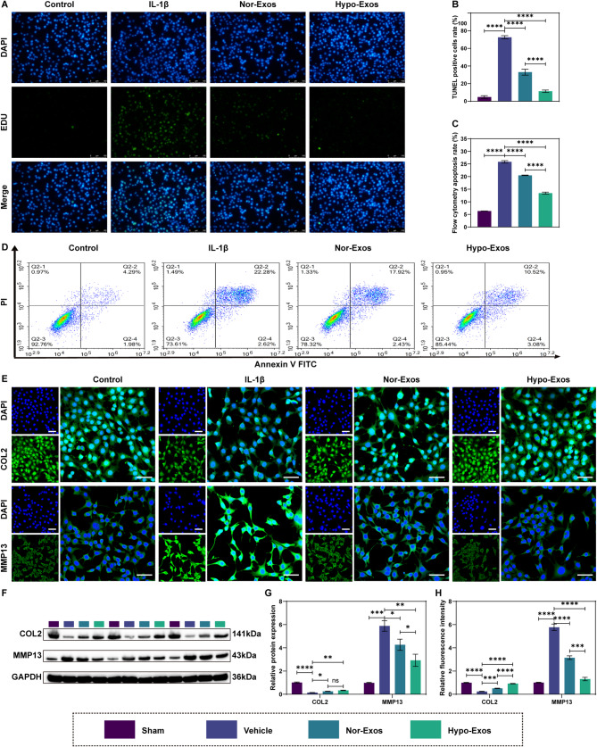
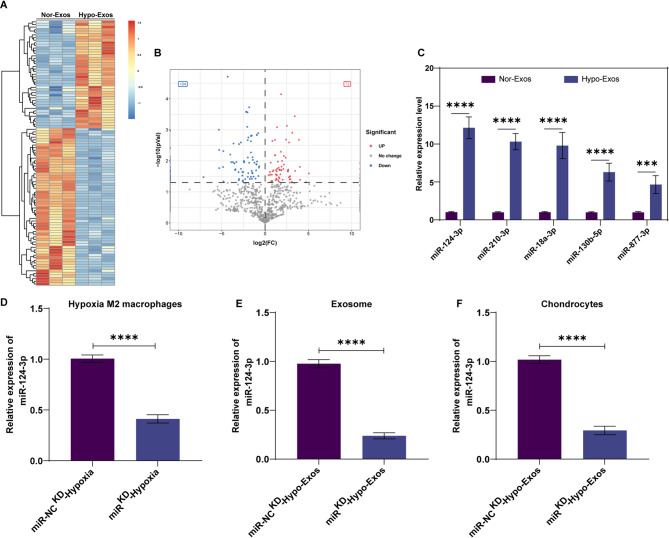
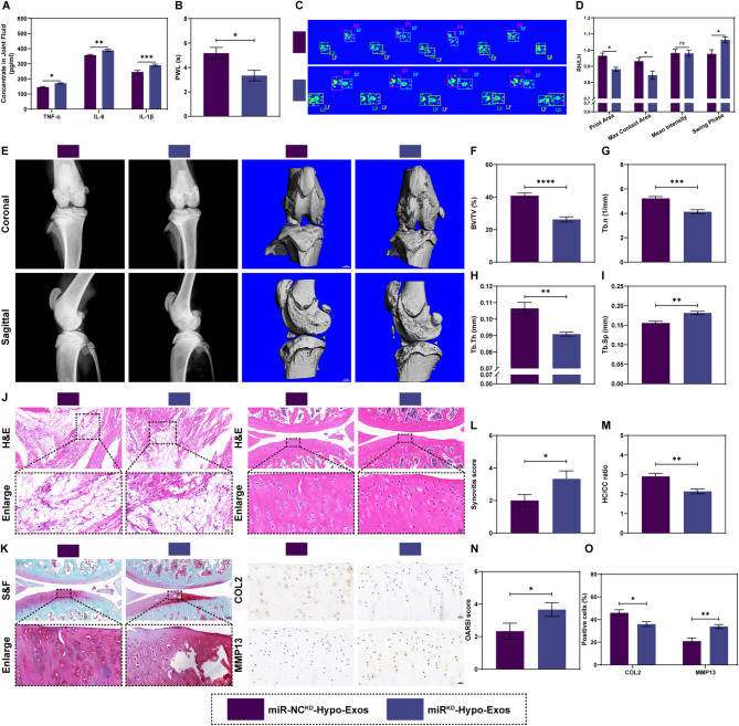
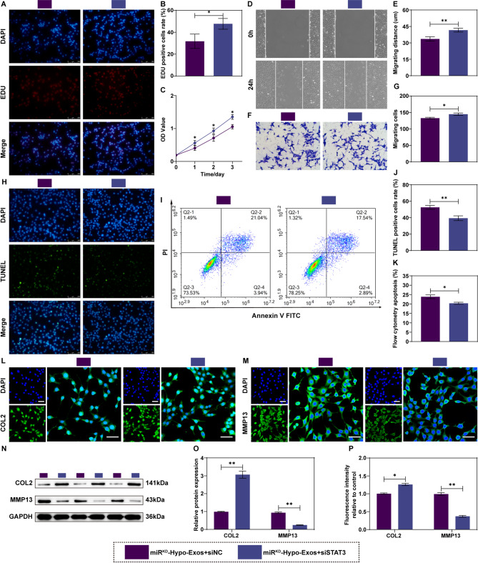
Methods: Exosomes were extracted from the supernatants of M2 macrophages cultured under normoxic or hypoxic conditions using low-temperature differential ultracentrifugation and were designated as Nor-Exos and Hypo-Exos. The exosomes were characterized by transmission electron microscopy, nanoparticle tracking analysis, and Western blotting. To evaluate the impact of hypoxic preconditioning on the biological effects of M2φ-Exos, the therapeutic effects of Nor-Exos and Hypo-Exos were assessed in an IL-1β-induced chondrocyte inflammation model and a rat knee OA model established by surgical intervention. Exosomal miRNAs with differential expression between Nor-Exos and Hypo-Exos were identified through exosomal miRNA sequencing. The miRNA with the highest upregulation in Hypo-Exos was selected for further functional validation. To investigate the role of this miRNA, miRNA inhibitors were used to knock down its expression in Hypo-Exos, and the subsequent impact of this change on Hypo-Exos activity was evaluated. Bioinformatic tools and dual-luciferase reporter assays were used to predict and verify the downstream target genes of the miRNA. Target gene expression was knocked down using small interfering RNA, and the effect of downregulating target gene expression on the inhibitory effect of low miRNA expression on Hypo-Exos was observed at the cellular level.

Results: Compared to Nor-Exos, Hypo-Exos exhibited more effective therapeutic effects in both inflammatory chondrocytes and OA rats. miR-124-3p was identified as the most upregulated miRNA in Hypo-Exos, and the suppression of miR-124-3p expression significantly inhibited the biological effects of Hypo-Exos. STAT3 was determined to be a downstream target gene of miR-124-3p. Further cellular experiments revealed that downregulation of STAT3 expression in chondrocytes successfully alleviated the inhibitory effect of low miR-124-3p expression on the biological effects of Hypo-Exos.

Conclusions: Hypoxic preconditioning enhances the biological effects of M2φ-Exos in the treatment of OA. The underlying molecular mechanism is associated with increased delivery of miR-124-3p to chondrocytes, which subsequently inhibits the post-transcriptional expression of STAT3. This provides a promising therapeutic strategy for the clinical intervention of OA.

Keywords: Exosomes; Hypoxic preconditioning; M2 macrophages; Osteoarthritis; STAT3; miR-124-3p.


Conflict of interest statement

Declarations. Ethics approval and consent to participate: The experimental procedures involving in animals was approved by the Ethics Committee of the Second Hospital of Shanxi Medical University (DW2024002). Consent for publication: Not applicable. Competing interests: The authors declare that they have no competing interests.

Figures

Fig. 1
Fig. 1
Identification of M2 macrophages and characterization of exosomes. (A) RT-qPCR to evaluate the mRNA expression levels of Cd68, Cd206, and Arg1 in RAW264.7 treated with IL-4 and IL-13. (B-C) Western blotting to detect the proteins expression levels of CD68, CD206, and ARG1 in RAW264.7 treated with IL-4 and IL-13. (D) Schematic diagram of exosomes extraction by ultracentrifugation. (E) Representative transmission electron microscopy images of two exosomes. Scale bar: 100 nm. (F) NTA to determine the size distribution of two exosomes. (G-H) Western blot analysis to detect the exosomal proteins expression of CD9, CD63, and TSG101 in two exosomes. (I) BCA assay to measure the protein concentration in two exosomes. (J) Cellular uptake of PKH26-labeled exosomes into chondrocytes. Chondrocytes were incubated with PKH26-lablled exosomes (red), chondrocytes were co-stained with phalloidin (green) and nuclei were stained with Hoechst 33,342 (blue). Scale bar: 25 μm. (K) Quantitative analysis of fluorescence intensity of uptake. Data are presented as the mean ± standard deviation (SD) (n = 3). ns, no significance, *P < 0.05, **P < 0.01, ***P < 0.005, ****P < 0.001
Fig. 2
Fig. 2
Inflammation, behavioral and imaging evaluation of Hypo-Exos on KOA rats in vivo. (A) Schematic diagram of in vivo evaluation. (B-D) ELISA to quantify inflammatory factors (IL-6, IL-1β, and TNF-α) in joint fluid. (E) Hot plate to measure paw withdrawal latency. (F) Representative images of Catwalk. (G-J) Quantitative analysis of gait indexes. (K) Representative images of X-ray and 3D reconstruction based on micro-CT. Scale bar: 1 mm. (L-O) Quantitative analysis of the region of interest in the subchondral bone of tibia. Data are presented as the mean ± SD (n = 5). ns, no significance, *P < 0.05, **P < 0.01, ***P < 0.005, ****P < 0.001
Fig. 3
Fig. 3
Histological evaluation of Hypo-Exos on KOA rats in vivo. (A) H&E staining of synovium to score synovial activation. Scale bar: 50 μm (20×), 20 μm (40×). (B) H&E staining of articular cartilage to observe the morphology and structure. Scale bar: 100 μm (10×), 20 μm (40×). (C) S&F staining of articular cartilage observe extracellular matrix and collagen fibers. Scale bar: 100 μm (10×), 20 μm (40×). (D) Immunohistochemical analysis for COL2 and MMP13 in articular cartilage. Scale bar: 20 μm. (E) Quantitative analysis of the synovial score in each group. (F) Quantitative analysis of the HC/CC ration in each group. (G) Quantitative analysis of the OARIS score in each group. (H) Quantitative analysis of COL2 and MMP13 in each group. Data are presented as the mean ± SD (n = 5). ns, no significance, *P < 0.05, **P < 0.01, ***P < 0.005, ****P < 0.001
Fig. 4
Fig. 4
Hypo-Exos promotes the proliferation and migration of chondrocytes in vitro. (A-C) EDU staining (red) and CCK-8 assays to observe the cell proliferation activity. Scale bar: 100 μm. (D-G) transwell assay and scratch test to measure the cell migration ability. Data are presented as the mean ± SD (n = 3). ns, no significance, *P < 0.05, **P < 0.01, ***P < 0.005, ****P < 0.001
Fig. 5
Fig. 5
Hypo-Exos inhibit the apoptosis and extracellular matrix catabolism of chondrocytes in vitro. (A-D) TUNEL staining (green) and Annexin V/FITC/PI double-staining flow cytometry to analyze the cell apoptosis rates. Scale bar: 100 μm. (E-H) Immunofluorescence and western blotting to detect the expression levels of proteins related to extracellular matrix anabolism (COL2) and catabolism (MMP13). Scale bar = 50 μm. Data are presented as the mean ± SD (n = 3). ns, no significance, *P < 0.05, **P < 0.01, ***P < 0.005, ****P < 0.001
Fig. 6
Fig. 6
The miR-124-3p is upregulated in Hypo-Exos and transferred to chondrocytes via exosomes. (A) Heat map of the differentially expressed miRNAs between Nor-Exos and Hypo-Exos. (B) Volcano map of the differentially expressed miRNAs between Nor-Exos and Hypo-Exos. (C) Verification of the top five elevated miRNAs in Hypo-Exos by RT-qPCR. (D) The expression level of miR-124-3p in hypoxia M2 macrophages after transfected with miR-124-3p inhabitor. (E) The expression level of miR-124-3p in exosomes derived from hypoxia M2 macrophages transfected with miR-124-3p inhabitor. (F) The expression level of miR-124-3p in chondrocytes treated with exosomes derived from hypoxia M2 macrophages transfected with miR-124-3p inhabitor. Data are presented as the mean ± SD (n = 3). ns, no significance, *P < 0.05, **P < 0.01, ***P < 0.005, ****P < 0.001
Fig. 7
Fig. 7
Knockdown of miR-124-3p in hypoxia M2 macrophages weakens the Hypo-Exos-mediated positive effect of chondrocytes in vitro. (A-C) EDU staining (red) and CCK-8 assays to observe cell proliferation activity. Scale bar: 100 μm. (D-G) Transwell and scratch test to measure the cell migration ability. (H-K) TUNEL staining (green) and Annexin V/FITC/PI double-staining flow cytometry to analyze the cell apoptosis rates. Scale bar: 100 μm. (L-P) Immunofluorescence and western blotting to detect the expression levels of proteins related to extracellular matrix anabolism (COL2) and catabolism (MMP13). Scale bar: 50 μm. Data are presented as the mean ± SD (n = 3). ns, no significance, *P < 0.05, **P < 0.01, ***P < 0.005, ****P < 0.001
Fig. 8
Fig. 8
Knockdown of miR-124-3p in hypoxia M2 macrophages attenuates the Hypo-Exos-mediated protective effect in vivo. (A) ELISA to quantify inflammatory factors (IL-6, IL-1β, and TNF-α) in joint fluid. (B) Hot plate to measure paw withdrawal latency. (C) Representative images of Catwalk. (D) Quantitative analysis of gait indexes. (E) Representative images of X-ray and 3D reconstruction based on micro-CT. Scale bar: 1 mm. (F-I) Quantitative analysis of the region of interest in the subchondral bone of tibia. (J) H&E staining of synovium and articular cartilage. Scale bar: 100 μm (10×), 50 μm (20×), 20 μm (40×). (K) S&F staining and immunohistochemical analysis in articular cartilage. Scale bar: 100 μm (10×), 20 μm (40×). (L) Quantitative analysis of the synovial score. (M) Quantitative analysis of the HC/CC ration. (N) Quantitative analysis of the OARIS score. (O) Quantitative analysis of COL2 and MMP13. Data are presented as the mean ± SD (n = 5). ns, no significance, *P < 0.05, **P < 0.01, ***P < 0.005, ****P < 0.001
Fig. 9
Fig. 9
The miR-124-3p-abundant Hypo-Exos play positive effect by targeting STAT3. (A-C) EDU staining (red) and CCK-8 assays to observe cell proliferation activity. Scale bar: 100 μm. (D-G) Transwell and scratch test to measure the cell migration ability. (H-K) TUNEL staining (green) and Annexin V/FITC/PI double-staining flow cytometry to analyze the cell apoptosis rates. Scale bar: 100 μm. (L-P) Immunofluorescence and western blotting to detect the expression levels of proteins related to extracellular matrix anabolism (COL2) and catabolism (MMP13). Scale bar: 50 μm. Data are presented as the mean ± SD (n = 3). ns, no significance, *P < 0.05, **P < 0.01, ***P < 0.005, ****P < 0.001
Fig. 10
Fig. 10
Schematic diagram of hypoxic M2 macrophage promotes proliferation and migration and inhabits apoptosis and ECM degradation in chondrocyte. Hypoxia mediates enhanced production of miR-124-3p, which is then transferred to chondrocyte where it exhibits its biological role and potential therapeutic effect in alleviating KOA via the miR124-3p/STATA3 axis

Similar articles

References

    1. Gelber AC. Knee osteoarthritis. Ann Intern Med. 2024;177(9):Itc129–44. - PubMed
    1. Minnig MCC, Golightly YM, Nelson AE. Epidemiology of osteoarthritis: literature update 2022–2023. Curr Opin Rheumatol. 2024;36(2):108–12. - PMC - PubMed
    1. Courties A, Kouki I, Soliman N, Mathieu S, Sellam J. Osteoarthritis year in review 2024: epidemiology and therapy. Osteoarthritis Cartilage. 2024;32(11):1397–404. - PubMed
    1. Tang S, Zhang C, Oo WM, Fu K, Risberg MA, Bierma-Zeinstra SM, et al. Osteoarthr Nat Rev Dis Primers. 2025;11(1):10. - PubMed
    1. Jenei-Lanzl Z, Zaucke F. Osteoarthritis year in review 2024: biology. Osteoarthritis Cartilage. 2025;33(1):58–66. - PubMed

MeSH terms

LinkOut - more resources