. 2025 Jul 10;23(1):778.
doi: 10.1186/s12967-025-06812-9.

USP5-Mediated PD-L1 deubiquitination regulates immunotherapy efficacy in melanoma

Affiliations

USP5-Mediated PD-L1 deubiquitination regulates immunotherapy efficacy in melanoma

Jiaheng Xie et al. J Transl Med. .

Abstract

Background: The role of post-translational modifications(PTMs) in PD-L1-mediated immune resistance and melanoma progression remains poorly understood.

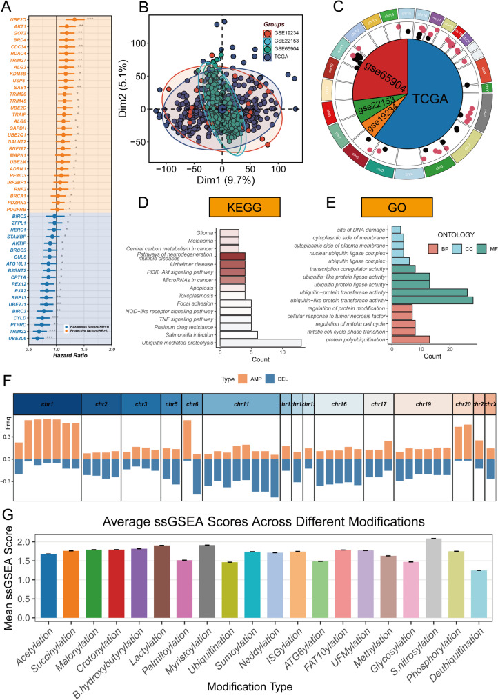
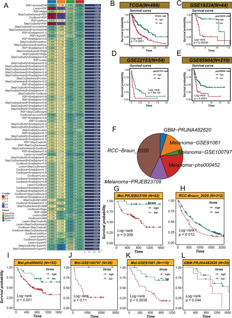
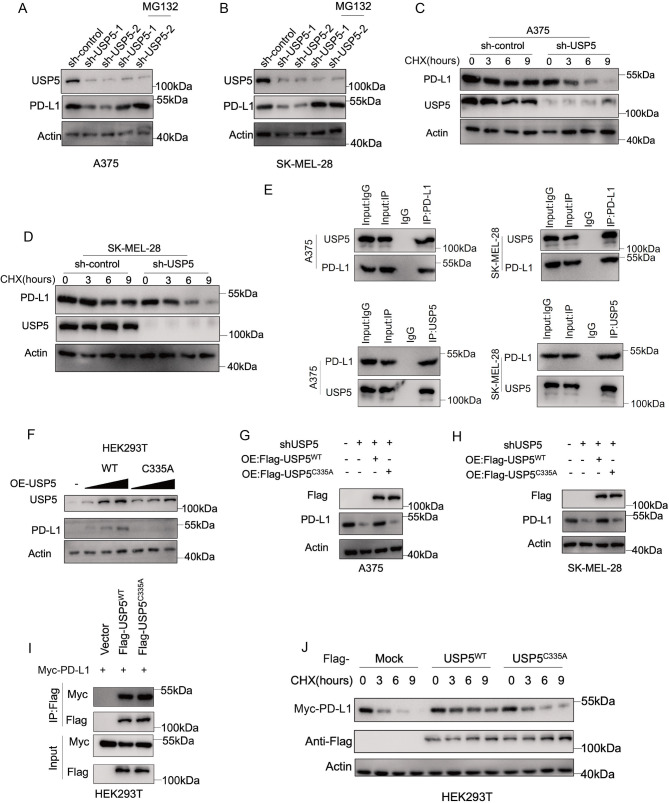
Methods: We conducted multi-omics analyses and constructed a prognostic model based on PTM-related genes using machine learning to identify key regulators in melanoma. In vitro and in vivo experiments, including cell culture, flow cytometry, and subcutaneous allografts models, were used to investigate USP5's function. Protein-protein interactions were validated using Western blotting and co-immunoprecipitation, while PD-L1 stability and ubiquitination were assessed using cycloheximide (CHX) chase and ubiquitination assays.

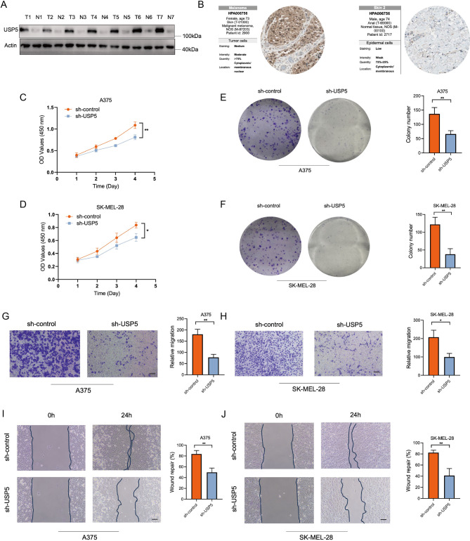
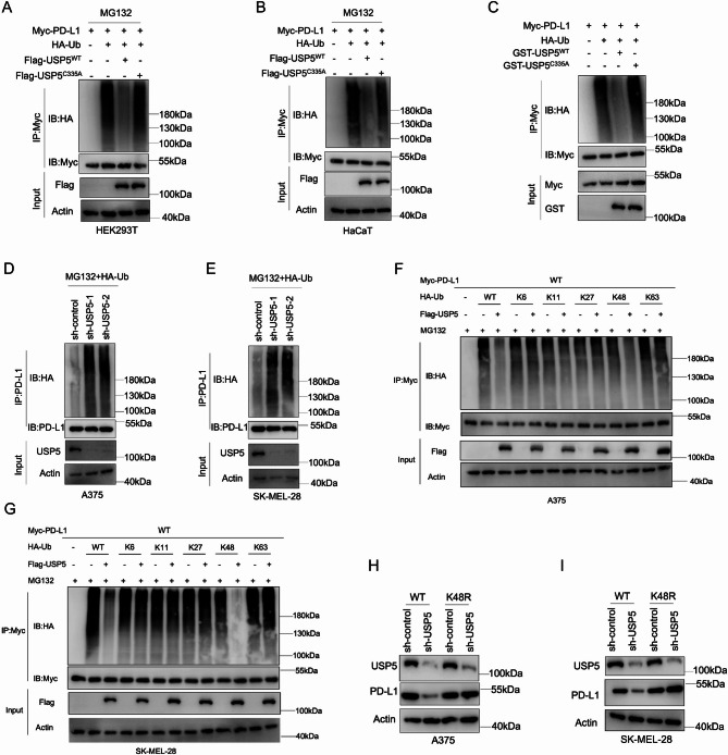
Results: USP5 was identified as a key DUB that specifically deubiquitinates K48-linked polyubiquitin chains on PD-L1, stabilizing its protein levels. USP5 knockdown reduced PD-L1 expression, enhanced CD8 + T-cell infiltration and activation, and suppressed melanoma progression in both in vitro and in vivo models. Furthermore, combining USP5 knockdown with anti-PD-1 therapy significantly improved therapeutic efficacy by reducing tumor burden and promoting T-cell activation.

Conclusion: USP5 promotes immune escape in melanoma by stabilizing PD-L1 through deubiquitination, representing a novel mechanism hindering the efficacy of ICIs. Targeting USP5 could enhance anti-PD-1 therapy and improve patient outcomes. These findings underscore the therapeutic potential of USP5 inhibition as a strategy to overcome immune resistance in melanoma.

Keywords: Immune escape; Immunotherapy; Melanoma; PD-L1; USP5.


Conflict of interest statement

Declarations. Ethics approval: The collection of specimens and animal handling for the study have been reviewed and approved by the Ethics Committee of the First Affiliated Hospital of Nanjing Medical University(2406070 and 2024-SR-949) and Ethics Committee of Xiangya Hospital of Central South University(2024030015). Competing interests: The authors declare no competing interests.

Figures

Fig. 1
Fig. 1
Identification and Functional Analysis of Prognostically Relevant PTM-Related Genes (PTMRGs) in Melanoma. (A) Volcano plot illustrating differentially expressed prognostic PTMRGs identified through TCGA and GTEx datasets. Red and blue dots represent upregulated and downregulated genes, respectively. (B) Batch effect correction applied to multiple datasets, demonstrating consistent gene expression profiles post-normalization. (C) Chromosomal distribution of PTMRGs visualized to indicate genomic localization. (D, E) KEGG and GO enrichment analyses highlighting significant pathways, including PI3K-Akt signaling and ubiquitin-like protein transferase activity. (F) Copy number variation (CNV) analysis of PTMRGs, revealing frequent genomic alterations. (G) ssGSEA analysis showing high activation of PTM-related pathways in melanoma samples
Fig. 2
Fig. 2
Development and Validation of PTMRM for Prognostic and Immunotherapy Applications. (A) Overview of machine learning algorithms tested for PTMRM construction, with RSF + survivalSVM selected as optimal. (BE) Kaplan-Meier survival curves in TCGA and validation cohorts stratified by PTMRM scores. (FL) Application of PTMRM scores in melanoma and pan-cancer immunotherapy datasets, demonstrating consistent prognostic trends and improved outcomes for low-score patients
Fig. 3
Fig. 3
PTMRM Performance Comparison and Immune Microenvironment Profiling. (A) C-index comparison of PTMRM with other melanoma prognostic models across four cohorts. (B) PCA of PTMRM gene expression levels, effectively stratifying melanoma patients. (C) ROC curve analysis validating PTMRM’s prognostic accuracy. (D) Representative H&E-stained tissue sections from TCGA high- and low-risk groups, showing higher lymphocyte infiltration in low-risk samples
Fig. 4
Fig. 4
Single-Cell Analysis of PTMRM Scores and Cellular Interactions. (A) Annotated major cell types from single-cell RNA sequencing of melanoma. (B) PTMRM score distribution across cell populations. (C) USP5 expression patterns in different cell types. (D, E) Proportional analysis of high- and low-PTMRM groups, showing enrichment of immune cells in the low-score group. (FI) Cellular interaction analyses reveal enhanced intercellular signaling in the high-PTMRM group, implicating pro-tumorigenic pathways
Fig. 5
Fig. 5
Functional Characterization of USP5 in Melanoma Progression. (A) Western blot analysis of USP5 protein levels in melanoma versus control samples. (B) Immunohistochemistry data from HPA confirming elevated USP5 expression. (CH) Functional assays (CCK8, colony formation, migration, and wound healing) demonstrating reduced proliferation and migration in USP5-knockdown melanoma cell lines
Fig. 6
Fig. 6
USP5 Regulates PD-L1 Stability via Ubiquitin-Proteasome Pathway. (A, B) Western blot showing reduced PD-L1 protein levels upon USP5 knockdown, rescued by MG132 treatment. (C, D) CHX chase experiments demonstrating reduced PD-L1 stability with USP5 knockdown. (E) Co-IP assays confirming USP5-PD-L1 interaction. (FJ) Overexpression and mutational analyses of USP5 reveal its catalytic activity is critical for stabilizing PD-L1
Fig. 7
Fig. 7
USP5 Deubiquitinates K48-Linked Chains on PD-L1 to Stabilize Its Protein Levels. (A, B) Ubiquitination assays showing reduced PD-L1 ubiquitination with USP5-WT overexpression. (C) In vitro deubiquitination assays confirming USP5-WT’s activity on PD-L1. (D, E) Increased PD-L1 ubiquitination in USP5-knockdown melanoma cells. (FG) Specific targeting of K48-linked ubiquitin chains by USP5. (H, I) Restoration of PD-L1 protein levels with K48-resistant ubiquitin in USP5-depleted cells
Fig. 8
Fig. 8
USP5 Silencing Enhances CD8 + T Cell Activation and Amplifies Anti-PD-1 Therapy. (A) Workflow of in vivo mouse model experiments with YUMM1.7 melanoma cells. (BE) Tumor volume and weight analyses showing synergistic effects of USP5 knockdown and PD-1 inhibition. (FH) Flow cytometry analyses of CD8 + T-cell infiltration and activation in tumor microenvironment, demonstrating enhanced immune response with combination therapy

References

    1. Carvalho LAD, Aguiar FC, Smalley KSM, Possik PA. Acral melanoma: new insights into the immune and genomic landscape. Neoplasia. 2023;46:100947. 10.1016/j.neo.2023.100947 - PMC - PubMed
    1. Samarkina A, Youssef MK, Ostano P, et al. Androgen receptor is a determinant of melanoma targeted drug resistance. Nat Commun. 2023;14(1):6498. 10.1038/s41467-023-42239-w. Published 2023 Oct 14. - PMC - PubMed
    1. Tagore M, Hergenreder E, Perlee SC, et al. GABA regulates electrical activity and tumor initiation in melanoma. Cancer Discov. 2023;13(10):2270–91. 10.1158/2159-8290.CD-23-0389 - PMC - PubMed
    1. Lazaroff J, Bolotin D. Targeted therapy and immunotherapy in melanoma. Dermatol Clin. 2023;41(1):65–77. 10.1016/j.det.2022.07.007 - PubMed
    1. Ozbay Kurt FG, Lasser S, Arkhypov I, Utikal J, Umansky V. Enhancing immunotherapy response in melanoma: myeloid-derived suppressor cells as a therapeutic target. J Clin Invest. 2023;133(13):e170762. 10.1172/JCI170762. Published 2023 Jul 3. - PMC - PubMed