. 2025 Jul 10;56(1):148.
doi: 10.1186/s13567-025-01582-0.

African swine fever virus infection of porcine peripheral blood monocyte-derived macrophages induces the formation of tunneling nanotube-connected large vesicle-like cell segments: a potential mechanism for intercellular ASFV trafficking

Affiliations

African swine fever virus infection of porcine peripheral blood monocyte-derived macrophages induces the formation of tunneling nanotube-connected large vesicle-like cell segments: a potential mechanism for intercellular ASFV trafficking

Brecht Droesbeke et al. Vet Res. .

Abstract

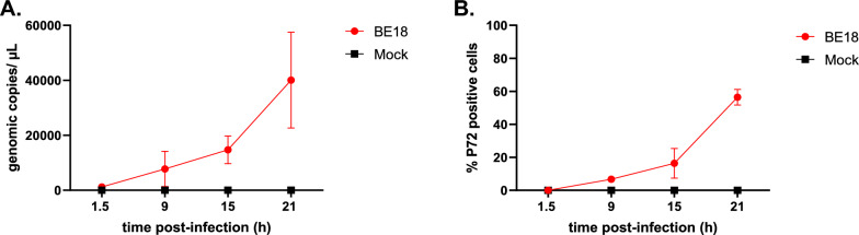
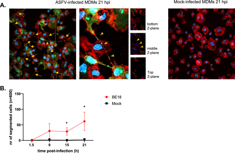
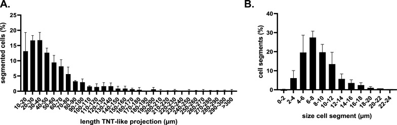
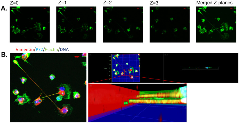
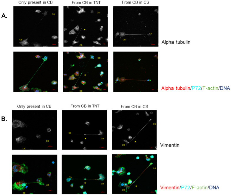
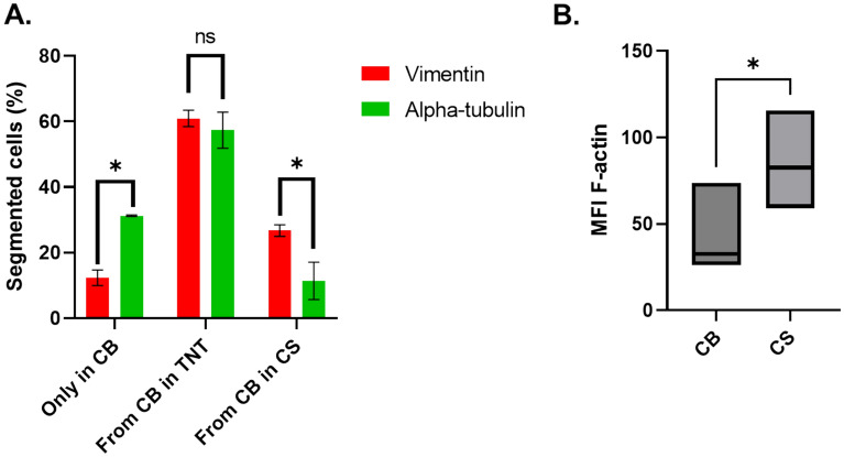
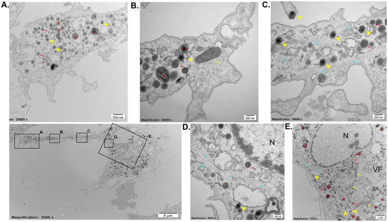
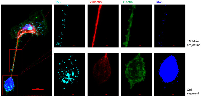
African swine fever (ASF) is a highly fatal viral disease in pigs, with mortality rates that can reach 100%. The causative agent, African swine fever virus (ASFV), primarily targets cells of the mononuclear phagocytic system (MPS), particularly monocyte-derived macrophages (MDMs). Despite the severity of the disease, there are currently no effective antiviral treatments available in Europe. A significant barrier to therapeutic development is the limited understanding of how ASFV interacts with its primary target cells. A deeper understanding of the morphological changes induced by ASFV in infected cells is crucial to this effort. To address this knowledge gap, we used conventional and confocal immunofluorescence microscopy, as well as transmission electron microscopy, to investigate ASFV-infected primary MDMs. Our analysis revealed that ASFV infection leads to the formation of large cellular protrusions, which are characterized by vesicle-shaped cellular segments (CSs) at their tips. These protrusions contain all major cytoskeletal components, showing characteristics similar to those of tunneling nanotubes (TNTs). In 84.93% of the cases, the nucleus remained in the cell body (CB) near the viral factory. In the remaining cases, the nucleus was found within these CSs, whereas the viral factory was present in the CB. Additionally, 57.6% of the cells were in contact with the CS and distant cells, suggesting a potential mechanism for ASFV transmission. These findings suggest that ASFV induces cellular segmentation linked by TNT-like structures. Further research is needed to better understand the biogenesis and functional significance of these segmented cells, which could inform future strategies for combating ASFV.

Keywords: African swine fever virus; cytopathic effect; tunneling nanotubes.


Conflict of interest statement

Declarations. Ethics approval and consent to participate: The activities in the BSL-3 laboratory were authorized by Brussels Environment under reference number SBB 219 2020/0493. Competing interests: The authors declare that they have no competing interests.

Figures

Figure 1
Figure 1
Infection of monocyte-derived macrophages with ASFV BE18 (MOI = 1). A At the indicated time points, the supernatants were collected, and the ASFV copy number was determined by RT‒qPCR. B Cells were fixed in 3% PFA, and the percentage of infected cells was determined by IF microscopy.
Figure 2
Figure 2
ASFV infection leads to enhanced formation of TNT-like projections. A Representative images of ASFV- or mock-infected cells at 21 hpi. The cells were stained for the presence of viral p72 (green) with a mouse monoclonal antibody (1BC11), F-actin (red) with Phalloidin-iFluor 594, and DNA with Hoechst 33342. Long, straight F-actin protrusions (yellow arrows) were observed in a fraction of the p72-positive cells. A close-up view of ASFV-infected MDMs reveals a TNT-like projection extending across the cell body of a neighbouring MDM at the apical side (middle z-plane, yellow arrowheads). Scale bar: 10 µm. B At 1.5, 9, 15, and 21 hpi, 600 mock-infected and 600 p72-positive cells were scored for the presence of one or more straight F-actin-containing cellular protrusions after immunofluorescence staining of the nuclei, actin cytoskeleton, and viral p72 antigen. Significantly more ASFV-infected cells presented with straight F-actin protrusions at 15 hpi (p < 0.01) and 21 hpi (p < 0.001).
Figure 3
Figure 3
ASFV-induced segmented cells can make contact with distant cells through the TNT-connected cell segment (CS), which, in certain cases, contains the cell nucleus. A At 18 hpi, 300 segmented cells were scored for the presence or absence of contact with distant cells and for the presence or absence of the cell nucleus in the CS. B Illustrative images of p72-positive cells (green) with F-actin-containing projections (red) at 18 hpi. Different configurations are presented on the basis of the presence or absence of contact with distant cells and the presence or absence of the cell nucleus (blue) in the CS. Scale bar: 10 µm.
Figure 4
Figure 4
High heterogeneity in the length of TNTs and size of the cell segments in ASFV-infected segmented cells was detected at 18 hpi. A The measured lengths of the TNTs varied from a minimum of 10 µm to over 300 µm (n = 2), with the most commonly observed length ranging between 30 and 40 µm (16.77% ± 2.55). B A similar distribution pattern was observed for the diameter of the CS (n = 300), which ranged from just under 2 µm to 22–24 µm (n = 3). The most common diameter ranged between 6 and 8 µm (27.44% ± 3.34).
Figure 5
Figure 5
ASFV-induced projections are TNT-like projections. A Representative confocal images of cell projections in ASFV-infected MDMs at 18 hpi, showing filamentous F-actin (green) after staining with phalloidin-iF 488 at different z-planes (z = 0–3). Both the cell segment (CS) and the cell body (CB) can be observed attached to the coverslip (z = 0). Fragments of the TNT-like projection can be seen in higher z-planes (z = 1 and z = 2, respectively) and in the merged z-planes after image acquisition. B Orthogonal projection of the same image presented in panel A, showing the presence of vimentin (red) with an anti-vimentin antibody (VI10), p72 (cyan) with anti-p72 (1BC11), F-actin (green) with phalloidin-iF 488, and nuclei (blue) with Hoechst 33342. Three-dimensional reconstruction and close-up visualization at the interface of both cellular projections confirmed that the projections cross each other without establishing contact with one another or with the surface (blue plane) of the coverslip on which the cells are seeded. Scale bar: 10 µm.
Figure 6
Figure 6
Illustrative confocal IF images of the three different phenotypes of segmented cells after image acquisition. A At 18 hpi, ASFV-infected MDMs were visualized for the presence of alpha-tubulin (red), p72 (cyan), F-actin (green), and DNA (blue) by IF staining. The upper row shows grayscale images of alpha-tubulin, clearly illustrating the different distributions of the three phenotypes. In the first phenotype, alpha-tubulin is present only in the cell body (CB) (left column). In the second phenotype, alpha-tubulin filaments extend from the CB into the TNT-like projection (middle column). In the third phenotype, alpha-tubulin extends from the CB into the cell segment (CS) (right column). When filamentous alpha-tubulin did not reach the CS (left and middle columns), aggregates of alpha-tubulin were observed in the CS. B ASFV-infected MDMs were visualized for the presence of vimentin (red), p72 (cyan), F-actin (green), and DNA (blue) by IF staining. The upper row shows grayscale images of vimentin, clearly illustrating the different distributions among the three phenotypes, similar to what was observed for alpha-tubulin. In certain cases, a spindle-shaped midbody (asterisk) was observed along the course of the TNT-like projection. Scale bar: 10 µm.
Figure 7
Figure 7
Distribution of cytoskeletal filaments in ASFV-segmented cells. A At 18 hpi, a total of 180 segmented cells were scored for the intracellular distribution of vimentin or alpha-tubulin after confocal image acquisition. Significantly more segmented MDMs exhibited alpha-tubulin, which was present only in the cell body (CB) (p < 0.01). The largest fraction of segmented MDMs showed vimentin or alpha-tubulin extending from the CB into the TNT-like projection. Significantly more cells presented with vimentin present in the CS than with filamentous alpha-tubulin extending into the CS (p < 0.05). B The mean fluorescence intensity (MFI) of F-actin in 180 cell segments (CSs) and their corresponding cell bodies (CBs) were determined after confocal imaging. The average MFI in the CSs was almost double that observed in the CB (p < 0.01).
Figure 8
Figure 8
Transmission electron microscopy image of an ASFV-segmented cell at 18 hpi. A, B Close-up visualization of the cell segment revealed the presence of lysosomes (red arrows), fully assembled virions (yellow arrowheads), vesiculated cellular material (red asterisk), vesiculated viral material (double red asterisks), and mitochondria (yellow arrows). C High-magnification view of the TNT-like projection showing bundled cytoskeletal filaments (blue arrows), fully assembled virions (yellow arrowheads), and lysosomes (red arrows). D, E Close-up of the base of the TNT-like projection demonstrating the presence of assembled virions (yellow arrowheads), lysosomes (red arrows), and bundled filaments extending from the VF along the nucleus towards the CS (blue arrows).
Figure 9
Figure 9
Double-positive ASFV virions (p72 + DNA +) colocalized with small actin-rich protrusions on the TNT-like projection surface and within the cell segment of an ASFV-segmented cell at 18 hpi. The upper row shows double-positive virions (p72 + DNA +) localized at small actin-rich protrusions on the TNT-like projection surface, whereas the bottom row highlights similar structures within the cell segment. The cytoskeleton was stained for vimentin (red), F-actin (green), cellular and viral DNA (blue), and the viral p72 antigen (cyan). Scale bar: 10 µm.

Similar articles

References

    1. Eustace Montgomery R (1921) On a form of swine fever occurring in British East Africa (Kenya Colony). J Comp Pathol Ther 34:159–191
    1. Gaudreault NN, Madden DW, Wilson WC, Trujillo JD, Richt JA (2020) African swine fever virus: an emerging DNA arbovirus. Front Vet Sci 7:215 - PMC - PubMed
    1. Venkateswaran D, Prakash A, Nguyen QA, Salman M, Suntisukwattana R, Atthaapa W, Tantituvanont A, Lin H, Songkasupa T, Nilubol D (2024) Comprehensive characterization of the genetic landscape of African swine fever virus: insights into infection dynamics, immunomodulation, virulence and genes with unknown function. Animals 14:2187 - PMC - PubMed
    1. Dixon LK, Chapman DAG, Netherton CL, Upton C (2013) African swine fever virus replication and genomics. Virus Res 173:3–14 - PubMed
    1. Andrés G, Charro D, Matamoros T, Dillard RS, Abrescia NGA (2020) The cryo-EM structure of African swine fever virus unravels a unique architecture comprising two icosahedral protein capsids and two lipoprotein membranes. J Biol Chem 295:1–12 - PMC - PubMed

MeSH terms

LinkOut - more resources