. 2025 Sep;32(3):243.
doi: 10.3892/mmr.2025.13608. Epub 2025 Jul 11.

Chimeric PD‑1 receptor redirects primary T cells against childhood solid tumors but not to PD‑1 ligand‑positive CD80‑coexpressing cells

Affiliations

Chimeric PD‑1 receptor redirects primary T cells against childhood solid tumors but not to PD‑1 ligand‑positive CD80‑coexpressing cells

Chansu Shin et al. Mol Med Rep. 2025 Sep.

Abstract

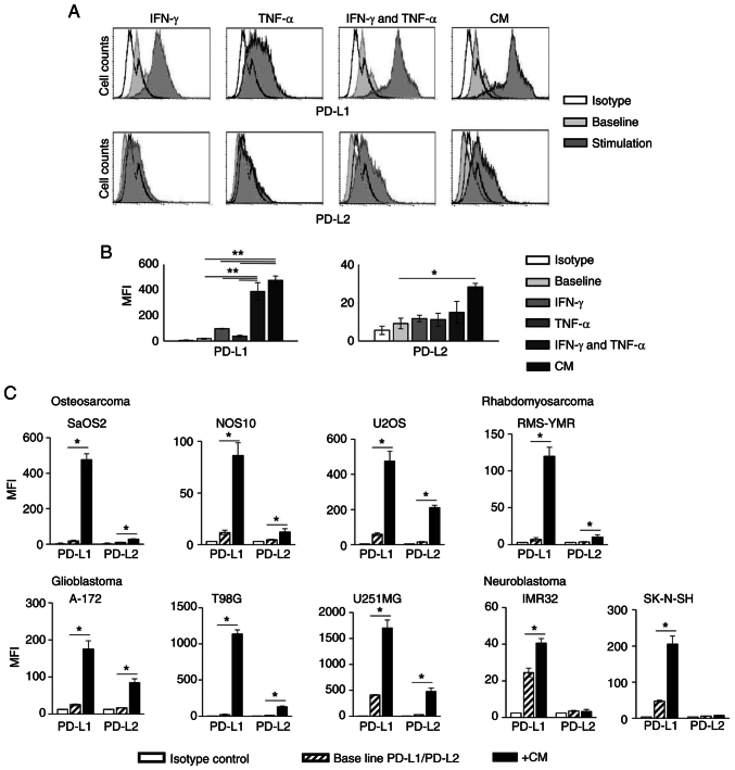
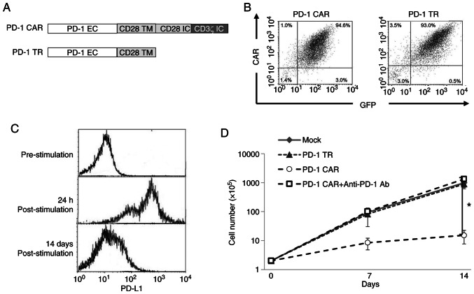
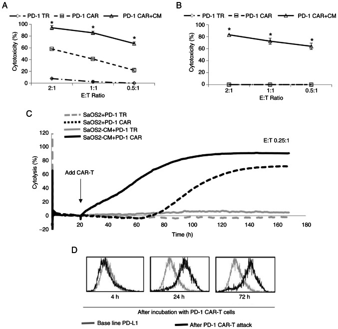
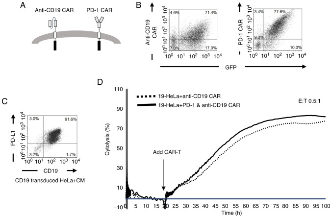
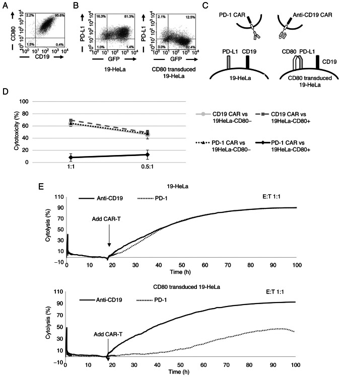
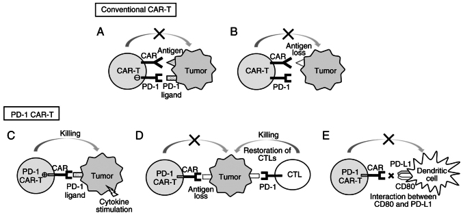
The clinical application of T cells engineered with chimeric antigen receptors (CARs) for solid tumors is challenging. A major reason for this involves tumor immune evasion mechanisms, including the high expression of immune checkpoint molecules, such as the programmed death 1 (PD‑1) ligands PD‑L1 and PD‑L2. The inducible expression of PD‑L1 in tumors has been observed after CAR‑T‑cell infusion, even in tumors natively not expressing PD‑L1. Furthermore, numerous types of pediatric cancer do not have suitable targets for CAR‑T‑cell therapy. Therefore, the present study aimed to develop novel CAR‑T cells that target PD‑L1 and PD‑L2, and to evaluate their efficacy against pediatric solid tumors. A novel CAR harboring the immunoglobulin V‑set domain of the human PD‑1 receptor as an antigen binding site (PD‑1 CAR‑T) was developed without using a single‑chain variable fragment. PD‑1 CAR‑T cells were successfully manufactured by adding an anti‑PD‑1 antibody, nivolumab, to the ex vivo expansion culture to prevent fratricide during the manufacturing process due to the inducible expression of PD‑L1 in activated human T cells. The expression of PD‑L1 (and PD‑L2 to a lesser extent) was revealed to be highly upregulated in various pediatric solid tumor cells, which displayed no or very low expression initially, on in vitro exposure to interferon‑γ and/or tumor necrosis factor‑α, which are cytokines secreted by tumor‑infiltrating T cells. Furthermore, PD‑1 CAR-T cells exhibited strong cytotoxic activity against pediatric solid tumor cells expressing PD‑L1 and PD‑L2. Conversely, the effect of PD‑1 CAR‑T cells was significantly attenuated against PD‑L1‑positive cells coexpressing CD80, suggesting that the toxicity of PD‑1 CAR‑T cells to normal immune cells, including antigen presenting cells, can be minimized. In conclusion, PD‑1 ligands are promising therapeutic targets for pediatric solid tumors. PD‑1 CAR‑T cells, either alone or in combination with CAR‑T cells with other targets, represent a potential treatment option for solid tumors.

Keywords: CD80; PD‑1; PD‑1 ligands; childhood solid tumors; fratricide.


Conflict of interest statement

CI reports patent royalties (patent title: Chimeric receptors with 4-1BB stimulatory signaling domain) from Juno Therapeutics and research funds and advisory fees from CURED, Inc. YS was an employee of CURED, Inc. from February 2022 to January 2024. The other authors declare that they have no competing interests.

Figures

Figure 1. Expression of (PD–1) ligands in pediatric tumor cell lines. (A) Histogram plots showing the expression of PD–L1 (upper panels) and PD–L2 (lower panels) in SaOS2 cells with or without treatme...
Figure 1.
Expression of (PD-1) ligands in pediatric tumor cell lines. (A) Histogram plots showing the expression of PD-L1 (upper panels) and PD-L2 (lower panels) in SaOS2 cells with or without treatment with cytokines or CM. (B) Expression of PD-L1 and PD-L2 in SaOS2 with or without treatment with cytokines or CM. (C) Expression of PD-L1 and PD-L2 in various tumor cell lines treated with CM. *P<0.05, **P<0.01. PD-1, programmed death 1; PD-L, programmed cell death 1 ligand; CM, conditioned medium; IFN-γ, interferon-γ; TNF-α, tumor necrosis factor-α; MFI, mean fluorescence intensity.
Figure 2. Generation of PD–1 CAR–T cells. (A) Gene constructs of PD–1 CAR. PD–1 TR represents a chimeric receptor lacking signaling domain to serve as a non–signaling control receptor. (B) Cell surfac...
Figure 2.
Generation of PD-1 CAR-T cells. (A) Gene constructs of PD-1 CAR. PD-1 TR represents a chimeric receptor lacking signaling domain to serve as a non-signaling control receptor. (B) Cell surface expression of PD-1 CAR and PD-1 TR in retrovirally transduced T cells. (C) Expression of programmed cell death 1 ligand 1 (PD-L1) in T cells before and after activation with CD3/CD28 beads. PD-L1 was not expressed before activation; however, expression was significantly upregulated 24 h after T cell activation and declined 14 days. (D) Absolute cell counts of CAR-T cells in the expansion culture. PD-1 CAR-T cells exhibited significantly poorer growth compared with mock-transduced T cells or PD-1 TR T cells that proliferated exponentially. By adding the anti-PD-1 Ab nivolumab to the culture during the first week, we were able to produce PD-1 CAR T cells with growth rates comparable to mock or PD-1 TR control T cells. *P<0.05 vs. PD-1, programmed death 1; CAR, chimeric antigen receptor; PD-1 TR, truncated PD-1 CAR; Ab, antibody; EC, extracellular domain; TM, transmembrane domain; IC, intracellular signaling domain; GFP, green fluorescent protein.
Figure 3. Cytotoxic effects of PD–1 CAR–T cells against tumor cell lines (A) 4–h cytotoxicity of PD–1 CAR–T cells against osteosarcoma cell line U2OS with modest expression of PD–1 ligands, with or wi...
Figure 3.
Cytotoxic effects of PD-1 CAR-T cells against tumor cell lines (A) 4-h cytotoxicity of PD-1 CAR-T cells against osteosarcoma cell line U2OS with modest expression of PD-1 ligands, with or without pretreatment with CM. (B) 4-h cytotoxicity of PD-1 CAR-T cells against osteosarcoma cell line SaOS2 that does not express PD-1 ligands, with or without pretreatment with CM. (C) Real-time monitoring of SaOS2 cytolysis. SaOS2 were incubated with PD-1 CAR-T at a low E:T ratio (E:T=0.25:1). SaOS2 cells were compared with SaOS2 cells with pretreatment with CM (SaOS2-CM). (D) Time series of histogram plots of PD-L1 expression in SaOS2 by flow cytometry. After co-culture with PD-1 CAR-T cells, PD-L1 expression of residual SaOS2 was gradually upregulated. *P<0.05 vs. PD-1 CAR. PD-1, programmed death 1; CAR, chimeric antigen receptor; CM, conditioned medium; E:T, effector-to-target; PD-L1, programmed cell death 1 ligand 1; PD-1 TR, truncated PD-1 CAR.
Figure 4. Dual targeting CAR–T cells. (A) The proposed scheme for dual–targeting CAR. PD–1 CAR and anti–CD19 CAR were transduced in the same T cells. (B) Surface expression of anti–CD19 CAR and PD–1 C...
Figure 4.
Dual targeting CAR-T cells. (A) The proposed scheme for dual-targeting CAR. PD-1 CAR and anti-CD19 CAR were transduced in the same T cells. (B) Surface expression of anti-CD19 CAR and PD-1 CAR in transduced T cells. T cells showed strong expression of both PD-1 CAR and anti-CD19 CAR. (C) Surface expression of CD19 and PD-L1 in HeLa cells retrovirally transduced with CD19 gene (19-HeLa). 19-Hela cells stimulated with CM expressed both CD19 and PD-L1. (D) Long-term cytotoxicity of dual-targeting CAR-T cells against 19-HeLa with pretreatment with CM. Anti-CD19 CAR-T cells were used as control. PD-1, programmed death 1; CAR, chimeric antigen receptor; CM, conditioned medium; PD-L1, programmed cell death 1 ligand 1; GFP, green fluorescent protein; E:T, effector-to-target.
Figure 5. CD80 inhibits PD–1 CAR binding to PD–L1 and attenuates cytotoxicity. (A) Surface expression of CD80 in 19–HeLa cells after transduction of CD80. (B) Surface expression of PD–L1 after treatme...
Figure 5.
CD80 inhibits PD-1 CAR binding to PD-L1 and attenuates cytotoxicity. (A) Surface expression of CD80 in 19-HeLa cells after transduction of CD80. (B) Surface expression of PD-L1 after treatment with CM in 19-HeLa cells and CD80-transduced 19-HeLa cells. Anti-PD-L1 antibody binding was modestly attenuated in CD80-expressing 19-HeLa cells. (C) Schematic diagram of the experiment. Cytotoxicity of anti-CD19 CAR-T cells or PD-1 CAR-T cells against CM-pretreated, CD80-transduced 19-HeLa (that simultaneously expresses CD80, CD19, and PD-L1) were evaluated. (D) Cytotoxic effects of PD-1 CAR-T cells against CM-pretreated 19-HeLa with or without CD80 overexpression. Cells were cocultured for 24 h. (E) Long-term cytotoxicity of PD-1 CAR-T cells and anti-CD19 CAR-T cells against CM-pretreated 19-HeLa cells with or without CD80 overexpression (lower and upper panels). PD-1, programmed death 1; CAR, chimeric antigen receptor; CM, conditioned medium; PD-L1, programmed cell death 1 ligand 1; GFP, green fluorescent protein; E:T, effector-to-target.
Figure 6. Proposed concept of the study. (A) Conventional CAR–T cells are inhibited by immune checkpoint molecules, such as PD–1 ligands. (B) Loss of the target antigen occurs after exposure to CAR–T ...
Figure 6.
Proposed concept of the study. (A) Conventional CAR-T cells are inhibited by immune checkpoint molecules, such as PD-1 ligands. (B) Loss of the target antigen occurs after exposure to CAR-T cells as one of immune evasion mechanisms by cancer cells. (C) PD-1 CAR-T cells can target PD-1 ligands. (D) Loss of PD-1 ligands in cancer cells occurring as an immune evasion mechanism after attack by PD-1 CAR-T cells leads to the restoration of the T-cell inhibiting microenvironment. (E) PD-1 CAR-T cells using the PD-1 extracellular domain developed in this study have minimal impact on normal immune cells that express both CD80 and PD-L1 (e.g. dendritic cells). PD-1, programmed death 1; CAR, chimeric antigen receptor; PD-L1, programmed cell death 1 ligand 1.

Similar articles

References

    1. Maude SL, Laetsch TW, Buechner J, Rives S, Boyer M, Bittencourt H, Bader P, Verneris MR, Stefanski HE, Myers GD, et al. Tisagenlecleucel in children and young adults with B-Cell lymphoblastic leukemia. N Engl J Med. 2018;378:439–448. doi: 10.1056/NEJMoa1709866. - DOI - PMC - PubMed
    1. Neelapu SS, Locke FL, Bartlett NL, Lekakis LJ, Miklos DB, Jacobson CA, Braunschweig I, Oluwole OO, Siddiqi T, Lin Y, et al. Axicabtagene ciloleucel CAR T-cell therapy in refractory large B-cell lymphoma. N Engl J Med. 2017;377:2531–2544. doi: 10.1056/NEJMoa1707447. - DOI - PMC - PubMed
    1. Rodriguez-Otero P, Ailawadhi S, Arnulf B, Patel K, Cavo M, Nooka AK, Manier S, Callander N, Costa LJ, Vij R, et al. Ide-cel or standard regimens in relapsed and refractory multiple myeloma. N Engl J Med. 2023;388:1002–1014. doi: 10.1056/NEJMoa2213614. - DOI - PubMed
    1. Xu X, Sun Q, Liang X, Chen Z, Zhang X, Zhou X, Li M, Tu H, Liu Y, Tu S, et al. Mechanisms of relapse after CD19 CAR T-Cell therapy for acute lymphoblastic leukemia and its prevention and treatment strategies. Front Immunol. 2019;10:2664. doi: 10.3389/fimmu.2019.02664. - DOI - PMC - PubMed
    1. Samur MK, Fulciniti M, Aktas Samur A, Bazarbachi AH, Tai YT, Prabhala R, Alonso A, Sperling AS, Campbell T, Petrocca F, et al. Biallelic loss of BCMA as a resistance mechanism to CAR T cell therapy in a patient with multiple myeloma. Nat Commun. 2021;12:868. doi: 10.1038/s41467-021-21177-5. - DOI - PMC - PubMed

MeSH terms