. 2025 Sep;56(3):140.
doi: 10.3892/ijmm.2025.5581. Epub 2025 Jul 11.

TNF‑α induces premature senescence in tendon stem cells via the NF‑κB and p53/p21/cyclin E/CDK2 signaling pathways

Affiliations

TNF‑α induces premature senescence in tendon stem cells via the NF‑κB and p53/p21/cyclin E/CDK2 signaling pathways

Hua Guo et al. Int J Mol Med. 2025 Sep.

Abstract

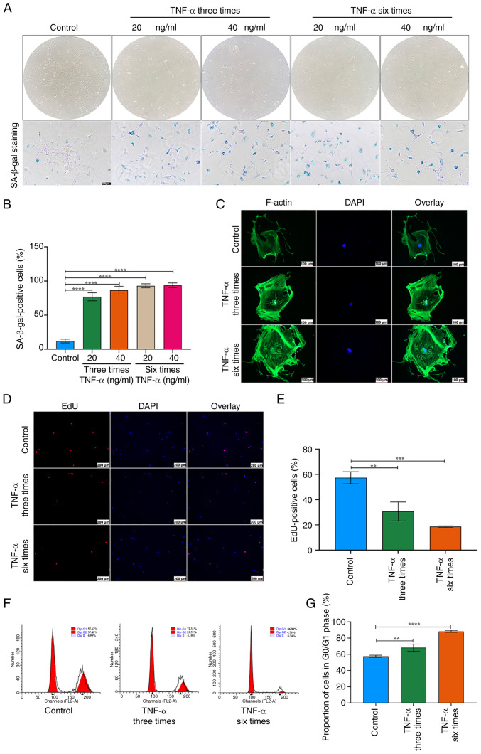
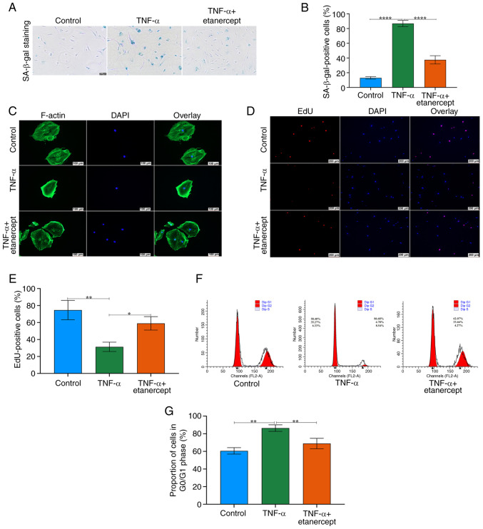
Achilles tendinitis (AT) is a complex disorder that affects tendon tissue and often responds poorly to non‑steroidal anti‑inflammatory drugs. Tumor necrosis factor‑α (TNF‑α), a proinflammatory cytokine involved in cell death and immune regulation, serves a central role in AT progression. The present study investigated the effects of TNF‑α on tendon stem cells (TSCs) and evaluated potential therapeutic strategies for AT. TNF‑α‑induced changes in TSCs were determined by investigating markers of cellular senescence, reactive oxygen species (ROS) activity, DNA damage and the expression of key transcription factors, including NF‑κB (phosphorylated‑p65, p65), p53, p21, cyclin E and CDK2. To determine whether TNF‑α‑induced senescence could be reversed, TSCs were treated with etanercept, a TNF‑α‑specific inhibitor. TNF‑α stimulation induced significant senescence in TSCs, as evidenced by increased ROS production, DNA damage and altered expression of senescence‑associated transcription factors. TNF‑α activated the NF‑κB and p53/p21/cyclin E/CDK2 signaling pathways, promoting TSC senescence. Etanercept treatment effectively reversed these effects, decreasing TSC senescence, suppressing inflammatory cell infiltration, decreasing TNF‑α protein expression and mitigating collagen fiber degradation. TNF‑α promotes TSCs senescence through specific signaling pathways and etanercept can counteract these deleterious effects. These results provide insights into the pathogenesis of AT and highlight TNF‑α inhibition as a promising therapeutic approach. Targeting TNF‑α may offer a novel treatment strategy for individuals with AT.

Keywords: Achilles tendinitis; DNA damage; senescence; tendon stem cell; tumor necrosis factor‑α.


Conflict of interest statement

The authors declare that they have no competing interests.

Figures

Figure 1
Figure 1
Identification of TSCs. (A) Alizarin red S, (B) ALP and (C) Oil red O staining of TSCs. (D) Chondrogenic differentiation of TSCs as detected by toluidine blue staining. (E) Immunofluorescence staining of Oct4 and Nanog in TSCs. Scale bar=100 µm. TSCs, tendon stem cells; ALP, alkaline phosphatase.
Figure 2
Figure 2
Effects of TNF-α on senescence in TSCs. (A) SA-β-gal staining following stimulation with TNF-α in TSCs. Scale bar=200 µm. (B) Quantitative analysis of SA-β-gal-positive cells in TSCs treated with TNF-α. (C) Immunofluorescence revealed aberrant distribution of F-actin in TSCs subjected to TNF-α treatment (20 ng/ml). Scale bar=100 µm. (D) Proliferation of TSCs decreased following stimulation with TNF-α, as assessed using the EdU staining assay. Scale bar=200 µm. (E) Quantitative analysis of EdU-positive cells indicating decreased proliferation in TNF-α-treated TSCs. (F) Cell cycle analysis with flow cytometry. Proportion of cells in the G0/G1 phase increased in TSCs exposed to TNF-α. (G) Quantitative summary showing an increased proportion of cells in the G0/G1 phase in TNF-α-treated TSCs. **P<0.01, ***P<0.001,****P<0.0001. TSCs, tendon stem cells; SA-β-gal, senescence-associated β-galactosidase; F-actin, filamentous-actin.
Figure 3
Figure 3
Effects of the NF-κB and p53/p21/cyclin E/CDK2 signaling pathways on senescence in TNF-α-treated TSCs. (A) ROS staining of TSCs using DCF fluorescence probe, showing intracellular ROS distribution. Scale bar=100 µm. (B) Quantitative analysis of DCF fluorescence intensity, demonstrating TNF-α-induced elevation of ROS levels. (C) Immunofluorescence staining of γ-H2A.X. Following stimulations with TNF-α (20 ng/ml, six times), the proportion of γ-H2A.X-positive TSCs exhibited a considerable increase. Scale bar=100 µm. (D) Quantitative analysis of γ-H2A.X-positive TSCs following TNF-α treatment. (E) Expression of γ-H2A.X, H2A.X, p-p65 and p65 following TNF-α stimulation as assessed by western blot. GAPDH was used as a control. (F) Bar groups showed the relative density of γ-H2A.X, H2A.X, p-p65 and p65. (G) Expression of p53, p21, cyclin E and CDK2 following TNF-α stimulation as assessed by western blot. GAPDH was used as a control. (H) Relative density of p53, p21, cyclin E and CDK2. (I) Immunofluorescence examination of p65, p53 and p21 expression was consistent with western blotting. Scale bar=100 µm. *P<0.05, **P<0.01, ***P<0.001, ****P<0.0001. ROS, reactive oxygen species; TSCs, tendon stem cells; p-, phosphorylation; ns, not significant.
Figure 4
Figure 4
Effects of etanercept on the NF-κB and p53/p21/cyclin E/CDK2 signaling pathways in tendon stem cell senescence. (A) ROS staining of TSCs. ROS generation was markedly elevated following repeated TNF-α stimulation and subsequently reduced after repeated administration of etanercept. Scale bar=100 µm. (B) Quantitative analysis of DCF fluorescence intensity. (C) Expression of γ-H2A.X, H2A.X, p53, p21, p-p65 and p65 after stimulation with TNF-α + etanercept as assessed by western blotting. GAPDH was used as a control. (D) Bar groups showed the relative density of γ-H2A.X, H2A.X, p53, p21, p-p65 and p65. (E) Immunofluorescence of γ-H2A.X and p65 yielded consistent results with those from western blotting. Scale bar=100 µm. (F) Immunofluorescence of p53 and p21 yielded consistent results with those from western blotting. Scale bar=100 µm.. **P<0.01, ***P<0.001, ****P<0.0001. ROS, reactive oxygen species; p-, phosphorylation; ns, not significant.
Figure 5
Figure 5
Effects of etanercept on senescence in TSCs. (A) Senescence of TSCs was analyzed by SA-β-gal staining. Scale bar=200 µm. (B) Quantitative analysis of SA-β-gal-positive cells in TSCs. (C) Aberrant distribution of F-actin in TSCs subjected to TNF-α treatment as revealed by immunofluorescence. Effects were reversed by etanercept. Scale bar=100 µm. (D) Substantial reduction in the proliferation rate of TNF-α-treated TSCs, as indicated by a EdU staining experiment. Proliferation recovered following treatment with etanercept. Scale bar=200 µm. (E) Quantitative analysis of EdU-positive TSCs. (F) Proportion of cells in the G0/G1 phase of the cell cycle increased following treatment with TNF-α, as detected using flow cytometry. This rise is subsequently reversed after treatment with etanercept. (G) Quantitative summary showing an decreased proportion of cells in the G0/G1 phase in etanercept-treated TSCs. *P<0.05, **P<0.01, ****P<0.0001. TSCs, tendon stem cells; SA-β-gal, senescence-associated β-galactosidase; F-actin, filamentous-actin.
Figure 6
Figure 6
Histological changes in rats with tendinitis. (A) Injection schedule of PBS, collagenase I and etanercept in Sprague-Dawley rats. (B) H&E and Masson's trichrome staining 2 weeks post-injection of PBS, collagenase I + PBS and collagenase I + etanercept into tendon tissues. Red denotes collagen fibers; blue denotes the collapse of the collagen matrix. (C) Immunostaining for TNF-α on PBS-treated tendon tissue sections revealed negligible staining. Collagenase I-treated tendon tissue sections exhibited substantial positive staining (brown). (D) The percentages of TNF-α positive cells in tendon tissue. (E) SA-β-gal staining reveals little positive staining in PBS and collagenase I + etanercept groups, but substantial staining in collagenase I + PBS tendon tissues. (F) SA-β-gal-positive area (%) of tendon tissues. (G) Co-localization of CD44 (green) with p53 (red) in tendon tissues. The nucleus inside the tendon tissues is labeled with DAPI (blue). Scale bar=50, 100, 200 µm. *P<0.05, ***P<0.001. H&E, hematoxylin and eosin, SA-β-gal, senescence-associated β-galactosidase.
Figure 7
Figure 7
Impact of TNF-α on TSCs in normal tendon tissues. Under physiological conditions, TSCs exhibit typical functionality, characterized by regular cell cycles, intact F-actin structures, low levels of ROS and normal expressions of transcription factors. Following stimulation by TNF-α, TSCs experience increased ROS production and DNA damage, activation of the NF-κB signaling pathway (resulting in elevated levels of p-p65 and p65, leading to p65 translocation to the nucleus) and modulation of the p53/p21/cyclin E/CDK2 signaling pathways (resulting in upregulation of p53 and p21 and downregulation of cyclin E and CDK2). These changes induce senescence in TSCs, characterized by alterations such as enlarged cell volume and disrupted F-actin structures. Etanercept, a TNF-α inhibitor, mitigates these effects by binding to TNF-α, thereby inhibiting the activation of signaling pathways. This inhibition leads to reduced ROS levels, mitigates DNA damage, decreases expression of p53, p21 and p-p65, normalizes cyclin E and CDK2 expression and ultimately reverses senescence in TSCs, thereby restoring normal cellular functions. Figure created using BioRender (app.biorender.com/illustrations). TSC, tendon stem cells; ROS, reactive oxygen species; F-actin, filamentous-actin; p-, phosphorylation; TNFR, tumor necrosis factor receptor; SA-β-gal, senescence-associated β-galactosidase.

Similar articles

References

    1. Iagnocco A, Riente L, Delle Sedie A, Filippucci E, Salaffi F, Meenagh G, Scirè CA, Grassi W, Montecucco C, Bombardieri S, Valesini G. Ultrasound imaging for the rheumatologist. XXII. Achilles tendon involvement in spondyloarthritis. A multi-centre study using high frequency volumetric probe. Clin Exp Rheumatol. 2009;27:547–551. - PubMed
    1. Alves EM, Macieira JC, Borba E, Chiuchetta FA, Santiago MB. Spontaneous tendon rupture in systemic lupus erythematosus: Association with Jaccoud's arthropathy. Lupus. 2010;19:247–254. doi: 10.1177/0961203309351729. - DOI - PubMed
    1. Järvinen TA, Kannus P, Paavola M, Järvinen TL, Józsa L, Järvinen M. Achilles tendon injuries. Curr Opin Rheumatol. 2001;13:150–155. doi: 10.1097/00002281-200103000-00009. - DOI - PubMed
    1. Lin YJ, Anzaghe M, Schülke S. Update on the pathomechanism, diagnosis, and treatment options for rheumatoid arthritis. Cells. 2020;9:880. doi: 10.3390/cells9040880. - DOI - PMC - PubMed
    1. Mobasheri A, Shakibaei M. Is tendinitis an inflammatory disease initiated and driven by pro-inflammatory cytokines such as interleukin 1β? Histol Histopathol. 2013;28:955–964. - PubMed

MeSH terms