. 2025 Mar 14;12(5):101601.
doi: 10.1016/j.gendis.2025.101601. eCollection 2025 Sep.

Chlorpyrifos induces spermatogenic dysfunction via ferroptosis in Sertoli cells

Affiliations

Chlorpyrifos induces spermatogenic dysfunction via ferroptosis in Sertoli cells

Yan Fu et al. Genes Dis. .

Abstract

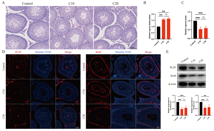
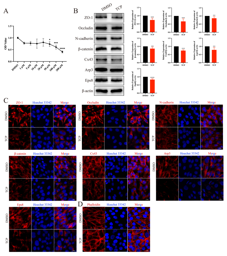
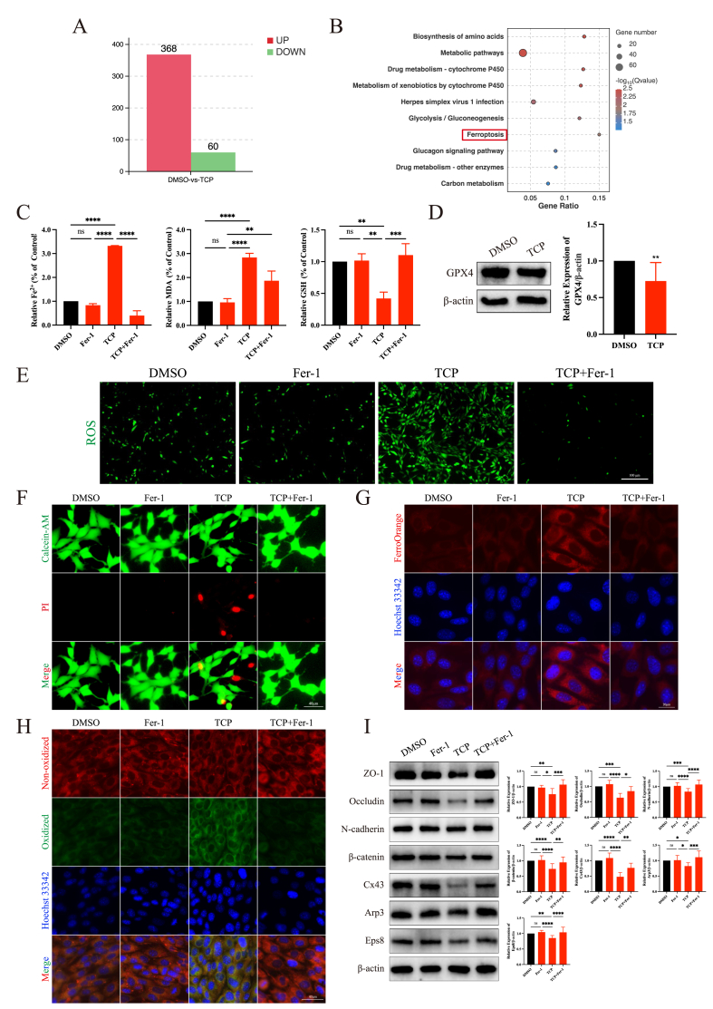
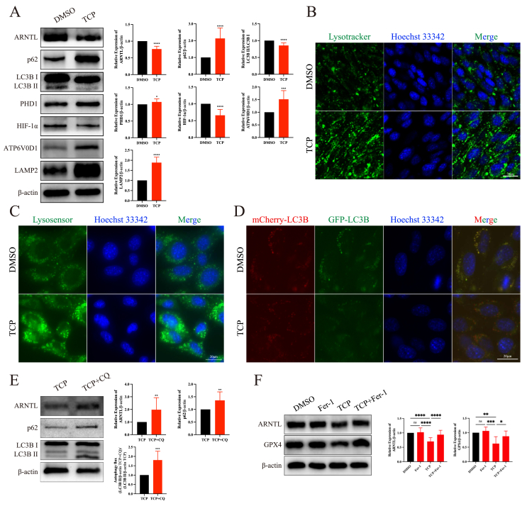
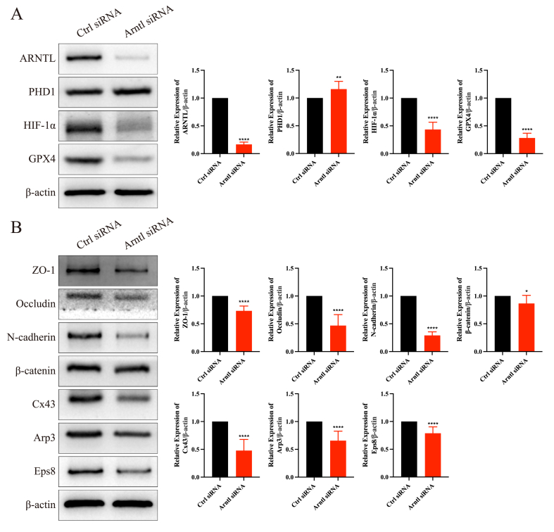
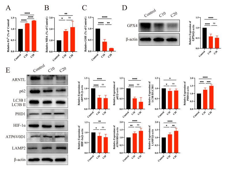
Chlorpyrifos (CPF), a widely used organophosphate pesticide, accumulates in the environment and affects human health. Its neurotoxicity has been extensively studied, and recent research has revealed that it can also lead to abnormal spermatogenesis. However, the factors and molecular mechanisms involved remain unclear. In this study, male Sprague-Dawley rats were gavaged with different concentrations of CPF for 30 days, resulting in a disrupted blood-testis barrier (BTB) and abnormal spermatogenesis. RNA sequencing analysis of Sertoli cells, the primary components of the BTB and key targets of environmental toxins, revealed that ferroptosis-related genes were predominantly among the differentially expressed genes. The expression of ferroptosis-related markers was up-regulated, malondialdehyde and Fe2+ levels were elevated, and glutathione levels were reduced in CPF-exposed testicular tissue and its metabolite TCP-exposed Sertoli cells, confirming that CPF exposure triggered ferroptosis in testes and Sertoli cells. Moreover, treatment with ferrostatin-1, a ferroptosis inhibitor, restored Sertoli cell junctional function. Given the important roles of clockophagy and the HIF-1α pathway in ferroptosis, we investigated the activity of clockophagy in testes and Sertoli cells. Unexpectedly, clockophagy activity was found to be enhanced by the significantly reduced expression levels of ARNTL and HIF-1α following CPF and TCP exposure. Notably, Arntl knockdown impaired Sertoli cell junctional function. Collectively, these findings strongly indicate that CPF induces ferroptosis in Sertoli cells through activating clockophagy, resulting in the decreased expression of HIF-1α and BTB-associated proteins; this ultimately leads to the disruption of BTB integrity and spermatogenesis dysfunction.

Keywords: Blood-testis barrier; CPF; Clockophagy; Ferroptosis; Sertoli cells.


Conflict of interest statement

The authors declare that they have no known competing financial interests or personal relationships that could have appeared to influence the work reported in this paper.

Figures

Figure 1
Figure 1
The toxic effects of chlorpyrifos exposure on spermatogenesis dysfunction. (A) The histological changes in the testes of rats in the three different groups. (B) The sperm deformation rate. (C) The number of sperm. (D) Immunofluorescence analysis of the expression of PLZF and Stra8 in the rat testes. (E) The protein expression levels of PLZF and Stra8 in the rat testes by Western blot. ∗∗∗p < 0.001 and ∗∗∗∗p < 0.0001.
Figure 2
Figure 2
Chlorpyrifos exposure disrupted the blood-testis barrier integrity. (A) The expression of blood-testis barrier-associated proteins and actin-binding proteins in the three different groups. (B) The localization of blood-testis barrier-associated proteins and actin-binding proteins in the three different groups. (C) Blood-testis barrier integrity analysis. ∗p < 0.05, ∗∗p < 0.01, ∗∗∗p < 0.001, and ∗∗∗∗p < 0.0001; ns, not significant.
Figure 3
Figure 3
TCP exposure disrupted the junctional function of Sertoli cells. (A) CCK-8 assays. (B) The expression of blood-testis barrier-associated proteins and actin-binding proteins in Sertoli cells following TCP exposure. (C) The localization of blood-testis barrier-associated proteins and actin-binding proteins in Sertoli cells following TCP exposure. (D) The cytoskeleton organization in Sertoli cells following TCP exposure. ∗p < 0.05, ∗∗p < 0.01, ∗∗∗p < 0.001, and ∗∗∗∗p < 0.0001.
Figure 4
Figure 4
TCP exposure disrupted junctional function by triggering ferroptosis in Sertoli cells. (A) The differentially expressed genes between the DMSO and TCP groups. (B) The results of the KEGG analysis. (C) The levels of Fe2+, malondialdehyde (MDA), and glutathione (GSH) in Sertoli cells in the four different groups. (D) The protein expression level of GPX4 after TCP exposure. (E) Reactive oxygen species (ROS) generation in the four different groups. (F) Calcein-AM/propidium iodine (PI) staining showed that ferrostatin-1 (Fer-1) treatment reversed the cell death induced by TCP exposure. (G) FerroOrange staining revealed that Fer-1 treatment decreased the level of ferrous ions induced by TCP exposure. (H) Fer-1 treatment reduced the level of lipid peroxidation induced by TCP exposure. (I) Fer-1 treatment reversed the decrease in the protein expression levels of blood-testis barrier-associated proteins after TCP exposure. ∗p < 0.05, ∗∗p < 0.01, ∗∗∗p < 0.001, and ∗∗∗∗p < 0.0001; ns, not significant.
Figure 5
Figure 5
TCP exposure enhanced clockophagy activity and lysosomal function in Sertoli cells. (A) The protein expression levels of markers of clockophagy and lysosomal function after TCP exposure. (B) LysoTracker staining showed that TCP exposure increased the number of lysosomes. (C) LysoSensor staining showed that TCP exposure enhanced lysosomal function. (D) TCP exposure enhanced autophagic flux in Sertoli cells. (E) Chloroquine (CQ) treatment inhibited clockophagy-mediated ARNTL degradation. (F) Ferrostatin-1 (Fer-1) treatment rescued the protein expression levels of ARNTL and GPX4, which decreased after TCP exposure. ∗p < 0.05, ∗∗p < 0.01, ∗∗∗p < 0.001, and ∗∗∗∗p < 0.0001; ns, not significant.
Figure 6
Figure 6
ARNTL regulated the junctional function of Sertoli cells. (A) The protein expression levels of ARNTL and its downstream genes after Arntl knockdown in Sertoli cells. (B) The protein expression levels of blood-testis barrier-associated proteins after Arntl knockdown in Sertoli cells. ∗p < 0.05, ∗∗p < 0.01, and ∗∗∗∗p < 0.0001.
Figure 7
Figure 7
Chlorpyrifos exposure promoted ferroptosis in the testes by activating clockophagy. (A) The level of Fe2+ in the three different groups. (B) The level of malondialdehyde (MDA) in the three different groups. (C) The level of glutathione (GSH) in the three different groups. (D) The protein expression level of GPX4 in the three different groups. (E) The protein expression levels of markers of clockophagy and lysosomal function in the three different groups. ∗p < 0.05, ∗∗p < 0.01, ∗∗∗p < 0.001, and ∗∗∗∗p < 0.0001; ns, not significant.

Similar articles

References

    1. Kermani M., Dowlati M., Gholami M., et al. A global systematic review, meta-analysis and health risk assessment on the quantity of malathion, diazinon and chlorpyrifos in vegetables. Chemosphere. 2021;270 - PubMed
    1. Jing Q., Liu J., Chen A., Chen C., Liu J. The spatial-temporal chemical footprint of pesticides in China from 1999 to 2018. Environ Sci Pollut Res Int. 2022;29(50):75539–75549. - PubMed
    1. Slotkin T.A. Guidelines for developmental neurotoxicity and their impact on organophosphate pesticides: a personal view from an academic perspective. Neurotoxicology. 2004;25(4):631–640. - PubMed
    1. Huang X., Cui H., Duan W. Ecotoxicity of chlorpyrifos to aquatic organisms: a review. Ecotoxicol Environ Saf. 2020;200 - PubMed
    1. Kim H.H., Lim Y.W., Yang J.Y., et al. Health risk assessment of exposure to chlorpyrifos and dichlorvos in children at childcare facilities. Sci Total Environ. 2013;444:441–450. - PubMed

LinkOut - more resources