

The clinical relevance of healthy neurodevelopmental connectivity in childhood and adolescence: a meta-analysis of resting-state fMRI
- PMID: 40641623
- PMCID: PMC12241014
- DOI: 10.3389/fnins.2025.1576932
The clinical relevance of healthy neurodevelopmental connectivity in childhood and adolescence: a meta-analysis of resting-state fMRI
Abstract
Background: In recent years, interest has grown in brain connectivity during infancy and adolescence, particularly in understanding neurodevelopment. Research is focusing on how brain network complexity evolves, providing insight into developmental neural connectivity. While some studies highlight key periods of brain maturation, findings remain inconsistent, leaving the neural correlates of typical development uncertain. This meta-analysis aims to identify brain regions and functional connectivity networks that show age-related activation patterns. Our goal is to clarify how neural wiring and complexity change with age, using seed-based d mapping (SDM) to analyze resting-state functional connectivity.
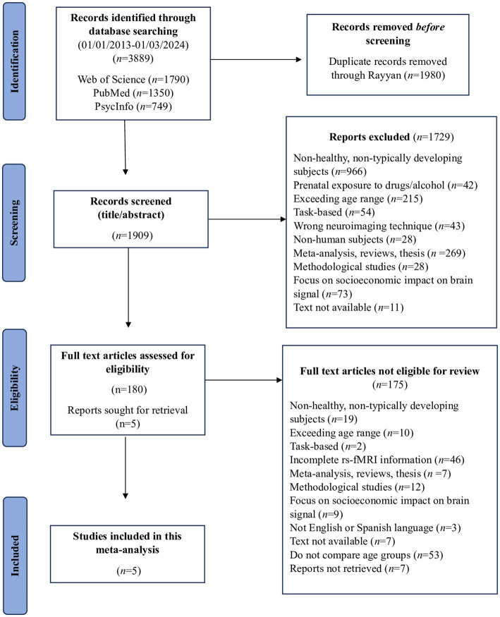
Methods: We reviewed studies employing resting-state functional magnetic resonance imaging (rs-fMRI) to examine brain connectivity in typically developing children and adolescents. After thoroughly application of the rigorous inclusion criteria, five studies published between 2013 and 2024 remained selected for this analysis. While this is a small number, this limitation reflects our unwavering commitment to methodological rigor and the current scarcity of available literature, ensuring that only high-quality studies were considered.
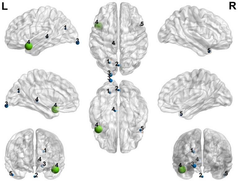
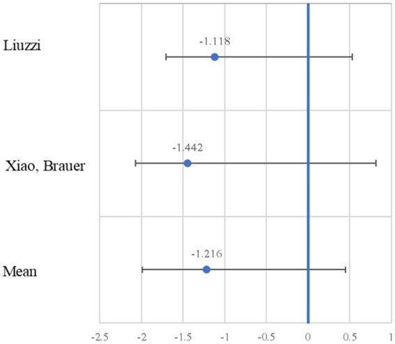
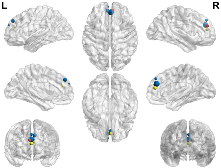
Results: Consistent increases in seed-based connectivity involving the left frontal and prefrontal cortices were observed, particularly the left superior frontal gyrus and bilateral anterior cingulate cortex. These areas showed increased connectivity in older compared to younger participants.
Conclusion: The left frontal and prefrontal cortices, which are critical for executive function, attention, and intelligence, appear to strengthen their connectivity during childhood and adolescence. These observations provide a preliminary glimpse into typical brain maturation. However, due to the small number of studies and heterogeneity in age comparisons. No clinical implications can be drawn at this stage, and further research is required to confirm these developmental trends.
Keywords: brain connectivity; fMRI; healthy neurodevelopment; meta-analysis; resting-state.
Copyright © 2025 Tapia Medina, Cosío-Guirado, Peró-Cebollero, Cañete-Massé, Villuendas-González and Guàrdia-Olmos.
Conflict of interest statement
The authors declare that the research was conducted in the absence of any commercial or financial relationships that could be construed as a potential conflict of interest.
Figures
References
-
- Barretto-García M., Grueschow M., Moisa M., Polania R., Ruff C. C. (2024). Causal evidence for a domain-specific role of left superior frontal sulcus in human perceptual decision making. eLife. doi: 10.7554/eLife.94576.1 - DOI
-
- Bernal B. (2022). Practical aspects of functional magnetic resonance in children. J. Pediatr. Neurol. 20, 83–96. doi: 10.1055/s-0041-1733853 - DOI
Publication types
LinkOut - more resources
Full Text Sources