Brainstorm: A Case of Granulomatous Encephalitis
- PMID: 40641815
- PMCID: PMC12169439
- DOI: 10.3138/jammi-2023-0036
Brainstorm: A Case of Granulomatous Encephalitis
Abstract
Background: Free-living amoebas (FLAs) can cause severe and fatal central nervous system infections that are difficult to diagnose.
Methods: We present the case of a 74-year-old immunocompetent woman admitted for focal neurological symptoms with enhancing lesions in the right cerebellar hemisphere. A first cerebral biopsy showed granulomatous inflammation, but no microorganisms were identified. After transient clinical improvement, she eventually deteriorated 4 months after initial presentation, with an MRI confirming multiple new masses affecting all cerebral lobes.
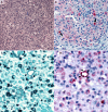
Results: A second brain biopsy revealed granulomatous and acute inflammation with organisms containing a large central nucleus with prominent karyosome, consistent with FLAs. Immunohistochemical and polymerase chain reaction assays performed at CDC were positive for Acanthamoeba spp, confirming the diagnosis of granulomatous amoebic encephalitis (GAE) caused by Acanthamoeba spp. The patient was treated with combination therapy recommended by CDC, but died a few days later. Upon histopathological rereview, amoebic cysts and trophozoites were identified by histochemical and immunohistochemical methods in the first cerebral biopsy.
Conclusion: FLA infections can be challenging to diagnose because of the low incidence, non-specific clinical and radiological presentation, lack of accessible diagnostic tools, and clinicians' unfamiliarity. This case highlights the importance of recognizing FLA as a potential cause of granulomatous encephalitis, even in the absence of risk factors, as early treatment might be associated with favourable outcomes in case reports. When suspected, CDC laboratories offer tests to confirm the diagnosis promptly.
Historique: Les amibes libres peuvent causer des infections du système nerveux central graves et fatales qui sont difficiles à diagnostiquer.
Méthodologie: Les auteurs présentent le cas d'une femme immunocompétente de 74 ans hospitalisée à cause de symptômes neurologiques focaux avec lésions rehaussantes dans l'hémisphère cérébelleux droit. Une première biopsie cérébrale a révélé une inflammation granulomateuse, mais aucun microorganisme n'a été décelé. Après une amélioration clinique transitoire, son état s'est détérioré quatre mois après la première consultation, et l'IRM a confirmé de multiples nouvelles masses touchant tous les lobes cérébraux.
Résultats: Une deuxième biopsie cérébrale a révélé une inflammation granulomateuse aiguë par des organismes dont les gros noyaux centraux et les caryosomes volumineux étaient évocateurs d'amibes libres. L'immunohistochimie et l'amplification en chaîne par polymérase effectuées aux CDC se sont avérés positives pour Acanthamoeba spp, ce qui a confirmé un diagnostic d'encéphalite amibienne granulomateuse causée par Acanthamoeba spp. La patiente a reçu une polythérapie recommandée par les CDC, mais est malheureusement décédée quelques jours plus tard. À la reprise de l'analyse histopathologique, des kystes amibiens et des trophozoïtes ont été décelés dans la première biopsie cérébrale par des méthodes histochimiques et immunohistochimiques.
Conclusion: Les infections par des amibes libres peuvent être difficiles à diagnostiquer en raison de leur faible incidence, de leur présentation clinique et radiologique non spécifique, de l'absence d'outils diagnostiques accessibles et de la méconnaissance des cliniciens. Ce cas renforce l'importance d'inclure les amibes libres dans les causes potentielles d'encéphalite granulomateuse, même en l'absence de facteurs de risque, car un traitement rapide a été associé à des résultats favorables dans certains rapports de cas. Lorsqu'on en soupçonne la présence, les laboratoires des CDC offrent des tests pour confirmer rapidement le diagnostic.
Keywords: Acanthamoeba; Balamuthia mandrillaris; free-living amoebas; granulomatous amoebic encephalitis.
© Association of Medical Microbiology and Infectious Disease Canada (AMMI Canada), 2024.
Conflict of interest statement
The findings and conclusions of this report are those of the authors and do not necessarily represent the official position of the Centers for Disease Control and Prevention (CDC).
Figures


References
Publication types
LinkOut - more resources
Full Text Sources
Miscellaneous
