. 2025 Jun 26:12:1616977.
doi: 10.3389/fmed.2025.1616977. eCollection 2025.

Tissue-engineered tubular substitutions for urinary diversion in a preclinical rabbit model

Affiliations

Tissue-engineered tubular substitutions for urinary diversion in a preclinical rabbit model

Qianliang Wang et al. Front Med (Lausanne). .

Abstract

Objective: To develop and evaluate tissue-engineered tubular constructs using homologous adipose-derived stem cells (ASCs), smooth muscle cells (SMCs), and decellularized fish swim bladder (DFSB) matrix for urinary diversion in a rabbit model.

Methods: Rabbit ASCs and SMCs were isolated and expanded in vitro; cultured cells were seeded onto bilateral surfaces of DFSB scaffolds followed by 7-day incubation; cell-seeded matrices were shaped into tubular constructs; constructs underwent 2-week in vivo pre-vascularization within omental pouches. Experimental group rabbits (n=24) underwent complete bladder resection with replacement by pre-vascularized constructs, while control group (n=6) received identical implantation of acellular DFSB tubes. Histological evaluations were conducted at postoperative weeks 2, 4, 8, and 16; intravenous urography (IVU) was performed at 16-week endpoint.

Results: All experimental animals survived until scheduled sacrifice with histological evidence of: (1) luminal multilayer urothelium, (2) organized smooth muscle tissue on abluminal surfaces, and (3) construct-wide neovascularization of varying diameters; IVU confirmed absence of urinary leakage, stricture, or obstruction. Conversely, all control animals died within 2 weeks post-operation; autopsy revealed urine leakage, extensive scar formation, and severe inflammation as mortality causes.

Conclusion: Tissue-engineered tubular constructs fabricated from homologous ASCs, SMCs, and DFSB scaffold demonstrate feasibility as a viable urinary diversion alternative in rabbit models, showing functional tissue regeneration and superior outcomes versus acellular controls.

Keywords: adipose-derived stem cells; decellularized fish swim bladder; epithelium; smooth muscle cells; tissue engineering; urinary diversion.


Conflict of interest statement

The authors declare that the research was conducted in the absence of any commercial or financial relationships that could be construed as a potential conflict of interest.

Figures
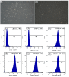
Figure 1
Figure 1
Morphological characterization and identification of ADSCs. (a) Primary culture after 3 days (x 100). (b) After incubation for 10 days, the cells proliferated rapidly and displayed a spindle fibroblastic appearance (x 100). There was no significant expression of (c) CD31 (1.90%), and (d) CD45 (1.60%). Flow cytometry analysis demonstrated expression of (e) CD29 (99.60%), (f) CD44 (98.20%), (g) CD90 (99.60%) and (h) CD105 (99.50%).
Figure 2
Figure 2
Morphological characterization and immunofluorescence staining of SMCs. (a) After 3 days of primary culture, the cells proliferated on the surfaces of Petri dish and reached about 30% confluence (x100). (b) After 7 days of culture, the cells reached 80–90% confluence and displayed a classic spindle-shaped morphology (x100). (c) a-SMA immunofluorescence staining (in green) of SMCs in combination with DAPI cell nuclei stain (in blue) (x200).
Figure 3
Figure 3
Characteristics of DFSB. (a) Fish swim bladder (gross appearance). (b) After decellularization procedure, the DFSB appeared as a translucent film (gross appearance). (c) H&E staining of DFSB (x400). (d) Masson’s trichrome staining of DFSB (x 400). (e) Sirius Red staining of DFSB (x400) (f) Scanning electron microscope of DFSB (x1000).
Figure 4
Figure 4
The feature of DFSB seeding. (a) DFSB with seeded cells. After seeding cells onto the DFSB, the cells fused well on the three-dimensional DFSB scaffold. (b) H&E staining of luminal side (x400). (c) H&E staining of outside side (x400). (d) Scanning electron microscope of luminal surface (x 2000). (e) Scanning electron microscope of outside surface (x2000). (f) Scanning electron microscope of luminal surface (x 2,500, longitudinal section).
Figure 5
Figure 5
Regeneration of epithelium and neovascularization after omental incubation. (a) TETEs. (b) TETSs were wrapped in the omentum (c) H&E staining of TETSs (x 400). (d) H&E staining of the unseeded DFSB (x 400). (e) Anti-AE1/AE3 immunohistochemistry staining displayed a one-layer epithelium structure (x200). (f) Anti-AE1/AE3 immunohistochemistry staining showed no obvious epithelium in the unseeded DFSB (x 200). (g) Anti-CD31 immunohistochemistry staining revealed neovascularization of TETSs (x 200). (h) Anti-a-SMA immunohistochemistry staining revealed vascular walls in TETSs (x 200).
Figure 6
Figure 6
Urinary diversion in rabbits. (a) The bilateral ureters were dissociated. (b) Bilateral ureters were anastomosed to the TETSs. (c) Gross observation after Urinary diversion in rabbits.
Figure 7
Figure 7
Histologic characteristics of TETSs in the experimental group after urinary diversion at 2, 4, 8, and 16 weeks. H&E staining (a–d) displayed the regeneration of epithelium layers of TETSs (x400). (e–p) Immunohistochemical staining of AE1/AE3, uroplakin IIIa, and ZO-1 revealed the regeneration of epithelium of TETSs, respectively (x 400).
Figure 8
Figure 8
Immunohistochemistry analysis for regeneration of smooth muscle and neovascularization at 16 weeks (x 200). (a) Anti-CD31 antibody positive showed well-developed angiogenesis. (b) Anti-a-SMA antibody positive revealed vessel wall.
Figure 9
Figure 9
Intravenous urography observation. (a) Intravenous urography at16 weeks postoperatively showed urinary tract was unblocked and that no leakage, stricture, or obstruction occurred in the TETSs and bilateral ureters. (b) Cystoscopy showed that the surface of the bladder cavity were smooth. (c) In the control group, stone formation after urinary diversion at 2 weeks.

Similar articles

References

    1. Biers SM, Venn SN, Greenwell TJ. The past, present and future of augmentation cystoplasty. BJU Int. (2012) 109:1280–93. doi: 10.1111/j.1464-410X.2011.10650.x, PMID: - DOI - PubMed
    1. Husmann DA, Rathbun SR. Long-term follow up of enteric bladder augmentations: the risk for malignancy. J Pediatr Urol. (2008) 4:381–5. doi: 10.1016/j.jpurol.2008.06.003 - DOI - PubMed
    1. Wu SY, Kuo HC. A real-world experience with augmentation enterocystoplasty-high patient satisfaction with high complication rates. Neurourol Urodyn. (2018) 37:744–50. doi: 10.1002/nau.23339, PMID: - DOI - PubMed
    1. Zi H, Liu MY, Luo LS, Huang Q, Luo PC, Luan HH, et al. Global burden of benign prostatic hyperplasia, urinary tract infections, urolithiasis, bladder cancer, kidney cancer, and prostate cancer from 1990 to 2021. Mil Med Res. (2024) 11:64. doi: 10.1186/s40779-024-00569-w, PMID: - DOI - PMC - PubMed
    1. Zhang Y, Atala A. Urothelial cell culture: stratified urothelial sheet and three-dimensional growth of urothelial structure. Methods Mol Biol. (2013) 945:383–99. doi: 10.1007/978-1-62703-125-7_23, PMID: - DOI - PubMed

LinkOut - more resources