. 2025 Jun 26:16:1550651.
doi: 10.3389/fimmu.2025.1550651. eCollection 2025.

The influence of H. pylori infection in HER2-positive gastric cancer cell lines: insights from Wnt/β-catenin pathway

Affiliations

The influence of H. pylori infection in HER2-positive gastric cancer cell lines: insights from Wnt/β-catenin pathway

Valli De Re et al. Front Immunol. .

Abstract

Introduction: The impact of H. pylori infection on the efficacy of trastuzumab in HER2-positive gastric cancer (GC) remains poorly understood, despite growing evidence that tumor microenvironment and host-pathogen interactions influence therapeutic outcomes. This study aimed to investigate how H. pylori strains of differing virulence, one high (HV-HP) and one low (LV-HP), affect GC cell behavior, particularly in the context of ERBB2 (HER2) amplification and Trastuzumab (TRAS)-resistance.

Methods: We used the HER2-amplified NCI-N87 GC cell line, alongside four non-HER2-amplified cell lines (AGS, SNU-1, SNU-16 and SNU-5), to examine the impact of infection. TRAS-resistant derivative cells (N87R) were generated by gradual exposure of the sensitive parental N87 cells (N87p) to increasing TRAS concentrations. Both N87R and N87p cells were infected with HV-HP and LV-HP strains and then treated with epidermal growth factor (EGF), TRAS or a combination of both. The infection was confirmed by confocal microscopy and downstream effects of gene expression were evaluated, focusing on Wnt-β-catenin signaling genes linked to metastasis and survival in HER2+ GC. HER2, PD-L1 and PD-L2 protein levels were assessed in all cell lines using multicolor flow cytometry (FACS) before and after HV-HP exposure.

Results: Our data revealed that HV-HP infection reduced MSH6 mRNA expression, which is indicative of impaired DNA repair, and up-regulated PDCD1LG2, suggesting enhanced immunosuppression. FACS analysis showed that HV-HP modulated PD-L2 expression in HER2-amplified N87 cells and to a lesser extent in SNU-16 and SNU-1 cells, while EGF administration increased PD-L1 expression. A strong correlation was observed between ERBB2 expression and TP53, but it was independent of HV-HP. A reduction of CDH1/SNAI ratio was associated with TRAS-resistance in N87 cells.

Discussion: These results suggest that virulent H. pylori in cell lines may contribute to altering tumor phenotype by downregulating the DNA repair machinery, and favouring immune evasion by inducing the expression of immunosuppressive signals, such as PDCD1LG2. Moreover, we found that HER2-targeted therapy may contribute to modulation of CD1/immune pathway. Further studies are warranted to determine whether these effects are common in HER2+ GC in vivo and whether the coexistence of H. pylori infection and TRAS treatment may influence response to immunotherapy.

Keywords: HER2; Helicobacter pylori; MSH6; PD-L1/PD-L2; TP53; Wnt; gastric cancer; trastuzumab.


Conflict of interest statement

The authors declare that the research was conducted in the absence of any commercial or financial relationships that could be construed as a potential conflict of interest. The author(s) declared that they were an editorial board member of Frontiers, at the time of submission. This had no impact on the peer review process and the final decision.

Figures

Figure 1
Figure 1
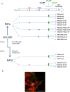
Molecular profiles of H. pylori according to virulence factors. (A) HV-HP profile (cagA+cagE+virB11+vacAs1i1m1+homB+), defined by the simultaneous presence of virB11 (located in the left half of the CagPAI), cagA and cagE (both located in the right half of the CagPAI), the vacA s1i1m1 haplotype, the presence of the homB gene and the absence of the homA gene in their loci. (B) LV-HP profile (cagA-cagE-virB11-vacAs2i2m2+homA+), defined by the concomitant deletion of virB11, cagA and cagE genes, the vacA s2i2m2 haplotype, the presence of homA gene and the absence of homB gene in their loci. The genes of interest are indicated by open rectangles. Crosses represent missing genes. The position of CagPAI and individual genes on the underlying partial physical map of H.pylori genome are indicated by grey and black arrows, respectively. The high-virulence strain was characterized by the concomitant presence of a stable Cag pathogenicity island (CagPAI), a vacA s1i1mx genotype, and homB, features associated with a strongly virulent phenotype, whereas the low-virulence strain lacked these markers (33). HV-HP, highly virulent H. pylori; LV-HP, low virulent H. pylori.
Figure 2
Figure 2
(A) Summary of the study design, including planned treatment. p, parental; R, resistant; EGF, Epidermal Growth Factor; TRAS, Trastuzumab; no-HP, no infection with H. pylori; HV-HP, highly virulent H. pylori; LV-HP, lowly virulent H. pylori;. (B) Representative image of confocal microscopy immunofluorescence of NCI-N87 cells infected with HV-HP. N-cadherin staining (red) was used to visualize the interaction of cells with Five-(and 6-) carboxyfluorescein diacetate, succinimidyl ester (CFDA-SE) stained bacteria (green). Thin white arrows indicate that bacteria interact with the cells near the plasma membrane; bold white arrow indicates bacteria inside the cells. Scale bar = 20 μm.
Figure 3
Figure 3
(A) Summary of gene expression changes in N87 gastric cancer cells infected with H. pylori. While ERBB2 levels remain stable upon Helicobacter pylori infection in N87p gastric cancer cells., PDCD1LG2 and CD274 is upregulated and MSH6 downregulated, highlighting infection may drive modulation of immune and genomic stability pathways. The increase of PDCD1LG2 is significant in most treated cells, except in N87p-EGF and trastuzumab-resistant N87R cells, where the increase did not reach a statistical significance. H. pylori infection reduces MSH6 mRNA levels in N87p gastric cancer cells, as well as in TRAS-resistant cells (N87R and N87R-EGF), suggesting impairment of DNA repair or stress-response pathways. Statistical significance (Student’s t-test): P < 0.05 (*), P < 0.01 (**), P < 0.001 (***). (B). Public dataset: Human Protein Atlas (HPA). Basal expression of HER2 and PD-L2 in gastric adenocarcinoma cell lines from the Human Protein Atlas (HPA) (40). RNA expression is shown as normalized transcripts per million (nTPM) for each cell line. Protein expression based on mass spectrometry data is indicated by circles. Data confirm constitutive ERBB2/HER2 expression in NCI-N87 cells and variable levels in other cell lines. White circles indicate absence of PD-L2 protein detection in N87, AGS, SNU1, SNU16, and SNU5 cells. Arrows mark the cell lines analyzed in the present study. (C). HER2 expression assessed by flow cytometry.N87p cells displayed a markedly higher HER2 fluorescence signal, as indicated by the median relative fluorescence intensity (MFI), compared to the other analyzed cell lines (AGS, SNU1, SNU16, SNU5). Median relative MFI = 2700 ± SEM1605. (D) PD-L2 expression assessed by flow cytometry. Boxplots ahow relative MFI values are shown for uninfected and HV-HP infected cell lines (AGS, N87, SNU-1, SNU-16 and SNU-5). Asterisks indicate statistically significant increases in median MFI in HV-HP-infected cells compared to uninfected controls in N87 (P < 0.001), SNU-16, and SNU-1 (p<0.05), as determined by Student’s t-test (performed in R).
Figure 4
Figure 4
Principal Component Analysis (PCA) of Wnt pathway-related mRNA expression in N87p cells. PCA score plot of mRNAs differentially expressed in untreated and treated N87p cells. Genes were selected based on differential expression analysis related to the Wnt/β-catenin pathway (volcano plot analysis). Red dots represent untreated N87p cells; grey and purple dots correspond to cells treated with EGF and TRAS, respectively; light blue dots indicate cells treated with both EGF and TRAS. Vectors indicate the contribution of each mRNA to the variance explained by the first two principal components (PC1 and PC2). Confidence ellipses, automatically generated by NanoString nSolver software, illustrate group clustering based on expression profiles.
Figure 5
Figure 5
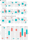
Effects of EGF and TRAS treatments on gene expression and surface marker levels in gastric cancer cell lines. (A) Boxplots showing mRNA expression levels (log2 normalized counts) of selected genes ERBB2, TP53, and CDH1/SNAI1, measured by NanoString technology in AGS, N87, and TRAS-resistant N87R cells following treatment with EGF or TRAS. Two types of statistical comparisons are indicated: light blue asterisks denote significant differences between HV-HP-infected and uninfected treated cells, while larger black asterisks indicate significant differences between treated conditions and the untreated, uninfected N87p control. (B) Boxplots showing the median fluorescence intensity (MFI), calculated as the signal from anti-target antibody minus isotype control, for HER2, PD-L1, and PD-L2. Data were obtained by flow cytometry (FACS) in AGS and N87 cells under untreated (NT) and EGF-treated conditions. Data represent the mean of three independent replicates (n = 3). Asterisks indicate statistically significant differences as determined by Student’s t-test in R (p < 0.05; p < 0.01; p < 0.001; ns, not significant).

References

    1. Malfertheiner P, Link A, Selgrad M. Helicobacter pylori: perspectives and time trends. Nat Rev Gastroenterol Hepatol. (2014) 11:628–38. doi: 10.1038/nrgastro.2014.99 - DOI - PubMed
    1. Uemura N, Okamoto S, Yamamoto S, Matsumura N, Yamaguchi S, Yamakido M, et al. Helicobacter pylori infection and the development of gastric cancer. N Engl J Med. (2001) 345:784–9. doi: 10.1056/NEJMoa001999 - DOI - PubMed
    1. Cancer Today. Available online at: https://gco.iarc.who.int/today/ (Accessed May 5, 2025).
    1. Chen Y-C, Malfertheiner P, Yu H-T, Kuo C-L, Chang Y-Y, Meng F-T, et al. Global prevalence of Helicobacter pylori infection and incidence of gastric cancer between 1980 and 2022. Gastroenterology. (2024) 166:605–19. doi: 10.1053/j.gastro.2023.12.022 - DOI - PubMed
    1. He F, Wang S, Zheng R, Gu J, Zeng H, Sun K, et al. Trends of gastric cancer burdens attributable to risk factors in China from 2000 to 2050. Lancet Reg Health West Pac. (2024) 44:101003. doi: 10.1016/j.lanwpc.2023.101003 - DOI - PMC - PubMed

MeSH terms