. 2025 Jun 26:12:1624016.
doi: 10.3389/fvets.2025.1624016. eCollection 2025.

Clinical and histopathological investigation of the possible occurrence of tracheobronchial disease in cats with chronic gingivostomatitis

Affiliations

Clinical and histopathological investigation of the possible occurrence of tracheobronchial disease in cats with chronic gingivostomatitis

Olga Lorida et al. Front Vet Sci. .

Abstract

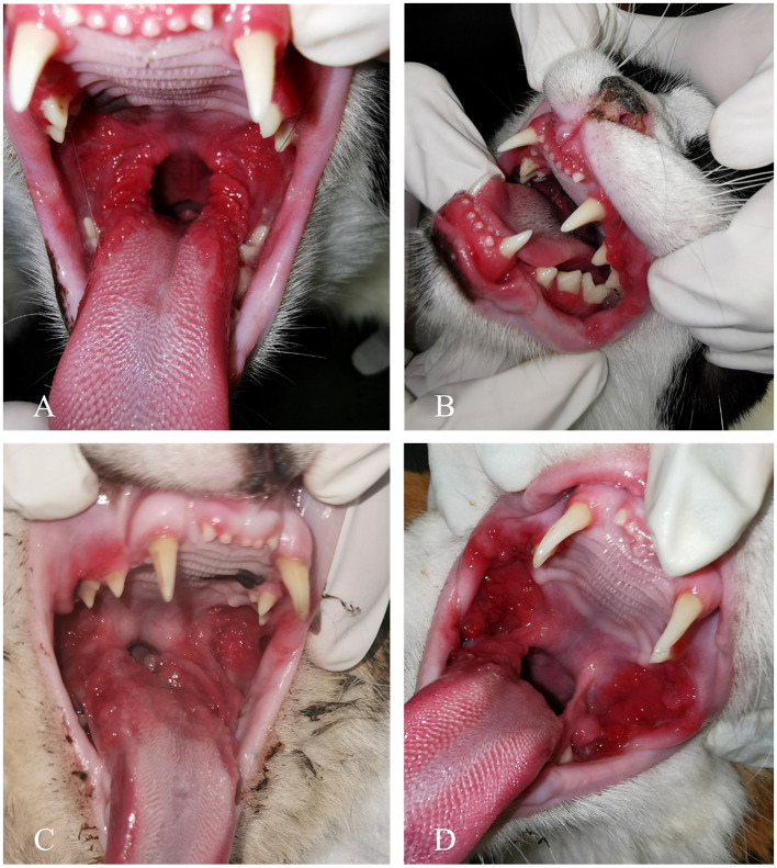
Introduction: Feline chronic gingivostomatitis (FCGS) is a debilitating and highly painful inflammatory disorder of the feline oral cavity. Evidence suggests that feline chronic gingivostomatitis (FCGS) induces systemic effects that extend beyond localized oral pathology, contributing to overall health decline in affected cats. The aim of this study was to investigate the potential impact of FCGS on the lower respiratory tract.

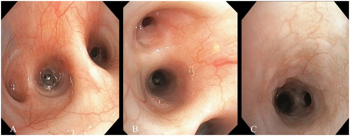
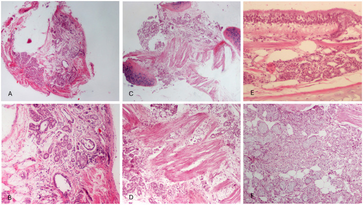
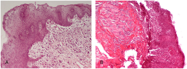
Methods: This is a prospective study, that included 42 cats with clinical signs of FCGS and five healthy control cats exhibiting no signs of oral disease. All cats underwent physical, oral, and endoscopic examinations of the lower respiratory tract. Radiological evaluation of the thorax was also performed. Lesions in the respiratory tract detected upon endoscopy and the oral cavity were recorded and scored. In cats with FCGS biopsies from bronchial mucosa were obtained from sites showing endoscopic evidence of inflammation.

Results: Respiratory lesions were identified in all FCGS cats included in the study. Specifically, secretions were detected in 42 out of 42 (100%) cats, bronchial mucosal edema in 33 out of 42 (78.6%), a granular appearance in 14 out of 42 (33.3%), and hyperemia in 11 out of 42 (26.2%). Histopathological examination revealed mucosal and submucosal inflammation in 30 out of 36 (83.3%) cats and mucosal edema in 25 out of 36 (69.4%). Additionally, fibrosis was observed in 25 out of 36 (69.4%) samples, hyperplasia, or dilatation of bronchial glands in eight out of 36 (22.2%), and vascular wall thickening in 11 out of 36 (30.5%). Bronchial smooth muscle hypertrophy was present in 22 out of 36 (61.1%) examined samples. An attempt to correlate oral and respiratory lesion severity found no statistically significant correlation between stomatitis index, tracheobronchoscopy, or histopathological scores.

Discussion: FCGS appears to coexist with lower respiratory tract disease. During FCGS management, it might be essential to address any underlying respiratory disorder, as it may favor the outcome of the primary disease, while remaining unattended it may increase the likelihood of FCGS recurrence.

Keywords: bronchial secretions; feline chronic gingivostomatitis; lower respiratory tract; oral inflammatory disease; tracheobronchial disease.


Conflict of interest statement

The authors declare that the research was conducted in the absence of any commercial or financial relationships that could be construed as a potential conflict of interest. The author(s) declared that they were an editorial board member of Frontiers, at the time of submission. This had no impact on the peer review process and the final decision.

Figures

Figure 1
Figure 1
Bronchoscopy images of cats with feline chronic gingivostomatitis. (A) Generalized hyperemia of the bronchial mucosa and increased bronchial secretions. (B) Edema of the bronchial mucosa. (C) Granularity of the bronchial mucosa.
Figure 2
Figure 2
Clinical manifestation of feline chronic gingivostomatitis (FCGS). (A) A patient with refractory ulcerative FCGS. (B) Concurrent inflammation of the gingiva, alveolar, and buccal mucosa. (C) Ulceration and proliferation affecting both palatoglossal folds and sublingual mucosa. (D) A case exhibiting both ulceration and proliferation, with notable proliferation of the buccal mucosa.
Figure 3
Figure 3
Histopathological findings of bronchial samples in cats with feline chronic gingivostomatitis. (A, B) Mild bronchial gland and smooth muscle hyperplasia, interstitial edema and inflammatory cell infiltration in the submucosa. Lymphocytes and plasmacytes predominate. Also a few glands show dilatation. (C, D) Moderate smooth muscle hyperplasia, with intersected bundles of muscle fibers and interstitial edema. Inflammatory cells, mostly lymphocytes as well as plasmacytes and neutrophils in the submucosa among bronchial gland acini and smooth muscle fibers. Dilated vessels are also observed. (E) Bronchial mucosa is characterized by intraepithelial and lamina proprial edema and mild mucosal and submucosal mononuclear cell infiltration. (F) Bronchial gland hyperplasia consisting mostly of foamy, mucous acinar cells with flattened nuclei. Foci with fibrosis, sometimes periacinar, and a few mononuclear cells are shown in the interstitial tissue. H-E, original magnification (A, C) 40×, (B, D, F) 100×, and (E) 200×.
Figure 4
Figure 4
Histopathological findings in cats with feline chronic gingivostomatitis. (A) Hyperplasia of squamous epithelium with features of degeneration, a few intraepithelial lymphocytes and presence of sparse neutrophils among epithelial cells. In the lamila propria edema and inflammatory cell infiltration of moderate degree, consisting mostly of plasmacytes and lymphocytes are also seen. (B) The gingiva shows focal ulceration and stratified squamous epithelium hyperplasia and degeneration. Among epithelial cells, numerous inflammatory cells (mostly neutrophils, lymphocytes, and macrophages) are observed. Severe inflammatory cell infiltration, mostly lymphocytic/plasmacytic, expands to the lamina propria, the submucosa, as well as the adjacent degenerated striated muscle. H-E, original magnification (A, B) 100×.

Similar articles

References

    1. Arzi B, Mills-Ko E, Verstraete FJM, Kol A, Walker NJ, Badgley MR, et al. Therapeutic efficacy of fresh, autologous mesenchymal stem cells for severe refractory gingivostomatitis in cats. Stem Cells Transl Med. (2016) 5:75–86. 10.5966/sctm.2015-0127 - DOI - PMC - PubMed
    1. Kouki MI, Papadimitriou SA, Psalla D, Kolokotronis A, Rallis TS. Chronic gingivostomatitis with esophagitis in cats. J Vet Intern Med. (2017) 31:1673–9. 10.1111/jvim.14850 - DOI - PMC - PubMed
    1. Nakanishi H, Furuya M, Soma T, Hayashiuchi Y, Yoshiuchi R, Matsubayashi M, et al. Prevalence of microorganisms associated with feline gingivostomatitis. J Feline Med Surg. (2019) 21:103–8. 10.1177/1098612X18761274 - DOI - PMC - PubMed
    1. Arzi B, Peralta S, Fiani N, Vapniarsky N, Taechangam N, Delatorre U, et al. A multicenter experience using adipose-derived mesenchymal stem cell therapy for cats with chronic, non-responsive gingivostomatitis. Stem Cell Res Ther. (2020) 11:115. 10.1186/s13287-020-01623-9 - DOI - PMC - PubMed
    1. Soltero-Rivera M, Goldschmidt S, Arzi B. Feline chronic gingivostomatitis current concepts in clinical management. J Feline Med Surg. (2023) 25:1098612X231186834. 10.1177/1098612X231186834 - DOI - PMC - PubMed

LinkOut - more resources