. 2025 Jun 26:16:1528067.
doi: 10.3389/fphys.2025.1528067. eCollection 2025.

An optimization method for hemi-diaphragm measurement of dynamic chest X-ray radiography during respiration based on graphics and diaphragm motion consistency criterion

Affiliations

An optimization method for hemi-diaphragm measurement of dynamic chest X-ray radiography during respiration based on graphics and diaphragm motion consistency criterion

Yingjian Yang et al. Front Physiol. .

Abstract

Introduction: Existing technologies are at risk of abnormal hemi-diaphragm measurement due to their abnormal morphology caused by lung field deformation during quiet breathing (free respiration or respiratory) interventions in dynamic chest radiography (DCR). To address this issue, an optimization method for hemi-diaphragm measurement is proposed, utilizing graphics and the consistency criterion for diaphragm motion.

Methods: First, Initial hemi-diaphragms are detected based on lung field mask edges of dynamic chest X-ray images abstracted from the DCR at respiratory interventions controlled by the radiologist's instructions. Second, abnormal hemi-diaphragms are identified, resulting from morphological deformation of the lung field during respiration. Lastly, these abnormal hemi-diaphragms are optimized based on the consistency criterion of diaphragm motion.

Results: Results show that the proposed optimization method can effectively measure the hemi-diaphragm, even in the presence of the inapparent cardiophrenic angle caused by abnormal deformations of the lung field morphology during respiration, reducing the mean error by 49.050 pixels (49.050 × 417 μm = 20,453.85 μm).

Discussion: Therefore, the proposed optimization method may become an effective tool for precision healthcare to find the pattern of diaphragm movement during respiratory interventions.

Keywords: convolutional neural network; diaphragm motion consistency criterion; dynamic chest radiography; graphics; hemi-diaphragm measurement; respiration.


Conflict of interest statement

Authors YY, JZ, PG, TW, and YL were employed by Shenzhen Lanmage Medical Technology Co., Ltd. Author QG was employed by Neusoft Medical System Co., Ltd. The remaining authors declare that the research was conducted in the absence of any commercial or financial relationships that could be construed as a potential conflict of interest.

Figures

FIGURE 1
FIGURE 1
Overall flowchart of the proposed method for optimizing hemi-diaphragm measurement in dynamic CXR images. The pink box: CXR images with an abnormal hemi-diaphragm that requires correction. The green box: CXR images with the normal hemi-diaphragm.
FIGURE 2
FIGURE 2
The schematic diagram for the hemi-diaphragm measurement based on left and right lung field mask edge images. (A) The initial hemi-diaphragm measurement (normal). (B) Optimized process of the abnormal hemi-diaphragm measurement. (b1) The initial hemi-diaphragm measurement (abnormal). (b2) Initialization of the abnormal left cardiophrenic angle. (b3) Optimization of abnormal left cardiophrenic angle.
FIGURE 3
FIGURE 3
Visual and statistical Euclidean distance error of abnormal left cardiophrenic angles measured by the previous method. (A) Visual Euclidean distance error of case 3. (B) Statistical Euclidean distance error of case 3. (C) Visual Euclidean distance error of case 4. (D) Statistical Euclidean distance error of case 4.
FIGURE 4
FIGURE 4
Visual and statistical length error of abnormal left hemi-diaphragms measured by the previous method. (A) Visual length error of case 3. (B) Statistical length error of case 3. (C) Visual length error of case 4. (D) Statistical length error of case 4.
FIGURE 5
FIGURE 5
Visual comparison of cases 3 and 4’s Euclidean distance error of left cardiophrenic angles and length error of left hemi-diaphragms measured by the previous and proposed method. (A) Visual comparison of case 3’s Euclidean distance error of left cardiophrenic angles measured by the previous and proposed methods. (B) Visual comparison of case 4’s Euclidean distance error of left cardiophrenic angles measured by the previous and proposed methods. (C) Visual comparison of case 3’s length error of left hemi-diaphragms measured by the previous and proposed methods. (D) Visual comparison of case 4’s length error of left hemi-diaphragms measured by the previous and proposed methods.
FIGURE 6
FIGURE 6
Visual and statistical Euclidean distance error of all costophrenic and cardiophrenic angles measured by the proposed method. (A) Visual Euclidean distance error of all costophrenic and cardiophrenic angles of all cases. (a1) Visual Euclidean distance error of all costophrenic and cardiophrenic angles of case 1. (a2) Visual Euclidean distance error of all costophrenic and cardiophrenic angles of case 2. (a3) Visual Euclidean distance error of all costophrenic and cardiophrenic angles of case 3. (a4) Visual Euclidean distance error of all costophrenic and cardiophrenic angles of case 5. (a5) Visual Euclidean distance error of all costophrenic and cardiophrenic angles of case 5. (B) Statistical Euclidean distance error of all costophrenic and cardiophrenic angles of all cases. (b1) Statistical Euclidean distance error of all costophrenic and cardiophrenic angles of case 1. (b2) Statistical Euclidean distance error of all costophrenic and cardiophrenic angles of case 2. (b3) Statistical Euclidean distance error of all costophrenic and cardiophrenic angles of case 3. (b4) Statistical Euclidean distance error of all costophrenic and cardiophrenic angles of case 4. (b5) Statistical Euclidean distance error of all costophrenic and cardiophrenic angles of case 5.
FIGURE 7
FIGURE 7
Visual and statistical length error of right and left hemi-diaphragms measured by the proposed method. (A) Visual length error of the right and left hemi-diaphragms of all cases. (a1) Visual length error of right and left hemi-diaphragms of case 1. (a2) Visual length error of right and left hemi-diaphragms of case 2. (a3) Visual length error of right and left hemi-diaphragms of case 3. (a4) Visual length error of right and left hemi-diaphragms of case 4. (a5) Visual length error of right and left hemi-diaphragms of case 5. (B) Statistical length error of right and left hemi-diaphragms of all cases. (b1) Statistical length error of right and left hemi-diaphragms of case 2. (b2) Statistical length error of right and left hemi-diaphragms of case 2. (b3) Statistical length error of right and left hemi-diaphragms of case 3. (b4) Statistical length error of right and left hemi-diaphragms of case 4. (b5) Statistical length error of right and left hemi-diaphragms of case 5. When statistically displaying the length error of right and left hemi-diaphragms, absolute value calculations were performed on these 30 pairs of length errors to avoid canceling positive and negative errors.
FIGURE 8
FIGURE 8
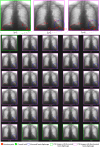
The optimization process of case 3’s left hemi-diaphragm visualizations. The pink box: CXR images with an abnormal hemi-diaphragm that requires correction. The green box: CXR images with the normal hemi-diaphragm.
FIGURE 9
FIGURE 9
The optimization process of case 4’s left hemi-diaphragm visualizations. The pink box: CXR images with an abnormal hemi-diaphragm that requires correction. The green box: CXR images with the normal hemi-diaphragm.

Similar articles

  • Hemi-diaphragm detection of chest X-ray images based on convolutional neural network and graphics.
    Yang Y, Zheng J, Guo P, Wu T, Gao Q, Zeng X, Chen Z, Zeng N, Ouyang Z, Guo Y, Chen H. Yang Y, et al. J Xray Sci Technol. 2024;32(5):1273-1295. doi: 10.3233/XST-240108. J Xray Sci Technol. 2024. PMID: 38995761
  • Systemic Inflammatory Response Syndrome.
    Baddam S, Burns B. Baddam S, et al. 2025 Jun 20. In: StatPearls [Internet]. Treasure Island (FL): StatPearls Publishing; 2025 Jan–. 2025 Jun 20. In: StatPearls [Internet]. Treasure Island (FL): StatPearls Publishing; 2025 Jan–. PMID: 31613449 Free Books & Documents.
  • Ventilator Management(Archived).
    Mora Carpio AL, Mora JI. Mora Carpio AL, et al. 2023 Mar 27. In: StatPearls [Internet]. Treasure Island (FL): StatPearls Publishing; 2025 Jan–. 2023 Mar 27. In: StatPearls [Internet]. Treasure Island (FL): StatPearls Publishing; 2025 Jan–. PMID: 28846232 Free Books & Documents.
  • Physical interventions to interrupt or reduce the spread of respiratory viruses.
    Jefferson T, Dooley L, Ferroni E, Al-Ansary LA, van Driel ML, Bawazeer GA, Jones MA, Hoffmann TC, Clark J, Beller EM, Glasziou PP, Conly JM. Jefferson T, et al. Cochrane Database Syst Rev. 2023 Jan 30;1(1):CD006207. doi: 10.1002/14651858.CD006207.pub6. Cochrane Database Syst Rev. 2023. PMID: 36715243 Free PMC article.
  • Interventions to reduce harm from continued tobacco use.
    Lindson-Hawley N, Hartmann-Boyce J, Fanshawe TR, Begh R, Farley A, Lancaster T. Lindson-Hawley N, et al. Cochrane Database Syst Rev. 2016 Oct 13;10(10):CD005231. doi: 10.1002/14651858.CD005231.pub3. Cochrane Database Syst Rev. 2016. PMID: 27734465 Free PMC article.

References

    1. Al-qaness M. A. A., Zhu J., Al-Alimi D., Dahou A., Alsamhi S. H., Abd Elaziz M., et al. (2024). Chest X-ray images for lung disease detection using deep learning techniques: a comprehensive survey. Arch. Comput. Methods Eng. 31, 3267–3301. 10.1007/s11831-024-10081-y - DOI
    1. Amin S. U., Taj S., Hussain A., Seo S. (2024). An automated chest X-ray analysis for COVID-19, tuberculosis, and pneumonia employing ensemble learning approach. Biomed. Signal Process. Control 87, 105408. 10.1016/j.bspc.2023.105408 - DOI
    1. Arias-Martínez P., Lafranca P. P. G., Mohamed Hoesein F. A. A., Vincken K., Schlösser T. P. C. (2025). Techniques for respiratory motion-resolved magnetic resonance imaging of the chest in children with spinal or chest deformities: a comprehensive overview. JCM 14, 2916. 10.3390/jcm14092916 - DOI - PMC - PubMed
    1. Badrinarayanan V., Kendall A., Cipolla R. (2017). SegNet: a deep convolutional encoder-decoder architecture for image segmentation. IEEE Trans. Pattern Anal. Mach. Intell. 39, 2481–2495. 10.1109/TPAMI.2016.2644615 - DOI - PubMed
    1. Boussuges A., Gole Y., Blanc P. (2009). Diaphragmatic motion studied by m-mode ultrasonography: methods, reproducibility, and normal values. Chest 135, 391–400. 10.1378/chest.08-1541 - DOI - PubMed

LinkOut - more resources