

A comparative analysis of CT angiography and echocardiography in the evaluation of chest findings in patients with interrupted aortic arch
- PMID: 40642309
- PMCID: PMC12240960
- DOI: 10.3389/fradi.2025.1616112
A comparative analysis of CT angiography and echocardiography in the evaluation of chest findings in patients with interrupted aortic arch
Abstract
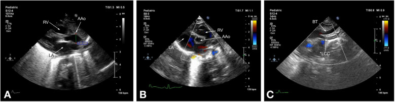
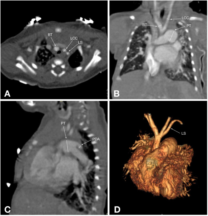
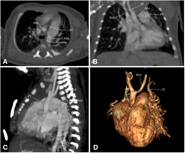
Interrupted aortic arch (IAA) is a rare congenital cardiovascular anomaly characterized by the absence of continuity between the ascending and descending aorta, often accompanied by congenital heart defects such as ventricular septal defects and patent ductus arteriosus. Accurate preoperative imaging is essential for surgical planning and patient management. This study aimed to compare the diagnostic accuracy of echocardiography and computed tomography angiography (CTA) in evaluating thoracic findings in patients with IAA. A retrospective analysis was conducted on 58 patients (median age: 18 days) diagnosed with IAA between September 2020 and January 2023 at the Heart Center, University Medical Center, Astana, Kazakhstan. Conventional echocardiography and multislice CTA were performed using standardized protocols. Sensitivity, specificity, and other diagnostic performance metrics were calculated. Statistical comparisons were made using McNemar's and Wilcoxon signed-rank tests, with p < 0.05 considered significant. Echocardiography correctly identified 91.4% of IAA cases, while CTA achieved 100% sensitivity and specificity. McNemar's test revealed a significant difference in diagnostic performance favoring CTA (p < 0.05). Measurements of the ascending aorta diameter showed no statistically significant difference between the two modalities (p = 0.09). IAA was predominantly type A (48.3%) and type B (46.6%), with hypoplastic ascending aorta identified in 34.5% of patients. Echocardiography remains a practical initial imaging modality for IAA, offering portability and cost-effectiveness. However, CTA demonstrated superior diagnostic accuracy and anatomical resolution, making it the preferred tool for detailed preoperative evaluation and surgical planning. Future studies with larger cohorts and additional modalities could further refine diagnostic strategies for IAA.
Keywords: computed tomography angiography; congenital heart defects; diagnostic imaging; echocardiography; interrupted aortic arch; surgical planning.
© 2025 Moldakhanova, Rakhimzhanova, Dautov, Bastarbekova, Kaliyev, Almussina, Zhankorazova and Zholshybek.
Conflict of interest statement
The authors declare that the research was conducted in the absence of any commercial or financial relationships that could be construed as a potential conflict of interest.
Figures
References
-
- Rea G, Valente T, Iaselli F, Urraro F, Izzo A, Sica G, et al. Multi-detector computed tomography in the evaluation of variants and anomalies of aortic arch and its branching pattern. Ital J Anat Embryol. (2014) 119:180–92. - PubMed
LinkOut - more resources
Full Text Sources