Review
. 2025 Jun 10;17(6):e85678.
doi: 10.7759/cureus.85678. eCollection 2025 Jun.

Supine Bridge Exercise in Degenerative and Functional Hip Disorders: A Biomechanical and Therapeutic Approach (Part III)

Affiliations
Review

Supine Bridge Exercise in Degenerative and Functional Hip Disorders: A Biomechanical and Therapeutic Approach (Part III)

Saverio Colonna et al. Cureus. .

Abstract

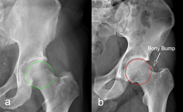
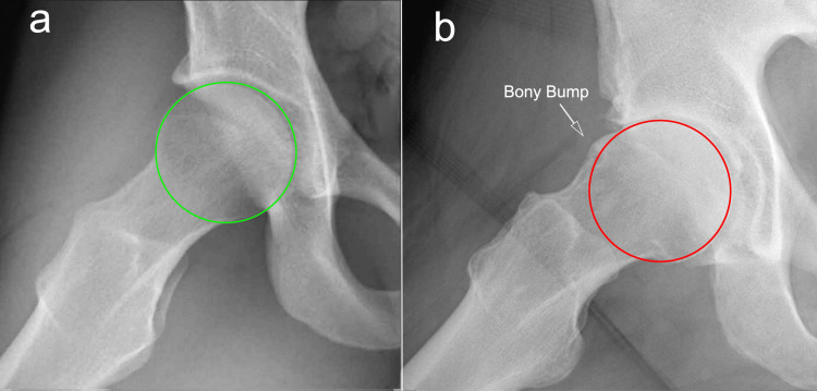
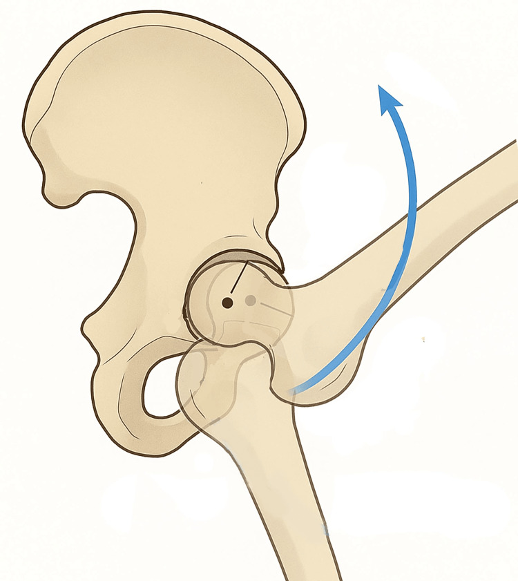
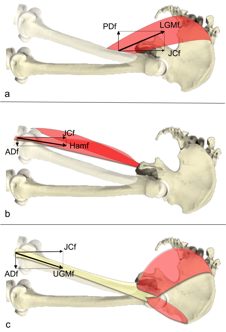
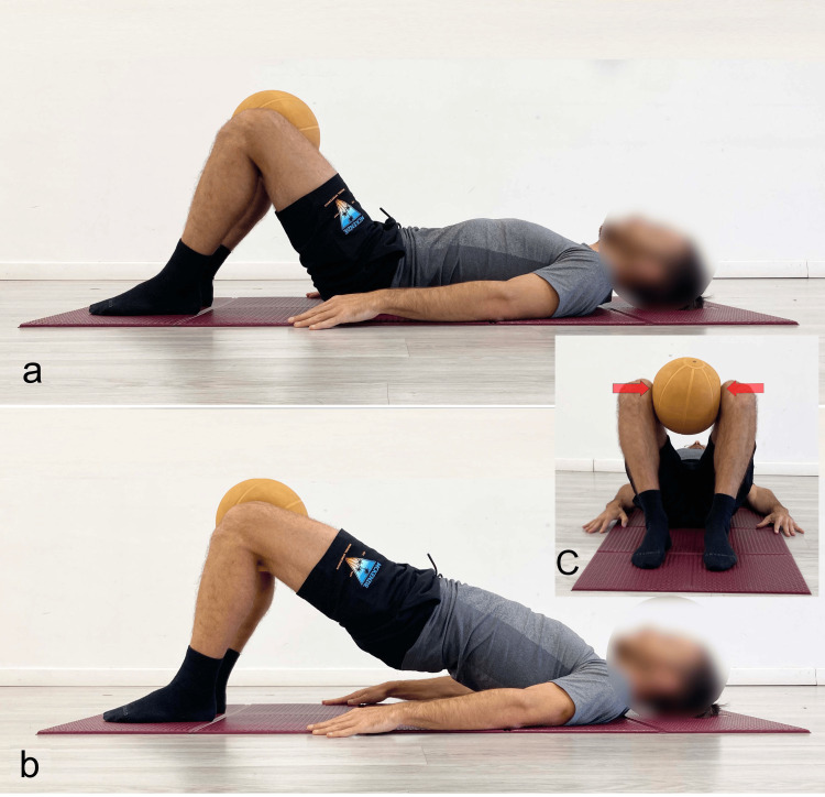
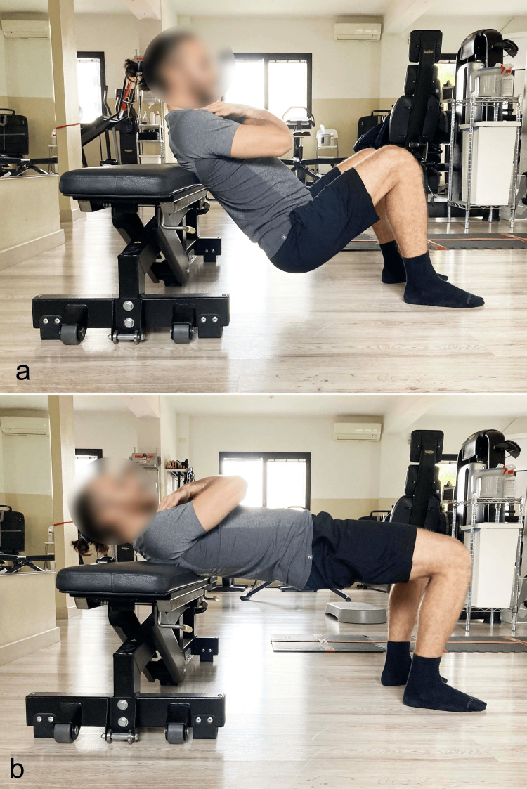
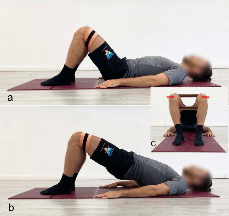
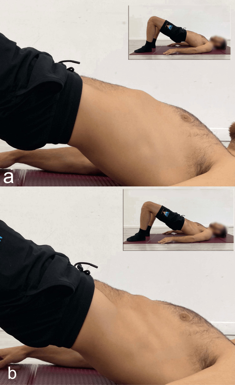
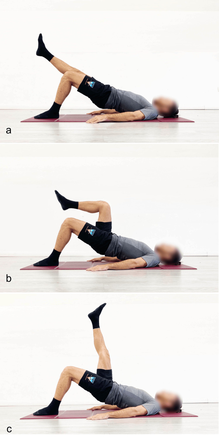
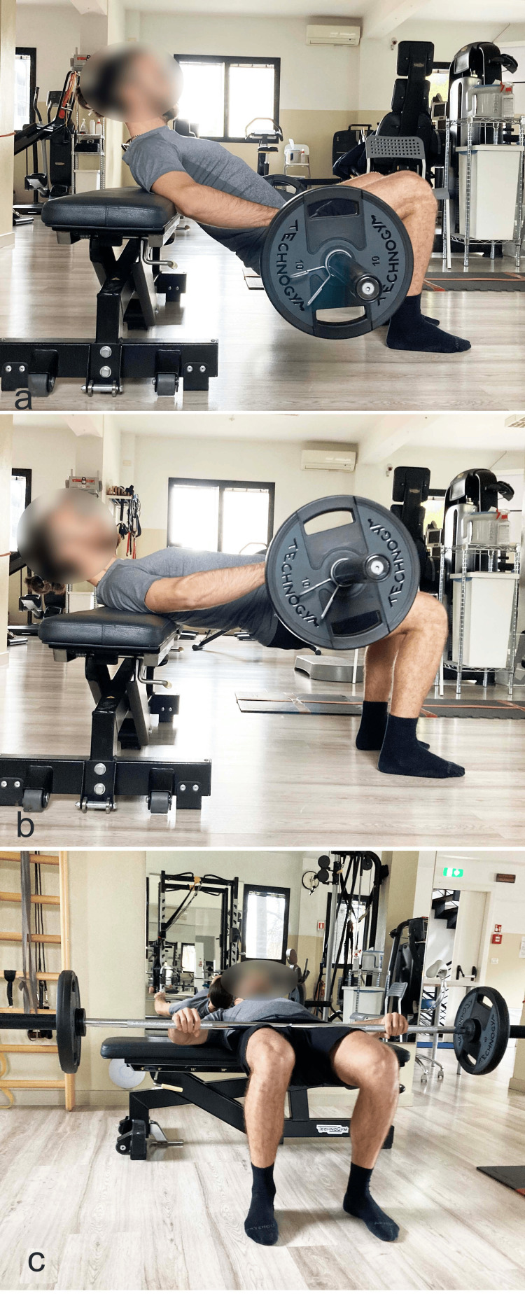
The supine bridge exercise (SBE) is widely recognized in rehabilitation for improving core stability and hip extensor strength. While its benefits in low back pain have been documented, its role in hip joint dysfunctions remains underexplored. This narrative review investigates the application of the SBE in degenerative and functional hip disorders, including femoroacetabular impingement (FAI), microinstability, and femoral anterior glide syndrome (FAGS). Particular attention is given to the biomechanical rationale behind gluteus maximus activation (especially the lower portion, or LGM) and the inhibition of synergistic dominance by the hamstrings Based on current evidence, specific SBE variations, including hip and ankle positioning, spinal alignment, and neuromuscular control strategies, may promote posterior femoral head translation and joint stability. Furthermore, the review highlights how in specific athletic populations, such as soccer players and dancers (where cam-type FAI alterations and restricted hip internal rotation are particularly prevalent), the inclusion of SBE sessions into preventive training programs could contribute to preserving hip joint health and mitigating degenerative processes. We argue that SBE when appropriately tailored, can become a fundamental therapeutic tool in both conservative management and functional retraining of hip dysfunctions.

Keywords: cam-type fai; femoral anterior glide syndrome; femoroacetabular impingement; gluteus maximus activation; hip disfunction; hip joint stability; hip pathologies; microinstability of the hip; synergistic dominance; upine bridge exercise.


Conflict of interest statement

Conflicts of interest: In compliance with the ICMJE uniform disclosure form, all authors declare the following: Payment/services info: All authors have declared that no financial support was received from any organization for the submitted work. Financial relationships: All authors have declared that they have no financial relationships at present or within the previous three years with any organizations that might have an interest in the submitted work. Other relationships: All authors have declared that there are no other relationships or activities that could appear to have influenced the submitted work.

Figures

Figure 1
Figure 1. Coronal plane radiographs of hip
a) Normal hip joint; b) hip joint showing a clear superior head-neck deformity, defined as a bony bump or pistol grip deformity. Image credit: Author, Saverio Colonna
Figure 2
Figure 2. Dunn view radiographs of hip
a) Normal hip joint; b) hip joint showing a clear anterior head-neck deformity, defined as a bony bump. Image credit: Author, Saverio Colonna
Figure 3
Figure 3. Schematic representation of hip flexion
In evidence physiological posterior glide of the center of rotation during hip flexion. Image credit: Author, Saverio Colonna
Figure 4
Figure 4. Biomechanical diagram of the force exerted by gluteus maximus and hamstrings
a) Lower gluteus maximus force (LGMf), divided into the force vectors expressed in the sagittal plane: JCf and PDf posteriorly directed force; b) hamstrings (Hamf), divided into the two force vectors expressed in the sagittal plane: JCf and ADf; c) upper gluteus maximus (UGMf), divided into the force vectors expressed in the sagittal plane: JCf and ADf. JCf: joint compression force; PDf: posteriorly directed force; ADf: anteriorly directed force Reprinted with permission from [42].
Figure 5
Figure 5. MRI image of the hip showing schematization of femoral head within the acetabulum
a) Physiological; b) dysfunctional anterior glide of the femoral head with increased tension of the anterior stabilizing structures. Image credit: Author, Saverio Colonna
Figure 6
Figure 6. Example of supine bridge exercise performed with high degrees of knee and hip flexion in the starting position
a) Starting position; b) end position. Reproduced from Colonna et al. [4], under a Creative Commons Attribution License (CC BY).
Figure 7
Figure 7. Example of supine bridge exercise performed with ankle dorsiflexion
a) Starting position; b) end position. Reproduced from Colonna et al. [4], under a Creative Commons Attribution License (CC BY).
Figure 8
Figure 8. Supine bridge exercise performed with different spinal angles
a) Neutral; b) hyperextension. Reproduced from Colonna et al. [4], under a Creative Commons Attribution License (CC BY).
Figure 9
Figure 9. Bridging with the addition of adductor muscle activation using a ball
a) Starting position; b) end position, lateral view; c) end position, foot-level view. Reproduced from Colonna et al. [4], under a Creative Commons Attribution License (CC BY).
Figure 10
Figure 10. Barbell hip thrust with dorsal support on a bench
a) Starting position; b) final position. Reproduced from Colonna et al. [4], under a Creative Commons Attribution License (CC BY).
Figure 11
Figure 11. Bridging exercise with added activation of the abductor muscles using an elastic band
a) Starting position; b) final position, lateral view; C) final position, foot-level view. Reproduced from Colonna et al. [4], under a Creative Commons Attribution License (CC BY).
Figure 12
Figure 12. Execution of supine bridge exercise
a) Traditional; b) with the abdominal drawing-in maneuver. Reproduced from Colonna et al. [4], under a Creative Commons Attribution License (CC BY).
Figure 13
Figure 13. Example of different execution modalities of the single-leg supine bridge exercise
a) The limb suspended in line with the trunk; b) hip and knee flexion; c) hip flexion with the knee extended. Reproduced from Colonna et al. [4], under a Creative Commons Attribution License (CC BY).
Figure 14
Figure 14. Barbell hip thrust with external load
a) Starting position with the barbell; b) end position; c) foot-level view of the end position. Reproduced from Colonna et al. [4], under a Creative Commons Attribution License (CC BY).

Similar articles

References

    1. An evidence-based framework for strengthening exercises to prevent hamstring injury. Bourne MN, Timmins RG, Opar DA, et al. Sports Med. 2018;48:251–267. - PubMed
    1. Rehabilitation exercise progression for the gluteus medius muscle with consideration for iliopsoas tendinitis: an in vivo electromyography study. Philippon MJ, Decker MJ, Giphart JE, Torry MR, Wahoff MS, LaPrade RF. Am J Sports Med. 2011;39:1777–1785. - PubMed
    1. Trunk muscle activity and ratio of local muscle to global muscle activity during supine bridge exercises under unstable conditions in young participants with and without chronic low back pain. Nakata A, Osuka S, Ishida T, et al. Healthcare (Basel) 2024;12 - PMC - PubMed
    1. Supine bridge exercise: a narrative review of the literature (part i) Colonna S, D'Alessandro A, Tarozzi R, Casacci F. Cureus. 2025;17:0. - PMC - PubMed
    1. Supine bridge exercise in low back pain: a fascial approach for movement impairment syndromes (part ii) Colonna S, D’Alezzandro A, Tarozzi R, Casacci F. Cureus. 2025;17:0. - PMC - PubMed

LinkOut - more resources