. 2025 Aug 5;13(8):e0333224.
doi: 10.1128/spectrum.03332-24. Epub 2025 Jul 11.

Characterization of a novel Phietavirus genus bacteriophage and its potential for efficient transfer of modified shuttle plasmids to Staphylococcus aureus strains of different clonal complexes

Affiliations

Characterization of a novel Phietavirus genus bacteriophage and its potential for efficient transfer of modified shuttle plasmids to Staphylococcus aureus strains of different clonal complexes

Łukasz Kałuski et al. Microbiol Spectr. .

Abstract

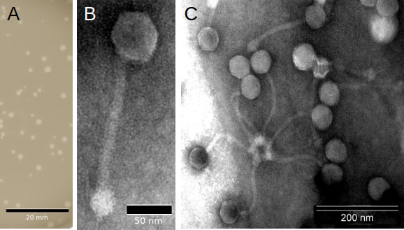
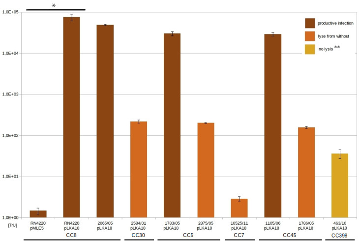
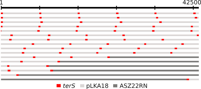
Staphylococcus aureus is a significant human pathogen responsible for various nosocomial and community-acquired infections, leading to considerable morbidity and mortality worldwide. Temperate bacteriophages contribute to its virulence and facilitate the dissemination of pathogenicity traits. We isolated a novel siphovirus of the Phietavirus genus, ASZ22RN, derived from a prophage of an S. aureus clonal complex 7 strain and capable of propagating in the prophage-free laboratory strain RN4220. ASZ22RN either productively infected or lysed from without all 47 tested S. aureus clinical strains across 12 clonal complexes (CCs), demonstrating its ability to puncture their cell envelopes. When ASZ22RN was propagated in RN4220 cells harboring an S. aureus-Escherichia coli plasmid replicating via theta mode, it transduced the plasmid to plasmid-free RN4220 with low frequency. The transduction frequency increased by nearly five orders of magnitude when the plasmid contained a fragment of ASZ22RN DNA (terS). Most terS+ plasmid-transducing particles carried plasmid concatamers, while some carried plasmid-phage DNA hybrids, as demonstrated by DNA sequencing. Strains from all tested CCs served as recipients for transduction, regardless of the presence of type I restriction-modification enzymes targeting plasmid/phage DNA, or prophages with lysis-lysogeny switch regions conferring superinfection immunity to ASZ22RN. Our results indicate that intracellular phage defense systems do not prevent phage-mediated plasmid transfer and demonstrate a simple method for introducing plasmids constructed in E. coli into clinical S. aureus isolates. Moreover, the presence of the ASZ22RN lysis-lysogeny switch region in 21% of tested ASZ22RN-resistant strains highlights superinfection exclusion as a dominant mechanism of resistance to siphoviruses in staphylococci.

Importance: This study highlights the capacity of a newly isolated staphylococcal Phietavirus, ASZ22RN, to transfer a low-copy-number shuttle Staphylococcus aureus-Escherichia coli plasmid to various S. aureus strains representing major clonal complexes from among clinical isolates. By increasing the plasmid transduction efficiency in an ASZ22RN-specific manner, we show that the primary factor determining a given strain's ability to be a recipient in transduction is the capacity of transducing phage to puncture the cell envelopes of this strain. This can be determined not only based on productive phage infection but also lysis from without. Major intracellular mechanisms protecting S. aureus from productive phage infection do not impede the transduction-mediated acquisition of plasmids. Moreover, the lack of phage DNA in most of the plasmid-transducing virions indicates the lack of phage contamination in most transductants. Our results offer a promising approach for developing efficient pipelines to introduce plasmids constructed in E. coli to clinical S. aureus isolates.

Keywords: Phietavirus; Staphylococcus aureus; horizontal gene transfer; plasmid; superinfection exclusion; temperate bacteriophage; transduction.


Conflict of interest statement

The authors declare no conflict of interest.

Figures

Fig 1
Fig 1
Morphology of plaques formed by ASZ22RN on a layer of S. aureus RN4220 strain cells (A) and transmission electron micrographs of ASZ22RN virions stained with uranyl acetate (B, C).
Fig 2
Fig 2
Taxonomic assignment of ASZ22RN bacteriophage. (A) Pairwise intergenomic similarities between genomes of phietaviruses calculated using Virus Intergenomic Distance Calculator with default parameters. Genome similarities (above the diagonal line) are indicated with color intensity, from white (no relation), through blue (somewhat similar) to dark green (identical). The aligned fractions for each genome (below the diagonal line) are marked using three indicator values for each compared pair from top to bottom: aligned fraction genome 1, genome length ratio, and aligned fraction genome 2. Orange-to-white and black-to-white color gradients emphasize alignments and genome length ratio, respectively, thus whiter colors correspond to genome pairs with higher similarity values. (B) Phylogenetic analysis of ASZ22RN and the most closely related phages based on whole-genome-wide sequence similarities calculated by tBLASTx with the use of ViPTree. The ASZ22RN phage is marked with red.
Fig 3
Fig 3
The alignment of genomic maps of ASZ22RN with the genomic maps of its selected relatives representing Phietavirus and Dubowvirus genus. Genes in the genomes of ASZ22RN relatives shown in the alignment were indicated based on the GenBank annotations corrected or supplemented according to the analysis performed as described in the Materials and Methods section. The re-annotated or newly identified genes of ASZ22RN relatives are indicated by asterisks. Their coordinates relative to the coordinates of their sequence annotations deposited in GenBank are listed in Table S5. The sequences of some phage genomes in the figure were reorganized for their alignment to set their start positions at the beginning of the small terminase subunit genes. Genes are colored according to the % of the identity of their products to the corresponding products of ASZ22RN genes (dark green: <70%, green: 70 > 40%, blue: 40 > 20%, brown: <20%. The alignment (gapped) was performed with the use of Geneious Prime. The scale length corresponds to the consensus sequence including gaps. The GenBank accession numbers of genomic sequences used for the re-annotations and alignment are provided in Table S5.
Fig 4
Fig 4
Efficiency of transduction (TrU) of pMLE5 plasmid and pLKA18 by phage ASZ22RN. *Student’s t-test P-value = 0.04. **Lysis from without was observed only with the concentrated phage suspension (1012/PFU/mL).
Fig 5
Fig 5
Composition of DNA molecules in pLKA18 plasmid-transducing particles of bacteriophage ASZ22RN. DNA of ASZ22RN for sequencing was isolated from phages propagated in RN4220/pLKA18. Fragments representing DNA of the pLKA18 plasmid and ASZ22RN phage are shown in light or dark gray, respectively, except for the terS gene, which is shown in red.
Fig 6
Fig 6
Alignment of the 163 bp DNA sequence of phage ASZ22RN region between genes immR and cro with the relevant prophage sequences identified in certain tested S. aureus strains resistant to productive infection with ASZ22RN.
Fig 7
Fig 7
Influence of cloned immR or immRA genes of phage ASZ22RN on the susceptibility of S. aureus RN4220 cells to ASZ22RN infection. Serially diluted lysates of phage ASZ22RN (initial titer: 10¹⁰ PFU/mL) were used in spot tests on lawns of S. aureus RN4220 cells harboring either the immR gene (left panel) or the immRA gene cluster (central panel), both including their upstream intergenic region cloned into the pMLE5 plasmid. Wild-type RN4220 cells served as a control (right panel). Plates were incubated overnight at 37°C prior to result assessment. To enhance lysis zone visibility, image colors were reversed and desaturated.

Similar articles

References

    1. Brüssow H, Canchaya C, Hardt W-D. 2004. Phages and the evolution of bacterial pathogens: from genomic rearrangements to lysogenic conversion. Microbiol Mol Biol Rev 68:560–602. doi: 10.1128/MMBR.68.3.560-602.2004 - DOI - PMC - PubMed
    1. Ingmer H, Gerlach D, Wolz C. 2019. Temperate phages of Staphylococcus aureus. Microbiol Spectr 7:7. doi: 10.1128/microbiolspec.GPP3-0058-2018 - DOI - PMC - PubMed
    1. Mašlaňová I, Doškař J, Varga M, Kuntová L, Mužík J, Malúšková D, Růžičková V, Pantůček R. 2013. Bacteriophages of Staphylococcus aureus efficiently package various bacterial genes and mobile genetic elements including SCCmec with different frequencies. Environ Microbiol Rep 5:66–73. doi: 10.1111/j.1758-2229.2012.00378.x - DOI - PubMed
    1. Penadés JR, Chen J, Quiles-Puchalt N, Carpena N, Novick RP. 2015. Bacteriophage-mediated spread of bacterial virulence genes. Curr Opin Microbiol 23:171–178. doi: 10.1016/j.mib.2014.11.019 - DOI - PubMed
    1. Haaber J, Penadés JR, Ingmer H. 2017. Transfer of antibiotic resistance in Staphylococcus aureus. Trends Microbiol 25:893–905. doi: 10.1016/j.tim.2017.05.011 - DOI - PubMed

MeSH terms

LinkOut - more resources