Review
. 2025 Jun 23;14(13):959.
doi: 10.3390/cells14130959.

Exosomes as Future Therapeutic Tools and Targets for Corneal Diseases

Affiliations
Review

Exosomes as Future Therapeutic Tools and Targets for Corneal Diseases

Joshua Gamez et al. Cells. .

Abstract

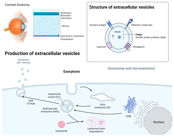
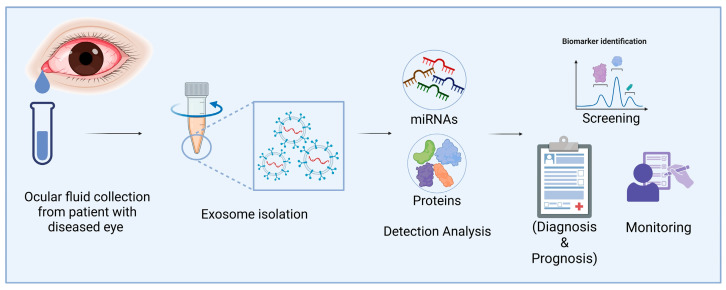
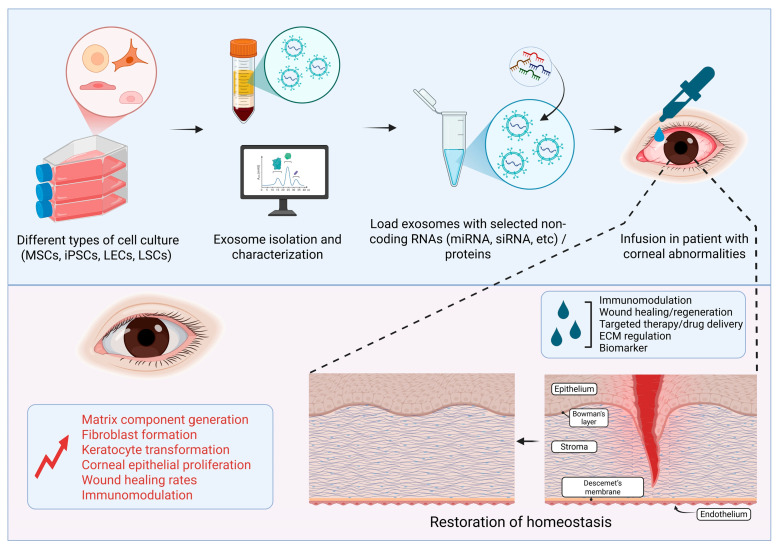
The therapeutic potential of exosomes (Exos), a subpopulation of extracellular vesicles (EVs) secreted by various cell types, has been broadly emphasized. Exos are endosome-derived membrane-bound vesicles 50-150 nm in size. Exos can be general or cell type-specific. Their contents enable them to function as multi-signaling and vectorized vehicles. Exos are important for maintaining cellular homeostasis. They are released into extracellular spaces, leading to uptake by neighboring or distant cells and delivering their contents to modulate cell signaling. Exos influence tissue responses to injury, infection, and disease by fusion with the target cells and transferring their cargo, including cytokines, growth and angiogenic factors, signaling molecules, lipids, DNA, mRNAs, and non-coding RNAs. They are implicated in various physiological and pathological conditions, including ocular surface events, such as corneal scarring, wound healing, and inflammation. Their biocompatibility, stability, low immunogenicity, and easy detectability in bodily fluids (blood, tears, saliva, and urine) make them promising tools for diagnosing and treating ocular diseases. The potential to engineer specific Exo cargos makes them outstanding therapeutic delivery vehicles. The objective of this review is to provide novel insights into the functions of Exo cargos and their applications as biomarkers and therapeutics, or targets in the cornea.

Keywords: biomarkers; cell–cell communication; cornea; crosstalk; exosome blockers; exosomes; miRNAs; therapeutic tools; therapeutics targets.


Conflict of interest statement

The authors declare no conflicts of interest.

Figures

Figure 1
Figure 1
A schematic overview of the corneal structures and biogenesis of Exos. The human cornea, the transparent outer part of the eye, consists of several distinct layers, including the epithelium, Bowman’s membrane, stroma, and Descemet’s membrane and endothelium. Exos carry nucleic acids, proteins, and lipids and are involved in intercellular communication. Their biogenesis involves the formation of early endosomes, leading to the formation of intraluminal vesicles (ILVs) through inward membrane budding and maturation into multivesicular bodies (MVBs). The MVBs can either be degraded by lysosomes or released as Exos outside the cell using Rab GTPase and SNARE complexes (image created in BioRender).
Figure 2
Figure 2
Applications of eye fluid-derived exosomal miRNA and proteins as diagnostic biomarkers of corneal diseases. Exos from eye fluids can be analyzed for omics data. These Exos may be useful for diagnosing corneal diseases after marker validation (Image created in BioRender).
Figure 3
Figure 3
Schematic showing the therapeutic potential of Exos derived from eye fluids or mesenchymal stem cells (MSCs), induced pluripotent stem cells (iPSCs), limbal epithelial cells (LECs), and limbal stromal cells (LSCs) in corneal repair and regeneration (Image created in BioRender).

Similar articles

References

    1. Dziasko M.A., Daniels J.T. Anatomical features and cell-cell interactions in the human limbal epithelial stem cell niche. Ocul. Surf. 2016;14:322–330. doi: 10.1016/j.jtos.2016.04.002. - DOI - PubMed
    1. Matic M., Petrov I.N., Chen S., Wang C., Dimitrijevich S.D., Wolosin J.M. Stem cells of the corneal epithelium lack connexins and metabolite transfer capacity. Differentiation. 1997;61:251–260. doi: 10.1046/j.1432-0436.1997.6140251.x. - DOI - PubMed
    1. Dogru M., Chen M., Shimmura S., Tsubota K. Corneal Surgery. 4th ed. Elsevier; Amsterdam, The Netherlands: 2009. Corneal epithelium and stem cells; pp. 25–31.
    1. Thomasy S.M., Leonard B.C., Greiner M.A., Skeie J.M., Raghunathan V.K. Squishy matters—Corneal mechanobiology in health and disease. Prog. Retin. Eye Res. 2024;99:101234. doi: 10.1016/j.preteyeres.2023.101234. - DOI - PMC - PubMed
    1. Zhang Y., Sun H., Liu Y., Chen S., Cai S., Zhu Y., Guo P. The limbal epithelial progenitors in the limbal niche environment. Int. J. Med. Sci. 2016;13:835–840. doi: 10.7150/ijms.16563. - DOI - PMC - PubMed