. 2025 Jun 30;14(13):1001.
doi: 10.3390/cells14131001.

Role of Nitric Oxide in Cardioprotection by Poloxamer 188

Affiliations

Role of Nitric Oxide in Cardioprotection by Poloxamer 188

Zhu Li et al. Cells. .

Abstract

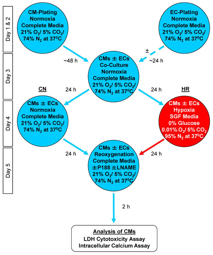
Poloxamer (P) 188 attenuates myocardial ischemia/reperfusion injury through cell membrane stabilization. Cell-cell interactions between endothelial cells (ECs) and cardiomyocytes (CMs) further protect CMs: co-cultures showed that, at an optimal density, ECs protected CMs against hypoxia/reoxygenation (HR) injury. The mechanism of interaction with P188 still requires exploration. We examined if N(ω)-nitro-L-arginine methyl ester (LNAME), a non-specific nitric oxide (NO) synthase inhibitor, abolishes protection in the presence or absence of P188 and/or ECs. We co-cultured mouse coronary artery ECs in an insert atop mouse CMs plated at confluency on the bottom of a well. Normoxic controls remained in complete media while HR groups were exposed to 24 h hypoxia at 0.01% O2 in serum- and glucose-free media, followed by 2 h reoxygenation in complete media. P188 (300 μM), LNAME (40 mM), or vehicle were administered upon reoxygenation. ECs at the used lower density did not decrease HR-triggered lactate dehydrogenase release or calcium overload in CMs by themselves. P188 reduced both indicators after HR by 16/18% without and by 22/25% with ECs, respectively. LNAME abrogated CM protection by P188. Neither intervention had an effect under normoxia. Our co-culture data indicates that P188 requires NO, not necessarily of endothelial origin, to elicit CM protection.

Keywords: NO; P188; calcium; cardiomyocyte; co-culture; co-polymer; cross-talk; endothelium; hypoxia reoxygenation; lactate dehydrogenase.


Conflict of interest statement

The authors declare no conflicts of interest.

Figures

Figure 1
Figure 1
Flow chart of the co-culture model. Cardiomyocytes (CMs) and endothelial cells (ECs) were cultured separately in complete media and under normoxic conditions. Next, either CMs continued to be cultured alone or EC inserts were placed atop the CM wells. This was followed by hypoxia in combination with serum- and glucose-free (SGF) media or by control/normoxia (CN) in complete media before the endpoint assays. HR: hypoxia/reoxygenation; LDH: lactate dehydrogenase.
Figure 2
Figure 2
Schematic of the insert with the endothelial cells (ECs) inside the well containing the cardiomyocytes (CMs) on the bottom. The semi-permeable barrier, indicated by a dotted line at the bottom of the EC insert, allows for cell–cell communication without mixing of media. ECs were plated at a lower density of 25,000 cells per insert and CMs at 300,000 per well. Modified from Li et al. [31].
Figure 3
Figure 3
Release of lactate dehydrogenase (LDH; panel (A)) and intracellular calcium (panel (B)) 2 h after reoxygenation following hypoxia (HR) vs. control/normoxia conditions (CN) in the absence (−) or presence (+) of the nitric oxide synthase inhibitor N(ω)-nitro-L-arginine methyl ester (LNAME; 40 mM) from isolated cardiomyocytes. Experiments were conducted in the absence (Con) or presence of co-cultured endothelial cells (EC; 25,000 per insert), Poloxamer (P) 188 (300 µM), or both. All data are shown as mean ± standard error of the mean normalized to 100% of CN conditions in the absence of ECs, P188, and LNAME; n = 5 to 6 per group, with at least 3 replicates per experiment. Statistics: One-Way Analysis of Variance followed by Student–Newman–Keuls post hoc comparisons, p < 0.05 (two-tailed), with * vs. Con, † vs. EC, and # vs. CN; n.s. = not significant.

Similar articles

References

    1. Lefer A.M., Tsao P.S., Lefer D.J., Ma X.L. Role of endothelial dysfunction in the pathogenesis of reperfusion injury after myocardial ischemia. FASEB J. 1991;5:2029–2034. doi: 10.1096/fasebj.5.7.2010056. - DOI - PubMed
    1. Szocs K. Endothelial dysfunction and reactive oxygen species production in ischemia/reperfusion and nitrate tolerance. Gen. Physiol. Biophys. 2004;23:265–295. - PubMed
    1. Perry M.O., Fantini G. Ischemia-reperfusion and cell membrane dysfunction. Microcirc. Endothel. Lymphat. 1989;5:241–258. - PubMed
    1. Cipolla M.J., Chan S.L., Sweet J., Tavares M.J., Gokina N., Brayden J.E. Postischemic reperfusion causes smooth muscle calcium sensitization and vasoconstriction of parenchymal arterioles. Stroke. 2014;45:2425–2430. doi: 10.1161/STROKEAHA.114.005888. - DOI - PMC - PubMed
    1. Kalogeris T., Baines C.P., Krenz M., Korthuis R.J. Ischemia/Reperfusion. Compr. Physiol. 2016;7:113–170. doi: 10.1002/j.2040-4603.2017.tb00741.x. - DOI - PMC - PubMed

MeSH terms

LinkOut - more resources