. 2025 Jul 7;14(13):1033.
doi: 10.3390/cells14131033.

Duchenne Muscular Dystrophy Patient iPSCs-Derived Skeletal Muscle Organoids Exhibit a Developmental Delay in Myogenic Progenitor Maturation

Affiliations

Duchenne Muscular Dystrophy Patient iPSCs-Derived Skeletal Muscle Organoids Exhibit a Developmental Delay in Myogenic Progenitor Maturation

Urs Kindler et al. Cells. .

Abstract

Background: Duchenne muscular dystrophy (DMD), which affects 1 in 3500 to 5000 newborn boys worldwide, is characterized by progressive skeletal muscle weakness and degeneration. The reduced muscle regeneration capacity presented by patients is associated with increased fibrosis. Satellite cells (SCs) are skeletal muscle stem cells that play an important role in adult muscle maintenance and regeneration. The absence or mutation of dystrophin in DMD is hypothesized to impair SC asymmetric division, leading to cell cycle arrest.

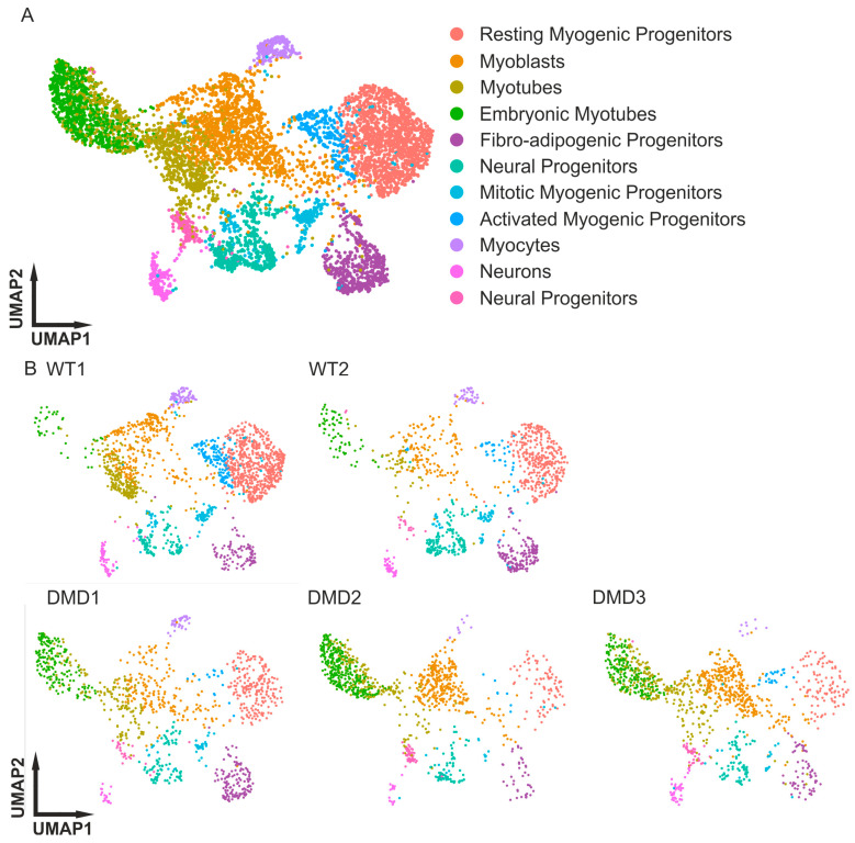
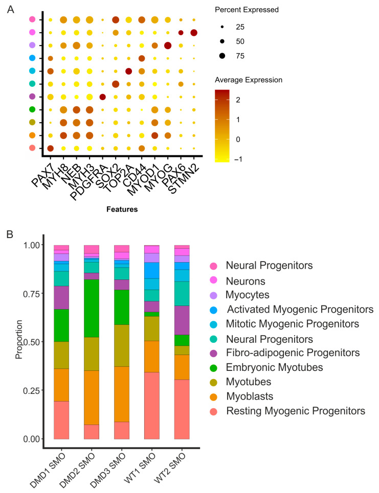
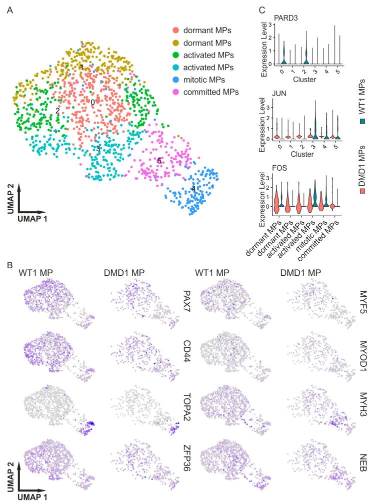
Methods: To overcome the limited availability of biopsies from DMD patients, we used our 3D skeletal muscle organoid (SMO) system, which delivers a stable population of myogenic progenitors (MPs) in dormant, activated, and committed stages, to perform SMO cultures using three DMD patient-derived iPSC lines.

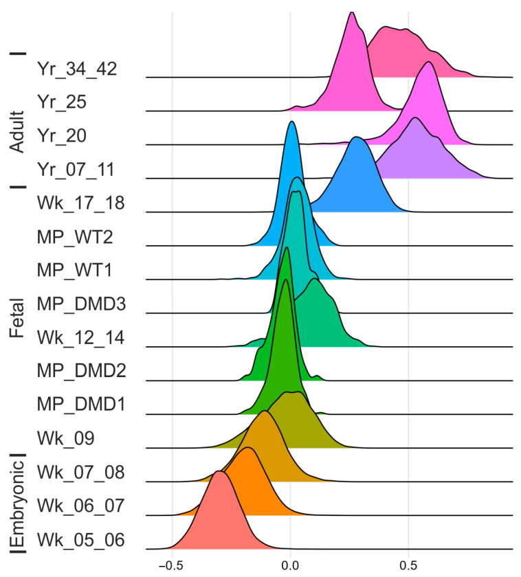
Results: The results of scRNA-seq analysis of three DMD SMO cultures versus two healthy, non-isogenic, SMO cultures indicate reduced MP populations with constant activation and differentiation, trending toward embryonic and immature myotubes. Mapping our data onto the human myogenic reference atlas, together with primary SC scRNA-seq data, indicated a more immature developmental stage of DMD organoid-derived MPs. DMD fibro-adipogenic progenitors (FAPs) appear to be activated in SMOs.

Conclusions: Our organoid system provides a promising model for studying muscular dystrophies in vitro, especially in the case of early developmental onset, and a methodology for overcoming the bottleneck of limited patient material for skeletal muscle disease modeling.

Keywords: Duchenne muscular dystrophy; FAPs; human-induced pluripotent stem cells; myogenesis; myogenic progenitors; organoids; satellite cells; scRNA-seq; skeletal muscle.


Conflict of interest statement

The authors declare no conflicts of interest.

Figures

Figure 1
Figure 1
Integrated scRNA-seq analysis of healthy control and DMD SMOs. (A) UMAP cluster of integrated scRNA-seq analysis showing cell type distribution in the SMOs. (B) UMAP analysis splitted into individual datasets to show cluster distribution between healthy and DMD SMOs. The DMD SMOs comprise a reduced population of resting myogenic progenitors and a greater pool of early embryonic myotubes compared to the healthy controls (DMD1, DMD2: [43]; DMD3: [44]; WT: [45]). SMOs = skeletal muscle organoids.
Figure 2
Figure 2
Identification of the types of cells within the organoid systems and altered distribution of clusters for diseased and healthy SMOs. (A) Dots showing marker genes for cluster identification. Clusters were identified via the upregulation of key markers (PAX7, MYH8, NEB, MYH3, PDGFRA, SOX2, TOP2A, CD44, MYOD1, MYOG, PAX6, and STMN2). (B) Distribution of clusters within the WT vs. DMD SMOs. The percent distribution of cells across the different datasets revealed increased clusters of myogenic progenitors in the WT SMOs. The number of myoblast clusters tends to be higher in DMD SMOs, and the number of embryonic (early) myotubes increases in all DMD SMOs.
Figure 3
Figure 3
Investigation of DMD-affected myogenic progenitors. (A) Results of an integrated scRNA analysis of organoid-derived myogenic progenitors of WT and DMD iPSCs showing up to 6 subclusters. (B) Feature plots of representative genes of dormant, activated, and committed myogenic progenitors (PAX7, CD44, TOP2A, ZFP36, MYF5, MYOD1, MYH3, and NEB). Clusters that expressed PAX7, MYF5, and CD44 were more prominent in the WT, while clusters expressing NEB and MYH3 were more prominent in the DMD MPs. (C) Example expression of the WT and DMD clusters of PARD3 (an asymmetric cell division marker), JUN, and FOS as markers for activated MPs (cluster numbers corresponding to subfigure A). MPs = myogenic progenitors.
Figure 4
Figure 4
Developmental scores of skeletal muscle organoid myogenic progenitors. The distribution of myogenic progenitors in the maturation score indicates delayed development of DMD1 and DMD2 MPs compared with WT1 and WT2 MPs across in vivo stages. DMD3 shows the same maturation grade as the WT MPs. The score is based on the difference between upregulated satellite cells and embryonic markers from human reference atlases for weeks (Wk) 5 to 18 in the embryonic and fetal stages and years (Yr) 7 to 42 for adult satellite cells and SMO-derived myogenic progenitors. The colours in the ridge plot were generated using the default settings of the ridgeplot function and are representing the different developmental maturation stages. MPs = myogenic progenitors.

Similar articles

References

    1. Bonne G., Rivier F., Hamroun D. The 2018 Version of the Gene Table of Monogenic Neuromuscular Disorders (Nuclear Genome) Neuromuscul. Disord. 2017;27:1152–1183. doi: 10.1016/j.nmd.2017.10.005. - DOI - PubMed
    1. Mercuri E., Bönnemann C.G., Muntoni F. Muscular Dystrophies. Lancet. 2019;394:2025–2038. doi: 10.1016/S0140-6736(19)32910-1. - DOI - PubMed
    1. Cohen E., Bonne G., Rivier F., Hamroun D. The 2022 Version of the Gene Table of Neuromuscular Disorders (Nuclear Genome) Neuromuscul. Disord. 2021;31:1313–1357. doi: 10.1016/j.nmd.2021.11.004. - DOI - PubMed
    1. Huml R.A. Muscular Dystrophy. Volume 31. Springer; Cham, Switzerland: 2015. Muscular Dystrophy: Historical Background and Types; pp. 5–7. - DOI
    1. Med D.D.-A.G. Undefined Recherches Sur La Paralysie Musculaire Pseudo Hypertrophique Ou Paralysie Myo-Sclerosique. P. Asselin; Paris, Frence: 1868.

MeSH terms

LinkOut - more resources