. 2025 Jul;106(7):002127.
doi: 10.1099/jgv.0.002127.

Translocating shRNA: a novel approach to RNA interference with Newcastle disease virus as viral vector

Affiliations

Translocating shRNA: a novel approach to RNA interference with Newcastle disease virus as viral vector

Wei Chin Koh et al. J Gen Virol. 2025 Jul.

Abstract

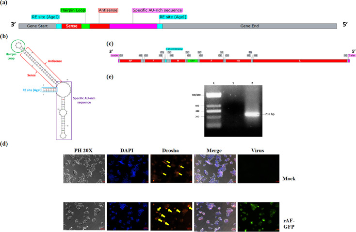
RNA interference is crucial in post-transcriptional gene silencing. Short hairpin RNA (shRNA) is particularly effective because it forms fully complementary matches with target mRNA, leading to its degradation. However, shRNA processing relies on nuclear microprocessors like Drosha, posing a challenge for RNA viral vectors that replicate exclusively in the cytoplasm. Although there have been reports of Drosha translocating to the cytoplasm upon viral infection, many RNA viruses, including Newcastle disease virus (NDV), remain inadequately studied in this context and, in some cases, fail to induce Drosha translocation for shRNA processing. In this study, we developed a novel approach to translocate an shRNA, expressed by NDV as an RNA viral vector, into the nucleus for Drosha processing. As a proof of concept, a recombinant NDV expressing the shRNA (rAF-shmcherry) with an AU-rich region at its 3' end in the expression cassette was constructed. This shRNA targets a constitutively expressed mCherry gene in a colorectal cancer cell line, SW620-mC. We confirmed the presence of the AU-rich shRNA in the nuclei of the rAF-shmcherry-infected SW620-mC using reverse transcription PCR (RT-PCR). The gene-silencing effect of the shRNA was then evaluated at mRNA and protein levels, showing ~90% downregulation of the mCherry transgene at 24 h post-infection and 70% downregulation of mCherry protein in SW620-mC at 48 h post-infection. This study marks the first exploration of NDV as an shRNA viral vector, presenting a promising approach for shRNA translocation that could be applicable to various RNA viruses.

Keywords: Drosha; Newcastle disease virus (NDV); ZC3H12D protein; short hairpin RNA; viral vector.


Conflict of interest statement

The authors declare that there are no conflicts of interest.

Figures

Fig. 1.
Fig. 1.. Construction of recombinant NDV (rAF-114shmCherry) and Drosha translocation determination upon NDV infection. (a) The complete shRNA sequence that targets mCherry consists of GS and GE of NDV, restriction enzyme sites (AgeI), the sense and antisense regions connected by a hairpin loop and a specific AU-rich domain at the 3′ end. The sequence was then verified using blast (https://blast.ncbi.nlm.nih.gov/Blast.cgi) for the recommended algorithms, G+C content (36–52 mol%), sense and antisense specificity [34]. The shRNA design follows the ‘rule of six’ of the Paramyxoviridae family, which meant that the number of nucleotides should be divisible by six to allow effective replication [35]. (b) Secondary structure of 114shmCherry predicted by mFold. 114shmCherry is able to form a hairpin structure after folding. The AU-rich region at its 3′ end can also form several loops after folding. (c) The genome of recombinant NDV, rAF-114shmCherry. (d) Drosha localization upon NDV infection. SW620-mC cells were treated without or with NDV (Mock and rAF-GFP), and immunofluorescent staining was performed after 24 h. Green fluorescence indicated the successful infection of the virus. The localization of Drosha (red) was determined by merging with the image of the nucleus, which was stained blue by DAPI, and images were taken using phase contrast at 20X. Scale bar: 50 µm. In the merged image of viral-infected cells, the red and blue colours overlapped with each other, producing a purple colour. This showed that Drosha remained in the nucleus. (e) Verification of ZC3H12D gene expression in the cell. RT-PCR on total RNA extracted from SW620-mC was carried out using ZC3H12D-specific primers to further confirm its expression in the cell used in the study. Agarose gel electrophoresis was run to visualize the presence of a band with the expected size of 232 bp. (L) TriDye ultraLow DNA ladder (NEB), (1) negative control (non-template control) and (2) RNA sample extracted from SW620-mC cell.
Fig. 2.
Fig. 2.. shmCherry localization, expression and functionality in target gene silencing. (a) The total RNA and nuclear RNA of uninfected/infected SW620-mC cells were extracted, followed by RT-PCR using NP and shmCherry-specific primers. The band size and intensity were shown in black and red, respectively. The lane was labelled ‘1, 2, 3, 4’, which indicated the harvest timepoint, ‘0, 24, 48, 72 h’, respectively. For the total RNA, the band of the NP gene was only present in the viral-infected cells, indicating the successful infection and replication of the viruses. The band of shmCherry was seen in the rAF-114shmCherry-infected cells, showing the generation of 114shmCherry over time (0–72 h). Besides, the NP gene of NDV is only present in the cytoplasm and can be used to evaluate the presence of cytoplasmic contamination in the nuclei content. For the nuclear RNA, the absence of a band of the NP gene in the figure indicates no cytoplasmic contamination during nuclei isolation (band shown in the figure was positive control of NP gene). The shmCherry, however, was detected in the rAF-114shmCherry-infected cells, suggesting the successful translocation into the nuclei due to the presence of the introduced AU-rich region in its 3′ end. (b) Absolute quantification of shmCherry in cells infected with rAF-GFP or rAF-114shmCherry was carried out by RT-qPCR. Viral infection was done at m.o.i. 1 and harvested upon reaching time points (0, 24, 48 and 72 h). RT-qPCR was carried out in technical replicates. The copy number was calculated based on the formula generated from the qPCR standard curve of the shRNA: X=(y−43.311)/–3.2364, where X is the log (shRNA copy number); y is the value mean Cq; 43.311 is the y-intercept value; −3.2364 is the slope of the standard curve. Data are presented as mean±sem from duplicate determinations. Statistically significant differences between the means were determined by one-way ANOVA followed by Dunnett multiple comparison tests to uninfected cells. Differences were considered significant when *P≤0.05. (c) Relative expression of shmCherry in cells infected with rAF-114shmCherry from 0 h to 72 h. Results of qPCR analysis are depicted as shmCherry expression at a particular timepoint relative to shmCherry expression at 0 h in infected cells. The expression of shmCherry produced by rAF-114shmCherry increased from 0 h to 72 h, indicating that shmCherry was continuously produced by the virus over time. (d) Relative expression of mCherry from 0 h to 24 h. Results of RT-qPCR analysis are depicted as mCherry expression at a particular timepoint relative to expression at 0 h in the respective group. Data are presented as mean±sem from duplicate determinations. Statistically significant differences between the means were determined by one-way ANOVA followed by Dunnett multiple comparison tests to uninfected cells. Differences were considered significant when *P≤0.05. At 24 h, rAF-114shmCherry downregulated the mCherry expression for around 90%, indicating that shmCherry produced by the virus was functional to cause gene silencing. (e) Western blot analysis on mCherry protein in SW620-mC. Viral infection was done at m.o.i. 1, and cell lysate was prepared at 48 h post-infection. Beta-actin was used as the housekeeping protein in this experiment. Upon the infection by rAF-114shmCherry, the mCherry protein in SW620-mC cells was successfully downregulated compared to the uninfected and rAF-GFP-infected cells. (f) Relative mCherry protein expression (%). The mCherry protein levels were normalized to beta-actin (housekeeping protein), and the relative mCherry expression in each sample was calculated compared to the uninfected sample. The results are presented as the mean±sem (n=3). Statistically significant differences between the means were determined by one-way ANOVA followed by Dunnett multiple comparison tests to uninfected cells. Differences were considered significant when *P≤0.05. rAF-114shmCherry caused significant mCherry protein downregulation (≈70%).

Similar articles

References

    1. Xu W, Jiang X, Huang L. Comprehensive Biotechnology. 2019. RNA interference technology; pp. 560–575. - DOI
    1. Lam JKW, Chow MYT, Zhang Y, Leung SWS. siRNA versus miRNA as therapeutics for gene silencing. Mol Ther Nucleic Acids. 2015;4:e252. doi: 10.1038/mtna.2015.23. - DOI - PMC - PubMed
    1. Roy S, Kundu TK. In: VII - Chemical genomics: understanding gene regulatory networks in differentiation and disease. Roy S, Kundu TK, editors. Academic Press; pp. 255–277. - DOI
    1. Xia XG, Zhou H, Xu Z. Multiple shRNAs expressed by an inducible pol II promoter can knock down the expression of multiple target genes. Biotechniques. 2006;41:64–68. doi: 10.2144/000112198. - DOI - PubMed
    1. Sheng P, Flood KA, Xie M. Short hairpin RNAs for strand-specific small interfering RNA production. Front Bioeng Biotechnol. 2020;8:940. doi: 10.3389/fbioe.2020.00940. - DOI - PMC - PubMed

LinkOut - more resources