. 2025 Jul 11;20(7):e0327621.
doi: 10.1371/journal.pone.0327621. eCollection 2025.

Computational discovery of potential therapeutic agents against brain-eating amoeba (Naegleria fowleri)

Affiliations

Computational discovery of potential therapeutic agents against brain-eating amoeba (Naegleria fowleri)

Jacopo Zattoni et al. PLoS One. .

Abstract

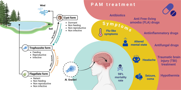
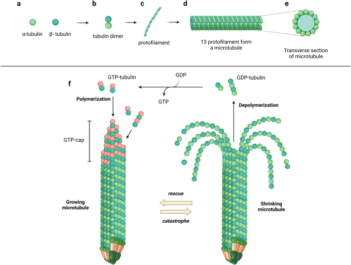
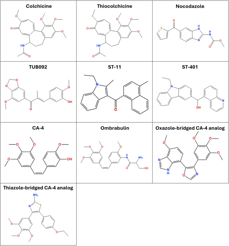
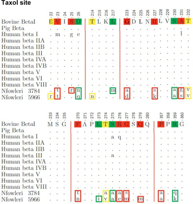
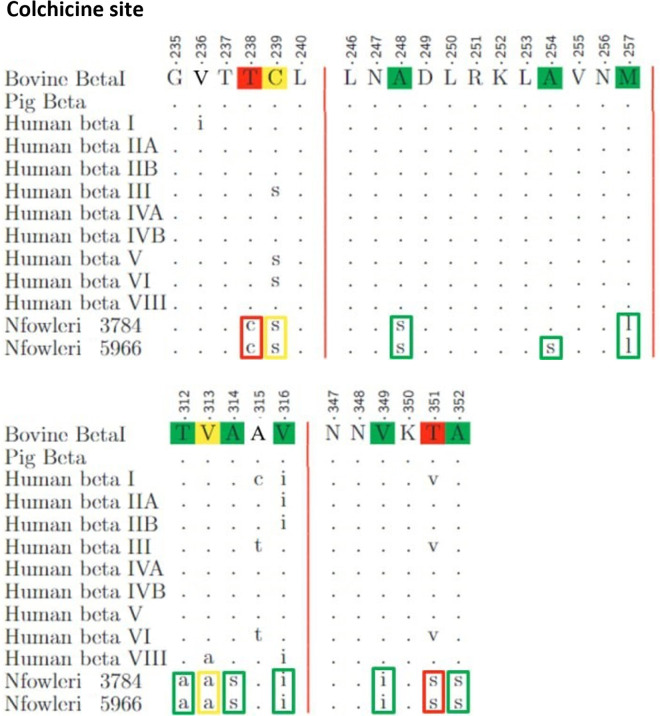
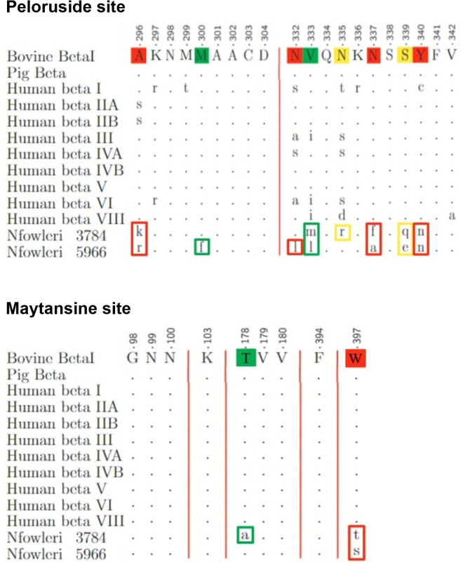
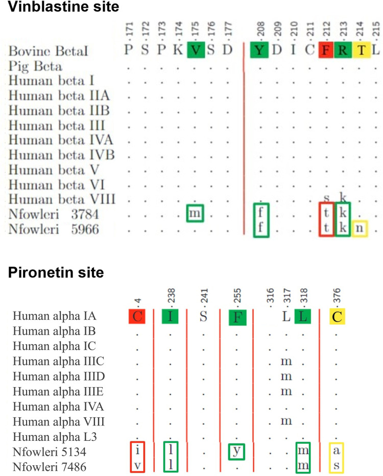
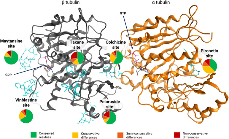
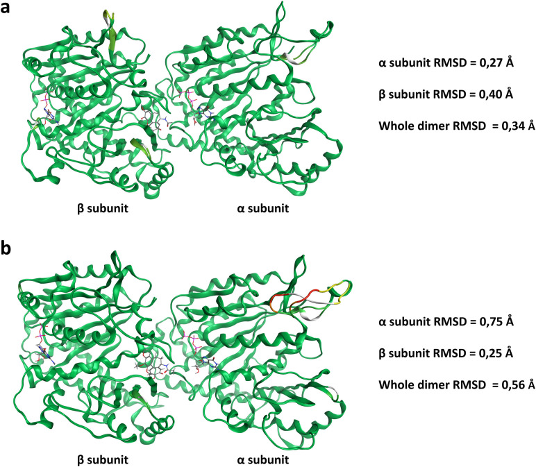
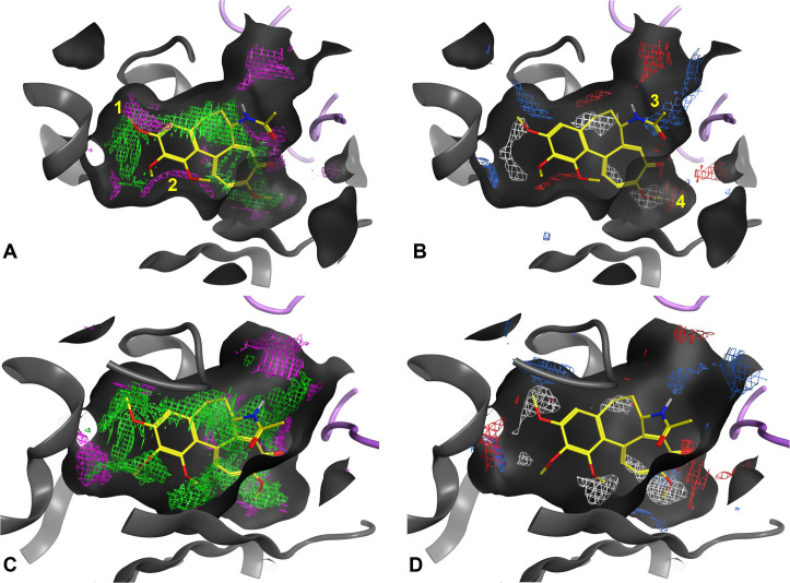
Naegleria fowleri is a human and animal pathogen well-known for its ability to digest neurons and astrocytes of the host's brain, causing a haemorrhagic and necrotizing inflammation called Primary Amoebic Meningoencephalitis. Although infections are rare, the mortality rate is over 97%, due to both the non-specificity of the symptoms and the absence of an effective treatment. In this work we employed bioinformatics tools to evaluate the possibility of treating the infection with tubulin-targeting compounds, which we regard as the most promising approach given the unclear view on the pathogenic factors in N. fowleri, the divergence of the amoeba's tubulins from the human counterparts, and how well-established microtubule-targeting therapies are in clinical practices. The amoeba's tubulin sequences were analyzed and compared to the human tubulins to conjecture the role of their differences in drugs resistance. The binding affinity of the compounds was computed for both species by performing docking simulations using Chemical Computing Group's MOE and CCSB's AutoDock4 and AutoDock Vina. The results were analyzed using a consensus method to increase their reliability. We found that the amoeba's mitotic tubulins show a significant number of changes that are expected to decrease their affinity for tubulin-targeting compounds. We identified the Colchicine binding site as the most suitable target, and propose that Colchicine analogs retain their ability to bind to the amoeba's tubulins in vivo. The selectivity of the compounds for the pathogen however remains an issue. The changes in the amino. acid sequences in the Colchicine site could create a template for designing novel derivatives with an improved selectivity for the parasite and a safer profile for the patient. We therefore believe that our results could be the starting point for a rational derivatization of the selected ligands, leading to the development of an effective treatment for Naegleria fowleri infection.


Conflict of interest statement

The authors have declared that no competing interests exist.

Figures

Fig 1
Fig 1. Infographic illustrating the life cycle, infection pathway, symptoms, and treatment options for the brain-eating amoeba (N. fowleri).
The life cycle begins with the cyst form, which is dormant and can survive for years outside of water. When conditions are favorable, cysts transform into trophozoites—the feeding, reproductive and infective form, which can in turn convert to the flagellate in absence of food. Trophozoites are responsible for animal and human infection. After reaching the frontal lobe of the host via nasal passage, they digest neurons and astrocytes causing a condition called Primary Amoebic Meningoencephalitis (PAM). Symptoms at the onset are flu-like, including headache, fever and altered mental state, and evolving within two weeks into seizures and coma. The current state-of-the-art treatment is a combination of drugs (antibiotics, anti-FLA drugs, antifungal and anti-inflammatory drugs), along with physical therapies commonly used to treat brain injuries. Nonetheless, the survival rate is estimated to be around 3%.
Fig 2
Fig 2. Assembly process and dynamic instability of microtubules.
(a) Tubulin dimer formation involves alpha and beta tubulin subunits (dark and light green) combining to form tubulin dimers. (b) Protofilament assembly sees these dimers align head-to-tail, extending into a protofilament. (c) Microtubule construction occurs as thirteen protofilaments come together in a cylindrical formation, held stable by lateral and longitudinal interactions. (d) GTP cap and stability are achieved when the growing microtubule end is capped with GTP-bound tubulin (red), which promotes elongation. (e) Hydrolysis and catastrophe happen as GTP is converted to GDP, causing instability that leads to rapid depolymerization (or ‘catastrophe’). (f) Rescue and regrowth occur when new GTP-tubulin is added, rescuing the structure by reestablishing the cap and allowing growth to resume, vital for microtubule functions in cellular processes. Image created using Biorender.com.
Fig 3
Fig 3. Colchicine site-targeting ligands selected for the docking simulations.
Fig 4
Fig 4. Alignment of the Taxane binding pocket sequences.
Fig 5
Fig 5. Alignment of the Colchicine binding pocket sequences.
Fig 6
Fig 6. Alignments of the Peloruside and Maytansine binding pocket sequences.
Fig 7
Fig 7. Alignments of the Vinblastine and Pironetin binding pocket sequences.
Fig 8
Fig 8. Pie charts depiction of the substitutions in each of the six tubulin binding sites of N. fowleri, compared to the animal sequences.
The tubulin dimer displayed corresponds to N. fowleri’s mitotic dimer 5134-5966. Each pie chart is placed in correspondence of the related binding site, identified by the bound ligands. The pie charts are color-coded according to the legend at the bottom of the Figure.
Fig 9
Fig 9. RMSD of the homology models of the two mitotic tubulin isotypes for N. fowleri in comparison to the template used (5EYP).
(a) Superimposition of the mitotic dimer 5134−5966 with 5EYP. (b) Superimposition of the mitotic dimer 7486−3784 with 5EYP. Both images were generated using MOE. White regions of the structures identify residues on the template that are matched with a gap in the N. fowleri sequences. The RMSD is color coded in shades of green; the darker the color, the lower the value. One may observe regions with yellow, orange and red residues on the α subunit of dimer 7476−3784. These identify regions with local RMSD greater than 3 Å. In particular, the yellow regions have an RMSD between 3.8 and 4.5 Å, the orange region and RMSD of 5.37 Å, and the red region an RMSD of 8.4 Å. The RMSD values for each dimer and its subunits can be observed on the right of the related image. Overall, the structure of the amoeba’s mitotic tubulins is highly conserved compared to the template used.
Fig 10
Fig 10. Comparison of the electrostatic and non-bonded interaction maps at the colchicine site between N. fowleri’s mitotic dimer 5134-5966 and PDB entry 5EYP.
The key changes influencing ligand interaction are highlighted with numbers 1 to 4. SubFigs A and C represent the non-bonded interaction maps, where green areas represent the most suitable locations for hydrophobic groups, while purple areas are those for hydrophilic groups. SubFigs C and D represent the electrostatic interaction maps, where white areas represent the most suitable locations for hydrophobic groups, blue areas those for H-bond donors and red areas those for H-bond acceptors. Although referring to similar properties, it is possible to observe slight differences between the two interactions profiles because of the way each is calculated. Overall, it is reasonable to say that they provide complementary rather than redundant information.

Similar articles

References

    1. Siddiqui R, Ali IKM, Cope JR, Khan NA. Biology and pathogenesis of Naegleria fowleri. Acta Trop. 2016;164:375–94. doi: 10.1016/j.actatropica.2016.09.009 - DOI - PubMed
    1. Maciver SK, Piñero JE, Lorenzo-Morales J. Is Naegleria fowleri an Emerging Parasite?. Trends Parasitol. 2020;36(1):19–28. doi: 10.1016/j.pt.2019.10.008 - DOI - PubMed
    1. Jahangeer M, Mahmood Z, Munir N, Waraich U-E-A, Tahir IM, Akram M, et al. Naegleria fowleri: Sources of infection, pathophysiology, diagnosis, and management; a review. Clin Exp Pharmacol Physiol. 2020;47(2):199–212. doi: 10.1111/1440-1681.13192 - DOI - PubMed
    1. Gharpure R, Bliton J, Goodman A, Ali IKM, Yoder J, Cope JR. Epidemiology and Clinical Characteristics of Primary Amebic Meningoencephalitis Caused by Naegleria fowleri: A Global Review. Clin Infect Dis. 2021;73(1):e19–27. doi: 10.1093/cid/ciaa520 - DOI - PMC - PubMed
    1. Güémez A, García E. Primary Amoebic Meningoencephalitis by Naegleria fowleri: Pathogenesis and Treatments. Biomolecules. 2021;11(9):1320. doi: 10.3390/biom11091320 - DOI - PMC - PubMed

MeSH terms

LinkOut - more resources