. 2025 Jul 11;11(28):eadj0006.
doi: 10.1126/sciadv.adj0006. Epub 2025 Jul 11.

Dynamically covalent lipid nanoparticles mediate CRISPR-Cas9 genome editing against choroidal neovascularization in mice

Affiliations

Dynamically covalent lipid nanoparticles mediate CRISPR-Cas9 genome editing against choroidal neovascularization in mice

Desheng Cao et al. Sci Adv. .

Abstract

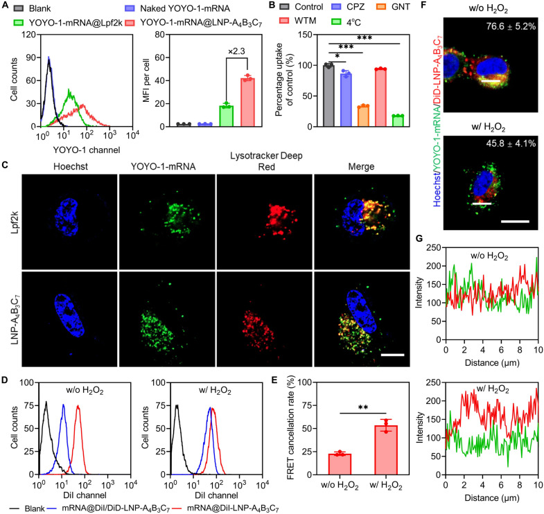
As an important modality for choroidal neovascularization (CNV) treatment, intravitreal injection of vascular endothelial growth factor A (VEGFA) inhibitors suffers from undesired response rate, low patient compliance, and ocular damage. Here, dynamically covalent lipid nanoparticles (LNPs) were engineered to mediate VEGFA gene editing and CNV treatment by codelivering Cas9 mRNA (mCas9) and single guide RNA (sgRNA) targeting VEGFA (sgVEGFA). A library of lipidoids bearing iminoboronate ester linkage was developed via facile "one-pot" synthesis, and the top-performing lipidoid-A4B3C7 was formulated into LNP-A4B3C7 with the highest mRNA transfection efficiency. Inside the diseased retinal pigment epithelial cells, LNPs were dissociated upon H2O2-triggered lipidoid degradation, facilitating mRNA/sgRNA release to potentiate the gene editing efficiency. In laser-induced CNV mice, mCas9/sgVEGFA@LNP-A4B3C7 after single intravitreal injection led to pronounced VEGFA disruption and CNV area reduction, outperforming the clinical anti-VEGF drug in eliciting sustained therapeutic effect. This study establishes a robust nonviral platform for mRNA delivery and genome editing and renders a promising strategy for CNV treatment.


Figures

Fig. 1.
Fig. 1.. Schematic illustration of dynamically covalent LNPs for delivering Cas9 mRNA (mCas9) and mediating VEGFA gene editing toward the treatment of CNV.
(A) The chemical structure of the top-performing lipidoid-A4B3C7 for mRNA delivery. (B) Formulation of mCas9/sgVEGFA@LNP-A4B3C7. (C) Intravitreally injected mCas9/sgVEGFA@LNP-A4B3C7 for VEGFA disruption in RPE cells in the laser-induced CNV mouse model. Lipidoid-A4B3C7 with iminoboronate ester structure can be hydrolyzed by the high-concentration H2O2 in the cytosol of laser-challenged RPE cells, which promotes the dissociation of LNPs and mCas9/sgVEGFA release to induce effective transfection and VEGFA disruption, ultimately leading to effective management of CNV.
Fig. 2.
Fig. 2.. Synthesis and screening of a library of dynamic covalent bond–based lipidoids.
(A) Synthetic route of diol-terminated lipid tails. (B) Synthetic route of iminoboronate ester–based lipidoids. The lipidoid was named Lipidoid-AxByCz, where A represents the serial number of amine groups, B represents the formylphenylboronic acids, and C represents the diol-terminated lipid tails. The formulated LNPs were named LNP-AxByCz. (C) Structures of amines, formylphenylboronic acids, and diol-terminated lipid tails used for lipidoid synthesis. (D) Transfection efficiencies of mLuc@LNPs in HeLa cells at 1 μg of mLuc/ml (n = 3). Results were represented as relative luminescence unit (RLU).
Fig. 3.
Fig. 3.. Acid- and H2O2-triggered lipidoid degradation, LNP dissociation, and mRNA release.
(A) Degradation mechanism of lipidoid-A4B3C7 upon acid and H2O2 treatment. 1H NMR (B) and 11B NMR (C) spectra of lipidoid-A4B3C7 in DMF-d7. (D) mRNA condensation by LNP-A4B3C7 at various N/P ratios as evaluated by gel electrophoresis. N represents naked mRNA. TEM images [(E); scale bar, 100 nm] and size distribution (F) of mLuc@LNP-A4B3C7 (N/P = 8). (G) Fluorescence emission spectra of mLuc@DiI/DiD-LNP-A4B3C7. (H) Cumulative release of mRNA from mLuc@LNP-A4B3C7 (n = 3). In the assessments [(B) to (H)], lipidoid-A4B3C7 or mLuc@LNP-A4B3C7 was treated with acid (pH 5.2), H2O2 (100 μM), or a combination thereof.
Fig. 4.
Fig. 4.. Intracellular delivery efficiency of mRNA@LNP-A4B3C7 in RPE cells and H2O2-triggered cytoplasmic mRNA release.
(A) Flow cytometric histograms and MFI per cell of H2O2-challenged ARPE-19 cells after 6-hour incubation with YOYO-1-mRNA-containing complexes (1 μg YOYO-1-mRNA/ml, n = 3). (B) Relative uptake level of YOYO-1-mRNA@LNP-A4B3C7 in H2O2-challenged ARPE-19 cells after 6-hour incubation at 4°C or in the presence of various endocytic inhibitors (n = 3). The uptake level of LNPs at 37°C in the absence of inhibitors served as 100% (control). (C) CLSM images of H2O2-challenged ARPE-19 cells following 6-hour incubation with YOYO-1-mRNA@Lpf2k or YOYO-1-mRNA@LNP-A4B3C7 (scale bar, 10 μm). Cell nuclei were stained with Hoechst 33342 and endolysosomes were stained with Lysotracker Deep Red. (D) Flow cytometric histograms of H2O2-challenged or unchallenged ARPE-19 cells after 6-hour incubation with mRNA@DiI/DiD-LNP-A4B3C7 or mRNA@DiI-LNP-A4B3C7. (E) The FRET cancellation rate calculated from (D) (n = 3). (F) CLSM images of H2O2-challenged or unchallenged ARPE-19 cells after 6-hour incubation with YOYO-1-mRNA@DiD-LNP-A4B3C7 (1 μg YOYO-1-mRNA/ml; scale bar, 20 μm). The colocalization ratios between YOYO-1-mRNA and DiD are listed (n = 20). (G) The fluorescence intensity of YOYO-1-mRNA (green) and DiD (red) in the white lines in (F) as measured by the ImageJ software.
Fig. 5.
Fig. 5.. H2O2-enhanced mRNA transfection and VEGFA genome editing in ARPE-19 cells.
(A) Transfection efficiencies of mLuc@Lpf2k or mLuc@LNP-A4B3C7 in H2O2-challenged or unchallenged ARPE-19 cells (n = 3). MFI per cell [(B), n = 3], percentage of EGFP-positive cells [(C), n = 3], and fluorescence images [(D); scale bar, 200 μm] of ARPE-19 cells with (w/) or without (w/o) H2O2 challenge after transfection with mEGFP@Lpf2k or mEGFP@LNP-A4B3C7. (E) mCas9/sgVEGFA@LNP-A4B3C7–induced indel mutations on VEGFA in H2O2-challenged ARPE-19 cells as determined by NGS. Frequencies of ≥0.1% were analyzed. (F) Relative VEGFA mRNA and VEGFA secretion levels in H2O2-challenged ARPE-19 cells after incubation with PBS, naked mCas9/sgVEGFA, mCas9/sgVEGFA@Lpf2k, or mCas9/sgVEGFA@LNP-A4B3C7 (n = 3).
Fig. 6.
Fig. 6.. Transport of intravitreally injected YOYO-1-mRNA@LNP-A4B3C7 to the fundus via Müller cell–mediated transcytosis.
(A) Representative CLSM images of mouse retina at different time points after intravitreal injection of naked YOYO-1-mRNA, YOYO-1-mRNA@Lpf2k, or YOYO-1-mRNA@LNP-A4B3C7 (0.5 μg of mRNA per eye; scale bar, 50 μm). RGC, retinal ganglion cell layer; INL, inner nuclear layer; ONL, outer nuclear layer; RPE, retinal pigmented epithelial layer. (B) Percentage of the injected naked YOYO-1-mRNA, YOYO-1-mRNA@Lpf2k, or YOYO-1-mRNA@LNP-A4B3C7 (0.5 μg of mRNA per eye) that was accumulated into the RCS complexes at 48 hours postinjection (n = 5). (C) Representative CLSM images of mouse retina at 6 hours post–intravitreal injection of naked YOYO-1-mRNA, YOYO-1-mRNA@Lpf2k, or YOYO-1-mRNA@LNP-A4B3C7 (0.5 μg of mRNA per eye; scale bar, 50 μm). Müller cells were stained with the Alexa Fluor 647–tagged glutamine synthetase (GS) antibody (red). Arrows point to the colocalization between GS and mRNA. (D) Internalization pathways of mRNA@LNP-A4B3C7 in MIO-M1 cells by monitoring the relative cell uptake level of YOYO-1-mRNA@LNP-A4B3C7 after 6-hour incubation at 4°C or in the presence of various endocytic inhibitors (n = 3). The uptake level after 6-hour incubation at 37°C without endocytic inhibitors served as 100%. (E) Representative CLSM images of MIO-M1 cells following 6-hour incubation with YOYO-1-mRNA@LNP-A4B3C7 (scale bar, 50 μm). Cell nuclei were stained with Hoechst 33342, endolysosomes were stained with Lysotracker Deep Red, endoplasmic reticula were stained with ER Tracker Red, and golgiosomes were stained with Golgi Tracker Red. (F) The fluorescence intensity of YOYO-1-mRNA (green) and organelle-specific probes (red) in the white lines in (E) as measured by the ImageJ software. (G) Exocytosis pathway of YOYO-1-mRNA@LNP-A4B3C7 in MIO-M1 cells by monitoring the remaining intracellular level of YOYO-1-mRNA@LNP-A4B3C7 after 6-hour incubation at 4°C or in the presence of various exocytic inhibitors (n = 3). The intracellular level after 6-hour incubation at 37°C without endocytic inhibitors served as 100%.
Fig. 7.
Fig. 7.. In vivo VEGFA gene editing efficiency of intravitreally injected mCas9/sgVEGFA@LNP-A4B3C7 in laser-induced CNV mice.
(A) Protocol of the in vivo gene editing study. (B) The indel mutations on VEGFA induced by mCas9/sgVEGFA@LNP-A4B3C7 in RPE cells collected from six mice. Frequencies of ≥0.1% were analyzed. (C) Deep-sequencing analysis of the on-target (On) and off-target (OT) effects in RPE cells collected from six mice at the VEGFA target site after treatment with mCas9/sgVEGFA@LNP-A4B3C7. Relative VEGFA mRNA (D) and protein (E) levels in the RCS complexes (n = 6).
Fig. 8.
Fig. 8.. Therapeutic efficacy of intravitreally injected mCas9/sgVEGFA@LNP-A4B3C7 in laser-induced CNV mice.
(A) Protocol of the efficacy study. (B) Representative infrared reflectance (IR), fluorescein fundus angiography (FFA), and indocyanine green angiography (ICGA) images of CNV regions. (C) Quantified mean intensities of CNV leakage according to FFA results in (B) (n = 6). (D) Clinical grade evaluation of the fluorescein leakage in CNV regions according to FFA results in (B) (n = 24). (E) Representative OCT images of CNV regions. BF represents bright-field images. Arrows point to CNV regions. (F) Relative CNV thicknesses calculated from (E) (n = 6). The CNV thicknesses of saline-treated mice served as 100%. (G) Representative CLSM images of choroid flat mounts (scale bar, 100 μm). Blue, nuclei; green, CNV lesions; red, endothelial cells. (H) Relative CNV areas calculated from (G) (n = 24). The CNV area of saline-treated mice served as 100%.
Fig. 9.
Fig. 9.. Long-term therapeutic efficacy of intravitreally injected mCas9/sgVEGFA@LNP-A4B3C7.
(A) Protocol of the efficacy study. (B) Representative IR, FFA, and ICGA images of CNV regions. (C) Quantified mean intensities of CNV leakage according to FFA results in (B) (n = 6). (D) Clinical grade evaluations of the fluorescein leakage in CNV regions according to FFA results in (B) (n = 24). (E) Relative CNV areas calculated from the ICGA results in (B) (n = 24). Relative VEGFA mRNA (F) and protein (G) levels in the RCS complexes (n = 6).

Similar articles

References

    1. Yim J., Chopra R., Spitz T., Winkens J., Obika A., Kelly C., Askham H., Lukic M., Huemer J., Fasler K., Moraes G., Meyer C., Wilson M., Dixon J., Hughes C., Rees G., Khaw P. T., Karthikesalingam A., King D., Hassabis D., Suleyman M., Back T., Ledsam J. R., Keane P. A., De Fauw J., Predicting conversion to wet age-related macular degeneration using deep learning. Nat. Med. 26, 892–899 (2020). - PubMed
    1. Wang S. V., Kulldorff M., Poor S., Rice D. S., Banks A., Li N., Lii J., Gagne J. J., Screening medications for association with progression to wet age-related macular degeneration. Ophthalmology 128, 248–255 (2021). - PubMed
    1. Doyle S. L., Ozaki E., Brennan K., Humphries M. M., Mulfaul K., Keaney J., Kenna P. F., Maminishkis A., Kiang A. S., Saunders S. P., Hams E., Lavelle E. C., Gardiner C., Fallon P. G., Adamson P., Humphries P., Campbell M., IL-18 attenuates experimental choroidal neovascularization as a potential therapy for wet age-related macular degeneration. Sci. Transl. Med. 6, 230–241 (2014). - PMC - PubMed
    1. Mulligan K., Seabury S. A., Dugel P. U., Blim J. F., Goldman D. P., Humayun M. S., Economic value of anti-vascular endothelial growth factor treatment for patients with wet age-related macular degeneration in the United States. JAMA Ophthalmol. 138, 40–47 (2020). - PMC - PubMed
    1. Campochiaro P. A., Low risk to retina from sustained suppression of VEGF. J. Clin. Invest. 129, 3029–3031 (2019). - PMC - PubMed

MeSH terms