. 2025 Jul 11;11(28):eadt8800.
doi: 10.1126/sciadv.adt8800. Epub 2025 Jul 11.

UFMylation maintains tumor suppressor pVHL stability by activating the deubiquitinase BAP1

Affiliations

UFMylation maintains tumor suppressor pVHL stability by activating the deubiquitinase BAP1

Xiao Yang et al. Sci Adv. .

Abstract

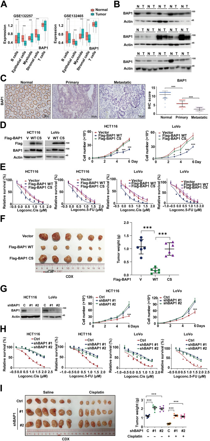
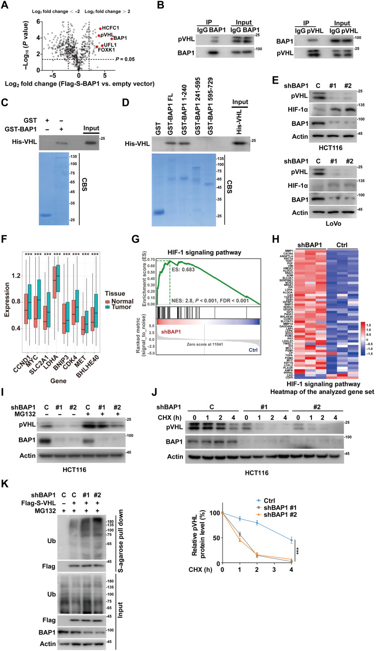
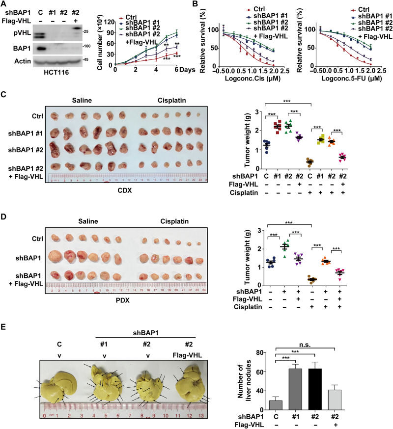
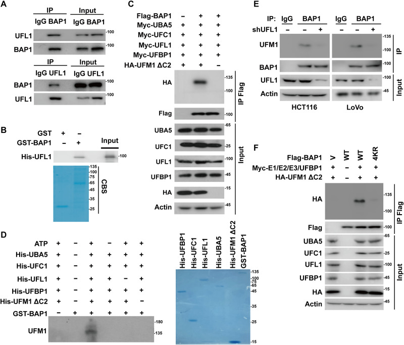
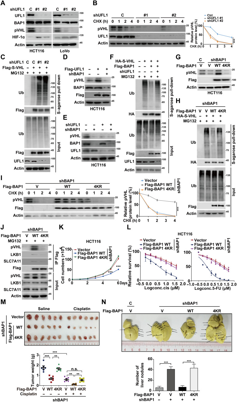
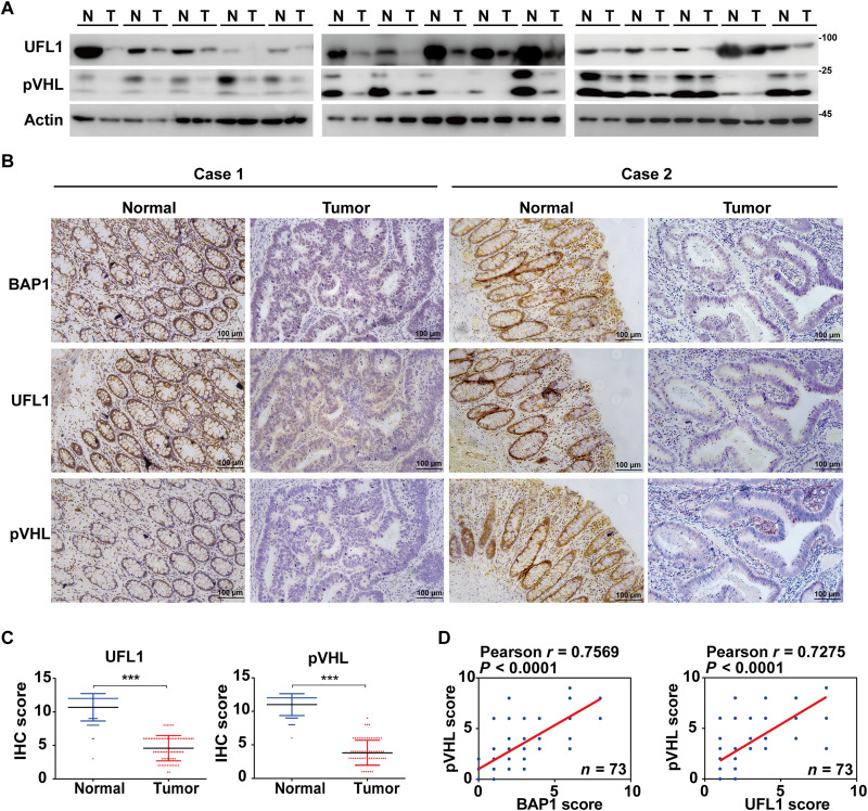
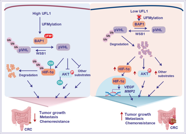
BRCA1-associated protein 1 (BAP1) can function as a tumor suppressor or oncogene depending on context, but its role in colorectal cancer (CRC) is not well understood. Here, we demonstrate that BAP1 suppresses CRC progression primarily by deubiquitinating and stabilizing von Hippel-Lindau tumor suppressor protein (pVHL). BAP1 undergoes covalent modification by ubiquitin-fold modifier 1 (UFM1) at Lys51, Lys61, Lys187, and Lys205, enhancing its interaction with pVHL and promoting pVHL stabilization. Loss of this modification through UFL1 depletion or reconstitution with a UFMylation-defective BAP1 mutant (4KR) impairs pVHL stabilization and promotes tumor progression in CRC cell line-based and patient-derived xenograft models. Clinically, down-regulation of UFL1 and BAP1 correlates with reduced pVHL level and poor prognosis in patients with CRC. These findings identify a previously unrecognized posttranslational mechanism regulating BAP1 activity and highlight UFMylation as essential for maintaining pVHL tumor-suppressive function. Targeting BAP1 UFMylation may represent a potential therapeutic strategy in CRC and other cancers with wild-type BAP1 and VHL.


Figures

Fig. 1.
Fig. 1.. BAP1 exerts a tumor-suppressive role in CRC.
(A) Reanalysis of BAP1 expression across five cell types in patients with CRC using scRNA-seq datasets (GSE132257 and GSE132465), comparing tumor and matched normal mucosa (n = 5), and in a larger cohort of CRC tumor (n = 23) and matched normal mucosa samples (n = 10). (B) BAP1 expressions were assessed in CRC and adjacent normal tissues (N, normal; T, tumor). (C) Representative immunohistochemical staining of BAP1 in normal (n = 73), primary (n = 38), and metastatic (n = 35) CRC tissues, with corresponding IHC score analysis. (D) HCT116 and LoVo cells stably expressing vector, Flag-BAP1 WT, or Flag-BAP1 C91S (CS) mutant were generated, and Western blotting was performed. Cell proliferation assay was performed. Data are shown as means ± SD (n = 3). (E) Cells in (D) were treated with cisplatin or 5-FU. Cell survival was determined. Data are shown as means ± SD (n = 4). (F) HCT116 cells (1 × 106) in (D) were subcutaneously implanted into nude mice (n = 6). Tumors were harvested at endpoint and weighed (means ± SD). (G) HCT116 and LoVo cells stably expressing control or BAP1 shRNAs were analyzed by Western blotting and subjected to proliferation assay. Data are shown as means ± SD (n = 3). (H) Cells in (G) were treated with cisplatin (Cis) or 5-FU. Cell survival was determined. Data are shown as means ± SD (n = 4) (I) HCT116 cells (1 × 106) in (G) were subcutaneously implanted into nude mice. Upon reaching about 100 mm3, mice were treated with saline or cisplatin (2 mg/kg weekly, n = 6). Tumors were weighed at endpoint and analyzed. Data are shown as means ± SD and were analyzed using one-way analysis of variance (ANOVA). *P < 0.05; ***P < 0.001.
Fig. 2.
Fig. 2.. BAP1 deubiquitinates and stabilizes pVHL.
(A) Volcano plot of BAP1-associated proteins identified by MS in HCT116 cells stably expressing Flag-S-BAP1 and treated with MG132 (10 μM) for 10 hours. (B) Co-immunoprecipitates (Co-IP) in HCT116 cells using immunoglobulin G (IgG), anti-BAP1, or anti-pVHL antibodies followed by immunoblotting with the indicated antibodies. (C) In vitro binding assay showing interaction between purified His-VHL and recombinant GST or GST-BAP1. CBS, Coomassie blue staining. (D) Mapping of BAP1 regions required for pVHL interaction using GST-fused full-length (FL) and truncated BAP1 proteins incubated with purified His-VHL in vitro. (E) Western blotting of HCT116 and LoVo cells stably expressing control or BAP1 shRNAs using the indicated antibodies. (F) Reanalysis of scRNA-seq data (GEO: GSE132465) from patients with CRC containing 23 primary CRC tumors and 10 matched normal mucosae. ***P < 0.001, Wilcoxon rank sum test, one-sided. (G) Gene set enrichment analysis (GSEA) of RNA-seq data from BAP1-depleted HCT116 cells showing significant enrichment of HIF-1 signaling pathway genes. NES, Normalized Enrichment Score. (H) Heatmap of HIF-1 target gene expression in BAP1-depleted versus control HCT116 cells by RNA-seq. (I) Western blotting of HCT116 cells stably expressing control or BAP1 shRNAs treated with vehicle or MG132 (10 μM) for 10 hours. (J) Cycloheximide pulse-chase assay to assess pVHL protein stability in cells as in (E). Data are shown as means ± SD (n = 3). h, hours. (K) Cells stably expressing control or BAP1 shRNAs were transfected with vector or pIRES-VHL (containing Flag and S tag), then treated MG132 for 10 hours. Cell lysates were pulled down by S-agarose, and the polyubiquitylated pVHL was measured by Western blotting with an anti-ubiquitin antibody. Data are shown as means ± SD and were analyzed by one-way ANOVA. ***P < 0.001.
Fig. 3.
Fig. 3.. BAP1 suppresses tumor progression through stabilizing pVHL.
(A) HCT116 cells stably expressing control or BAP1 shRNAs were transfected with vector or Flag-VHL. The protein levels of BAP1 and pVHL were determined by Western blotting, and cell proliferation was measured. Results represent the means ± SD of three independent experiments. (B) Cells as in (A) were treated with the indicated concentrations of cisplatin or 5-FU, and cell survival was determined. Results represent the means ± SD of four independent experiments. (C) HCT116 cells expressing control or BAP1 shRNAs and stably reconstituting vector or Flag-VHL were injected subcutaneously in nude mice. When tumor volume reached 100 mm3, mice were treated with saline or cisplatin (2 mg/kg once a week), respectively (n = 6 per group). Tumors were collected at 6 weeks, and tumor weights were measured and analyzed. (D) CRC patient-derived xenografts (PDXs) were subcutaneously implanted into nude mice. Xenograft tumors were injected with lentivirus expressing the indicated constructs when tumor volume reached 30 mm3. Mice were then treated with saline or cisplatin (2 mg/kg once a week), respectively (n = 6 per group). Tumors were collected, and tumor weights were measured and analyzed. Results represent the means ± SD from six mice. (E) HCT116 cells expressing control or BAP1 shRNAs were stably reconstituted with vector or Flag-VHL and injected into the spleen of nude mice (n = 6 per group). Mice were euthanized, and metastatic nodules on liver were counted. Representative images and quantitative analysis of liver metastatic nodules were showed. Results represent the means ± SD from six mice. Data are shown as means ± SD and were analyzed by one-way ANOVA. **P < 0.01; ***P < 0.001. n.s., not significant.
Fig. 4.
Fig. 4.. UFL1 interacts with and UFMylates BAP1.
(A) HCT116 cell lysates were subjected to immunoprecipitation with control IgG, anti-BAP1, or anti-UFL1 antibodies. The immunoprecipitates were then blotted with the indicated antibodies. (B) Purified recombinant GST, GST-BAP1, and His-UFL1 were incubated in vitro, and the direct interaction between BAP1 and UFL1 was examined. CBS, Coomassie blue staining. (C) UFMylation of BAP1 was analyzed by Western blotting using an anti-HA antibody in HEK293T cells transfected with Flag-BAP1 and the components of the UFMylation system (Myc-UBA5, Myc-UFC1, Myc-UFL1, Myc-UFBP1, and HA-UFM1 ΔC2). (D) In vitro UFMylation of BAP1. Purified UFMylation components (His-UBA5, His-UFC1, His-UFL1, His-UFBP1, and His-UFM1 ΔC2) were bacterially produced and incubated with GST or GST-BAP1 in UFMylation buffer at 30°C for 90 min. The reaction was stopped by the addition of SDS sample buffer containing 5% mercaptoethanol and boiled at 95°C for 10 min. Samples were subjected to Western blotting with an anti-UFM1 antibody. (E) UFMylation of endogenous BAP1 was analyzed by immunoprecipitation with an anti-BAP1 antibody, followed by Western blotting using an anti-UFM1 antibody in HCT116 and LoVo cells stably expressing control or UFL1 shRNA. (F) UFMylation assay of the Flag-BAP1 WT and 4KR was performed in HEK293T cells expressing the indicated components of the UFMylation system.
Fig. 5.
Fig. 5.. UFL1-mediated UFMylation activates BAP1 activity toward pVHL in CRC.
(A) Western blotting of HCT116 and LoVo cells stably expressing control or UFL1 shRNAs. (B) Cycloheximide pulse-chase assay from (A) assessing pVHL half-life (means ± SD, n = 3). (C) Cells from (A) were transfected with vector or pIRES-VHL (Flag/S tagged), treated MG132 for 10 hours, and subjected to S-agarose pull-down and anti-ubiquitin blotting. (D) HCT116 cells stably expressing Flag-UFL1 were infected with lentivirus expressing control or BAP1 shRNA. Western blot was performed. (E) Western blotting of HCT116 cells with BAP1, UFL1 or double knockdown using indicated antibodies. (F) Cells were transfected as indicated, followed by S-agarose pull-down and anti-ubiquitin blotting. (G) HCT116 cells were transfected with indicated plasmids, and Western blotting was performed. (H) Cells stably expressing BAP1 shRNA were transfected as indicated, treated MG132, and examined polyubiquitylated pVHL using an anti-ubiquitin antibody. (I) Cycloheximide pulse-chase assay in cells from (G) assessing pVHL half-life (means ± SD, n = 3). (J) Cells transfected as indicated were treated with MG132 and subjected to anti-Flag immunoprecipitation to assess BAP1 interaction with pVHL, LKB1, or SLC7A11. (K) Cell proliferation assays were performed on cells from (G) (means ± SD, n = 3). (L) Cells from (G) were treated with cisplatin or 5-FU, and cell survival was determined (means ± SD, n = 4). (M) HCT116 cells stably expressing vector, Flag-BAP1 WT, or 4KR mutant were injected subcutaneously into nude mice. Upon reaching about 100 mm3, mice were treated with saline or cisplatin (2 mg/kg weekly, n = 6). Tumors were weighed at endpoint. n.s., not significant. (N) Cells as in (M) were injected into the spleen of nude mice. Metastatic nodules on liver were counted at endpoint. Results represent the means ± SD from six mice and were analyzed by one-way ANOVA. **P < 0.01; ***P < 0.001. h, hours.
Fig. 6.
Fig. 6.. BAP1 and UFL1 expressions are positively correlated with pVHL expression in CRC.
(A) UFL1 and pVHL expressions in 15 pairs of fresh CRC tissues and adjacent normal tissues were measured by Western blotting. N, normal tissues; T, tumor tissues. (B) Representative IHC staining of BAP1, UFL1, and pVHL in CRC (n = 73) and adjacent normal tissues (n = 73). (C) The IHC scores of UFL1 and pVHL expression in (B) were analyzed. (D) The correlation of UFL1 and BAP1 expression with pVHL expressions in CRC tissues. Statistical analysis was performed with the chi-square test. r, the Pearson’s correlation coefficient. Data are presented as the means ± SD and were analyzed by one-way ANOVA. ***P < 0.001.
Fig. 7.
Fig. 7.. The experimental model of this study.
Schematic representation of the experimental model of this study, summarizing the proposed mechanisms by which UFL1-mediated UFMylation of BAP1 regulates pVHL stability and tumor-suppressive activity in CRC.

Similar articles

Cited by

  • iCAFs control chemoresistance.
    Lloyd L. Lloyd L. Nat Rev Urol. 2025 Aug;22(8):495. doi: 10.1038/s41585-025-01068-w. Nat Rev Urol. 2025. PMID: 40659812 No abstract available.

References

    1. Sung H., Ferlay J., Siegel R. L., Laversanne M., Soerjomataram I., Jemal A., Bray F., Global cancer statistics 2020: GLOBOCAN estimates of incidence and mortality worldwide for 36 cancers in 185 countries. CA Cancer J. Clin. 71, 209–249 (2021). - PubMed
    1. Zhou H., Liu Z., Wang Y., Wen X., Amador E. H., Yuan L., Ran X., Xiong L., Ran Y., Chen W., Wen Y., Colorectal liver metastasis: Molecular mechanism and interventional therapy. Signal Transduct. Target. Ther. 7, 70 (2022). - PMC - PubMed
    1. Testa J. R., Cheung M., Pei J., Below J. E., Tan Y., Sementino E., Cox N. J., Dogan A. U., Pass H. I., Trusa S., Hesdorffer M., Nasu M., Powers A., Rivera Z., Comertpay S., Tanji M., Gaudino G., Yang H., Carbone M., Germline BAP1 mutations predispose to malignant mesothelioma. Nat. Genet. 43, 1022–1025 (2011). - PMC - PubMed
    1. Gerlinger M., Horswell S., Larkin J., Rowan A. J., Salm M. P., Varela I., Fisher R., McGranahan N., Matthews N., Santos C. R., Martinez P., Phillimore B., Begum S., Rabinowitz A., Spencer-Dene B., Gulati S., Bates P. A., Stamp G., Pickering L., Gore M., Nicol D. L., Hazell S., Futreal P. A., Stewart A., Swanton C., Genomic architecture and evolution of clear cell renal cell carcinomas defined by multiregion sequencing. Nat. Genet. 46, 225–233 (2014). - PMC - PubMed
    1. Harbour J. W., Onken M. D., Roberson E. D., Duan S., Cao L., Worley L. A., Council M. L., Matatall K. A., Helms C., Bowcock A. M., Frequent mutation of BAP1 in metastasizing uveal melanomas. Science 330, 1410–1413 (2010). - PMC - PubMed

MeSH terms

Substances