. 2025 Aug:118:105848.
doi: 10.1016/j.ebiom.2025.105848. Epub 2025 Jul 10.

Genotype and transcript processing of the tumour necrosis factor receptor TNFRSF1A in epithelial cells: implications for survival in cystic fibrosis

Collaborators, Affiliations

Genotype and transcript processing of the tumour necrosis factor receptor TNFRSF1A in epithelial cells: implications for survival in cystic fibrosis

Alexander Uden et al. EBioMedicine. 2025 Aug.

Abstract

Background: Cystic fibrosis is caused by mutations of the cystic fibrosis transmembrane conductance regulator, CFTR, an epithelial anion transport protein, responsible for, inter alia, sputum viscoelasticity in the lung. We previously identified the TNF receptor superfamily 1A TNFRSF1A (TNFR1) as a genetic modifier of CFTR function and disease severity in the CF twin and sibling study population. We aimed to replicate our findings in independent cohorts, assess the role of TNFR1 for patient survival and identify functional changes associated with TNFR1 polymorphisms.

Methods: We incorporated data from three independent long-term mono- and multicentric cohorts of people with cystic fibrosis (pwCF) to confirm the previously described association of TNFR1 with CFTR function and to extend our study to include survival data for our local cohort and a pan-European cohort of pwCF. We studied TNFR1 transcripts obtained from primary airway epithelia grown as air-liquid interface cultures to address possible mechanisms involved in up-stream and down-stream effects of TNFR1.

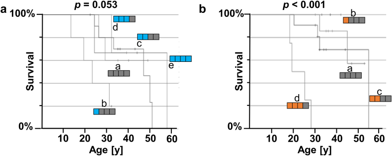
Findings: Survival differed by more than a decade when comparing carriers of contrasting TNFR1 genotypes among unrelated pwCF as well as among CF siblings pairs. The presence of the TNFR1 transcript variant TNFR1delEx2 in primary airway epithelia was associated with TNFR1 genotype.

Interpretation: The association of the TNFR1 transcript variant TNFR1delEx2 associates with the TNFR1 genotype, possibly mediating the genotype-survival association we found regarding TNFR1 genotype and patient survival in cystic fibrosis.

Funding: Supported by the German Ministry for Education and Research (BMBF) (82DZL009B1 to MAM and 82DZL002A1, to GH, BT, AMD, FS) and the Mukoviszidose Institut gGmbH (MI-2002, to LN, AMD, FS).

Keywords: Alternative transcript; Cystic fibrosis; Genetic association study; Patient survival; TNFRSF1A; Tumour necrosis factor receptor.


Conflict of interest statement

Declaration of interests The following authors state no conflict of interest with respect to this manuscript’s content and in accordance with the ICMJE guidelines: Alexander Uden (AU); Inga Dunsche (ID); Longhua Feng (LF); Stephanie Tamm (ST); Silke Hedtfeld (SH); Gesa Stege (GS); Kirstin Jahn (KJ); Julia Kontsendorn (JK); Nadine Alfeis (NA); Ines Kühbandner (IK); Rebecca Minso (RM); Christian Dopfer (CD); Matthias Griese (MG); Gesine Hansen (GH); Frauke Stanke (FS). Sabina Janciauskiene (SJa); Simon Gräber (SG); Olaf Sommerburg (OS); Felix C. Ringshausen (FR); Lutz Nährlich (LN); Peter Braubach (PB); Marcus A Mall (MM); Burkhard Tümmler (BT) and Anna-Maria Dittrich (AMD) disclose institutional or personal grants, consulting fees or payment honoraria received within the past 36 months from: the European Union (FR); the German Centre for Lung Research-DZL (SJa); the Helmholtz centre for infection research (FR, BT); the German Innovation Fund (MM); the German CF foundation (SG, FR, LN); the Christiane-Herzog-Foundation (AMD); the Paul-Ehrlich-Institute (AMD); the German Kartagener Syndrome and Primary Ciliary Dyskinesia Patient Advocacy Group (FR); the Austrian Society for Rare Disease (AMD); the VW-Stiftung (BT); University Hospital Frankfurt (FR); University Hospital Hamburg (FR); University Dundee (FR); Social Court Cologne (FR); Vertex Pharmaceuticals Incorporated (SG, OS, FR, LN, MM, BT, AMD); I!DE Werbeagentur GmbH (FR); AstraZeneca (FR, PB); Arcturus (FR); Bayer (FR); Boehringer Ingelheim (FR, PB, MM); Chiesi GmbH (SG); Danone (OS), Enterprise Therapeutics (MM); Glaxo Smith-Kline (AMD); Grifols (FR, SJa); Hogrefe Publishing Company (AMD); InfectoPharm (FR); Insmed Germany (FR); Interkongress GmbH (FR); Kither Biotec (MM); Novartis (FR, AMD); Pari (OS, MM); Parion (FR); ReCode (FR); Sanofi (FR); Splisense (MM); Zambon (FR). The following authors participate on data safety monitoring or advisory boards of: Boehringer Ingelheim (FR, MM); Chiesi (SG, FR); Enterprise Therapeutics (MM); Grifols (FR); Insmed (FR); Kither Biotec (MM); Pari (MM); Trial Steering Committee for CF STORM (LN); Vertex Pharmaceutical Incorporated (SG, OS, BT). The following authors serve a leadership or fiduciary role in other board, society, committee or advocacy group: Coordinator of the ERN-LUNG Bronchiectasis Core Network (FR); Chair of the German Bronchiectasis Registry PROGNOSIS (FR); Member of the SteerCo of the European Bronchiectasis Registry EMBARC (FR); Member of the SteerCo of the European Nontuberculous Mycobacterial Pulmonary Disease Registry EMBARC-NTM (FR); Co-Speaker of the Medical Advisory Board of the German Kartagener Syndrome and PCD Patient Advocacy Group (FR); PI of the German Centre for Lung Research (FR); Member of the Protocol Review Committee of the PCD-CTN (FR); Medical lead of the German CF-registry (LN); Pharmacovigilance study manager of the European Cystic Fibrosis Society Patient Registry (LN); European Respiratory Society (MM); Fellow of the European Respiratory Society (MM); Christiane-Herzog-Foundation (BT); German CF patient advocacy board (AMD): German CF Clinical Trial Network ExecCommittee (AMD); European Cystic Fibrosis Society Clinical Trials network ExecCommittee (AMD). The consortium of CF teams was established through outreach efforts, during which caregivers of individuals with cystic fibrosis were contacted via telephone. Caregivers, physicians and other CF professionals who agreed to participate contributed survival data of participants of EUCFTSib and those contributors are listed here as CF teams from the European CF twin and sibling study.

Figures

Fig. 1
Fig. 1
TNFR1 genotype distributions among F508del-CFTR homozygous pwCF subclassified by year of birth. DNA was collected for CFTR mutation analysis in 1990–1994 from 174 F508del-CFTR homozygous pwCF born between 1959 and 1994, seen regularly at the CF centre of the Hannover Medical School.TNFR1 genotypes are visualised as combinations of two alleles shown as two rectangles side-by-side in blue (D12S889-10), orange (D12S889-13) and white (unclassified other TNFR1 variant). a. Kaplan–Meier plots of survival are shown for subsamples of pwCF defined as overlapping cohorts of 60 individuals each for survival analysis (see Supplemental Fig. S1, three cohorts I for distribution of year of birth). Subgroups for survival analysis were defined by D12S889 genotype 10-10 (blue/blue; blue survival curve), 10–13 (blue/orange; grey survival curve) and 13-13 (orange/orange; orange survival curve) and analysed for the event “death” or “lung transplant”. Mean survival is shown as a vertical, dotted line in orange, blue and grey, respectively. The 95% confidence interval of median survival is visualised with shaded rectangles surrounding the vertical dotted line marking mean survival for each of the three groups defined by D12S889 genotype. Numbers at risk and numbers of censored patients are disclosed as source data for Fig. 1a as well as the Kaplan–Meier–Survival plot of the entire MHHSurv cohort irrespective of year of birth stratification. b. D12S889 genotype frequencies were monitored in non-overlapping subsamples of 35 pwCF each (see Supplemental Fig. S1, three cohorts II for distribution of year of birth). Genotype distributions were compared using Monte-Carlo simulation on chi-squared test statistics. While survival analysis (a.) was carried out with three subgroups classified by D12S889 genotype, i.e., carriers of D12S889 genotypes 10-10, 10–13, and 13-13, genotype frequency distribution (b.) included also rare TNFR1 variants as genotype “heterozygous D12S889-10/other” and “heterozygous D12S889-13/other” to accommodate D12S889 alleles such as D12S889-8, D12S889-9, D12S889-12, D12-889-14 and D12S889-15 which are summarised as “other”. Moreover, while the year-of-birth-distributions of the three cohorts I for survival analysis (a.) and cohorts II for genotype distribution analysis (b.) do target matching year-of-birth spans in the left, middle and right subfigures (see Supplemental Fig. S1), the strategy for subsampling differs. For survival analysis, (a.) overlapping subsamples were necessary to accommodate the rareness of the risk allele D12S889-13 in the earlier birth year subsamples. In contrast, genotype distributions (b) were informative in non-overlapping subcohorts, however, to define these subcohorts of equal size, pwCF were sorted according to their exact date of birth, resulting in subsampling pwCF born in 1977 in two adjacent subcohorts II.
Fig. 2
Fig. 2
Survival of pwCF within the sibling cohort of EUCFTSib. As sibling pairs share on average half of their genes by default, sib-pair dependency of TNFR1 genotype data was accounted for by counting the number of D12S889-10 per pair (blue squares, a) or reciprocally the number of D12S889-13 per pair (orange squares, b). As a consequence, each sibling pair was observed in group “a” (none of four TNFR1 genes of the two siblings as specified), “b” (one of four TNFR1 genes of the two siblings as specified), “c” (two of four TNFR1 genes of the two siblings as specified), “d” (three of four TNFR1 genes of the two siblings as specified) or “e” (both siblings homozygous for the specified TNFR1 variant). Please note that pairs of two siblings, both homozygous for D12S889-13, were not observed (b). The phenotype of pairs was taken into account with an index case strategy whereby either the older sib (a) or the younger sib (b) was taken as a measure of the pair’s survival phenotype. Kaplan–Meier charts were constructed using data from 27 F508del-CFTR homozygous sibling pairs.
Fig. 3
Fig. 3
Replication study on pwCF participating in ORKAMBIfacts. We enrolled 46 pwCF from ORKAMBIfacts with a valid NPD prior to start of therapy and retrieved parental DNA from 40 parents, which we genotyped for markers rs1860545-rs4149581-rs4149580-rs4149577-D12S889-rs4149576 in intron 1 of TNFR1. FAMHAP, was used to inquire for TNFR1 variant distribution comparing subsamples of pwCF with CFTR-mediated residual chloride conductance in NPD to a reference subsample who did not display chloride conductance. The set of 46 families was provided as a training set to FAMHAP in all comparisons. (a) Allele distribution among 46 pwCF enrolled into the study. Frequency of TNFR1 chromosomes carrying 2-marker-haplotypes 10-1 (blue), 13-2 (orange) and other D12S889-rs4149581 variants (white) in the entire set of 43 families with pwCF. (b) TNFR1 genotype distribution for subsamples with (12 pwCF) and without (14 pwCF) CFTR-mediated residual chloride conductance in nPD using the stringent definition of > |5 mV| response to the sum of responses to chloride-free solution and isoproterenol (Pbest = 0.041 observed for marker combination D12S889-rs4149581, dipcc algorithm within program FAMHAP). TNFR1 genotypes are visualised as combinations of two alleles shown as two rectangles side-by-side in blue (D12S889-10), orange (D12S889-13) and white (unclassified other TNFR1 variant).
Fig. 4
Fig. 4
Expression of TNFR1delEx2 in primary ALI epithelia. a. Expression of TNFR1 wild-type transcript (TNFR1 wt), aldolase (Aldolase) as a housekeeping gene and TNFR1 alternative transcript specific for the deletion of TNFR1 exon 2 (TNFR1delEx2) in selected six out of 64 primary respiratory epithelia. Signals shown were obtained after 35 cycles of polymerase chain reaction (PCR) upon which PCR products using primers specific for TNFR1 wt (technical duplicates), aldolase (technical duplicates) and TNFR1delEx2 (technical triplicates) were loaded on agarose gels and visualised with SYBR Green. Arrows on left side of gel denote the position of a positive control amplicon provided as a size marker for each analysis. Three representative samples each of ALI epithelia derived from donors with D12S889 genotype 10-10 or 13-13, respectively, were selected to reflect presence (13-13) or absence (10-10) of TNFR1delEx2 in concordance with the association of D12S889 genotype to TNFR1transcript phenotype shown in more detail in b. TNFR1 genotypes are visualised as combinations of two alleles shown as two rectangles side-by-side in blue (D12S889-10), and orange (D12S889-13). For the entire set of 64 ALI epithelia, an ALI sample was counted as “positive for TNFR1delEx2” if one or more out of the three technical replicates were positive for a signal of the expected size. wtTNFR1 and aldolase was observed in all 64 samples. b. Dependency of TNFR1 transcript repertoire on TNFR1 genotype among primary respiratory epithelia cultured at air-liquid-interface from 64 independent donors. ALI biomaterial was genotyped for D12S889 and phenotyped as visualised in a. TNFR1 genotype distribution is shown conditional on D12S889 alleles 10 and 13, displaying frequencies for homozygous genotypes 10-10 and 13-13 as well as heterozygous for 10–13. Alleles D12S889-12 and D12S889-14, differing one repeat unit from D12S889-13, were considered equivalent to D12S889-13 as well as D12S889-11, differing one repeat unit from D12S889-10, were considered equivalent to D12S889-10. TNFR1 genotypes are visualised as combinations of two alleles shown as two rectangles side-by-side in blue (D12S889-10), and orange (D12S889-13) or white (other). Genotypes of two samples could not be assigned to these genotype categories: one sample with D12S889 genotype 14–15 (negative for TNFRdelEx2 transcript), one sample with D12S889 genotype 11–17 (positive for TNFRdelEx2 transcript). As 10-10 homozygotes, the following ALI were pooled: 10-10 (13 ALI epithelia), 10–11 (one ALI epithelium), 8–10 (one ALI epithelium); as 10–13, the following ALI were pooled: 10–12 (one ALI epithelium), 10–13 (16 ALI epithelia), 10–14 (four ALI epithelia), 11–14 (one ALI epithelium), 8–12 (one ALI epithelium), 8–13 (three ALI epithelia), 8–14 (one ALI epithelium), 9–13 (one ALI epithelium). As 13-13 homozygotes, the following ALI were pooled: 12–13 (9 ALI epithelia), 13-13 (eight ALI epithelia), 13–14 (four ALI epithelia). The algorithms of Sham and Curtis were used to compare the TNFR1 genotype distribution as a 3 × 2 table between ALI epithelia negative for TNFR1delEX2 transcript and ALI epithelia positive for TNFR1delEx2 transcript. Comparison was done using Monte-Carlo simulation on chi-squared test statistics. Pbest = 0.04 was observed comparing TNFR1 genotype distributions from ALI epithelia negative for TNFR1delEx2 transcript to ALI epithelia positive for TNFR1delEx2 transcripts whereby pooling was done as follows: genotypes with no D12S889 risk allele 13 were tested against pooled heterozygotes and homozygotes for D12S889 risk allele 13. Chi-square from this comparison was 6.688 and reached 42 times in 1000 MC simulations (p = 0.042; uncorrected p value from chi-squared distribution with 1 df: p = 0.0097; program CLUMP26).

References

    1. Chen G., Goeddel D.V. TNF-R1 signaling: a beautiful pathway. Science. 2002;296:1634–1635. - PubMed
    1. Silke J. The regulation of TNF signalling: what a tangled web we weave. Curr Opin Immunol. 2011;23:620–626. - PubMed
    1. Gómez M.I., Lee A., Reddy B., et al. Staphylococcus aureus protein A induces airway epithelial inflammatory responses by activating TNFR1. Nat Med. 2004;10:842–848. - PubMed
    1. Gómez M.I., O’Seaghdha M., Magargee M., Foster T.J., Prince A.S. Staphylococcus aureus protein A activates TNFR1 signaling through conserved IgG binding domains. J Biol Chem. 2006;281:20190–20196. - PubMed
    1. Classen A., Kalali B.N., Schnopp C., et al. TNF receptor I on human keratinocytes is a binding partner for staphylococcal protein A resulting in the activation of NF kappa B, AP-1, and downstream gene transcription. Exp Dermatol. 2011;20:48–52. - PubMed

Substances