Case Reports
. 2025 Jul 9;30(18):103910.
doi: 10.1016/j.jaccas.2025.103910.

Delayed Diagnoses of Cardiac Amyloidosis

Affiliations
Case Reports

Delayed Diagnoses of Cardiac Amyloidosis

Cooper B Kersey et al. JACC Case Rep. .

Abstract

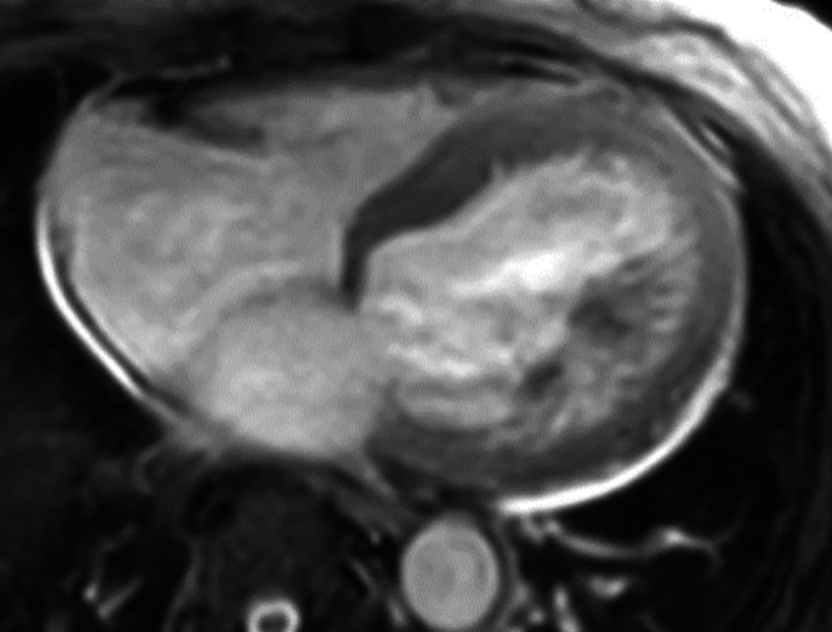
Cardiac sarcoidosis (CS) can be challenging to accurately diagnose and relies on a complex diagnostic framework because of the limited sensitivity of endomyocardial biopsy. Frequently, the diagnosis of CS depends on cardiac 18F-fluorodeoxyglucose positron emission tomography, which has limited specificity for sarcoidosis. We report 3 cases of individuals who were initially diagnosed and treated for isolated CS based on multimodality cardiac imaging and later definitively reclassified to wild-type transthyretin cardiac amyloidosis from histopathology obtained by endomyocardial biopsy.

Keywords: cardiac amyloidosis; cardiac sarcoidosis; case report; multimodality imaging; nonischemic cardiomyopathy.


Conflict of interest statement

Funding Support and Author Disclosures The authors have reported that they have no relationships relevant to the contents of this paper to disclose.

Figures

None
Graphical abstract
Figure 1
Figure 1
Echocardiogram Images for Patient 1 (A) Patient 1 had reduced global longitudinal strain on the echocardiogram. (B) Transthoracic echocardiogram demonstrating mild symmetrical increase in left ventricular wall thickness (1.3 cm) for patient 1.
Figure 2
Figure 2
Cardiac Magnetic Resonance Imaging of Patient 1 Showing Patchy Late Gadolinium Enhancement
Figure 3
Figure 3
Electrocardiogram of Patient 2 Showing First-Degree Atrioventricular Block and a Right Bundle Branch Block
Figure 4
Figure 4
Patient 2 Had Reduced Global Longitudinal Strain on the Echocardiogram With Apical Sparing
Figure 5
Figure 5
Cardiac Magnetic Resonance Imaging of Patient 2 Showing Late Gadolinium Enhancement of the Basal Midinferolateral, Basal Midlateral, and Basal Inferior Wall
Figure 6
Figure 6
Electrocardiogram of Patient 3 Showing Low Voltage in Limb Leads With Pseudoinfarct Pattern
Figure 7
Figure 7
Echocardiogram Images for Patient 3 (A) Patient 3 had reduced global longitudinal strain with apical sparing pattern on transthoracic echocardiogram. (B) Mitral inflow Doppler signal consistent with restrictive filling pattern for patient 3.
Figure 8
Figure 8
Cardiac Magnetic Resonance Imaging for Patient 3 Showing Patchy Late Gadolinium Enhancement
Figure 9
Figure 9
FDG PET Images, Cardiac Magnetic Resonance Images, and Histology for Patients 1 to 3 18F-fluorodeoxyglucose positron emission tomography (18F FDG PET) images, cardiac magnetic resonance (CMR) images, and histology slides for patients 1 to 3 (left to right). 18F FDG PET images show FDG uptake, CMR images demonstrate delayed gadolinium enhancement, and histology with Congo red stain reveals amyloid deposition.

References

    1. Kandolin R., Lehtonen J., Airaksinen J., et al. Cardiac sarcoidosis: epidemiology, characteristics, and outcome over 25 years in a nationwide study. Circulation. 2015;131(7):624–632. doi: 10.1161/circulationaha.114.011522. - DOI - PubMed
    1. Terasaki F., Azuma A., Anzai T., et al. JCS 2016 guideline on diagnosis and treatment of cardiac sarcoidosis—digest version. Circ J. 2019;83(11):2329–2388. doi: 10.1253/circj.CJ-19-0508. - DOI - PubMed
    1. Divakaran S., Stewart G.C., Lakdawala N.K., et al. Diagnostic accuracy of advanced imaging in cardiac sarcoidosis. Circ Cardiovasc Imaging. 2019;12(6) doi: 10.1161/circimaging.118.008975. - DOI - PMC - PubMed
    1. Lal M., Chen C., Newsome B., et al. Genetic cardiomyopathy masquerading as cardiac sarcoidosis. J Am Coll Cardiol. 2023;81(1):100–102. doi: 10.1016/j.jacc.2022.10.021. - DOI - PubMed
    1. Cheng R.K., Kittleson M.M., Beavers C.J., et al. Diagnosis and management of cardiac sarcoidosis: a scientific statement from the American Heart Association. Circulation. 2024;149(21):e1197–e1216. doi: 10.1161/cir.0000000000001240. - DOI - PubMed

Publication types