. 2025 Jul 11;15(1):25158.
doi: 10.1038/s41598-025-10290-w.

Multomic analysis reveals the potential of LAG3 as a prognostic and immune biomarker and its validation in osteosarcoma

Affiliations

Multomic analysis reveals the potential of LAG3 as a prognostic and immune biomarker and its validation in osteosarcoma

Qianqian Cao et al. Sci Rep. .

Abstract

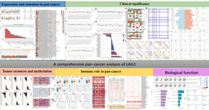
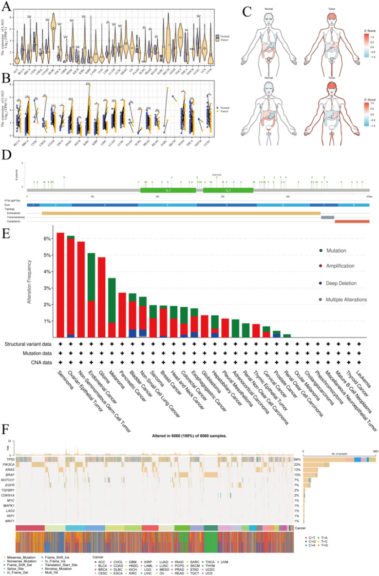
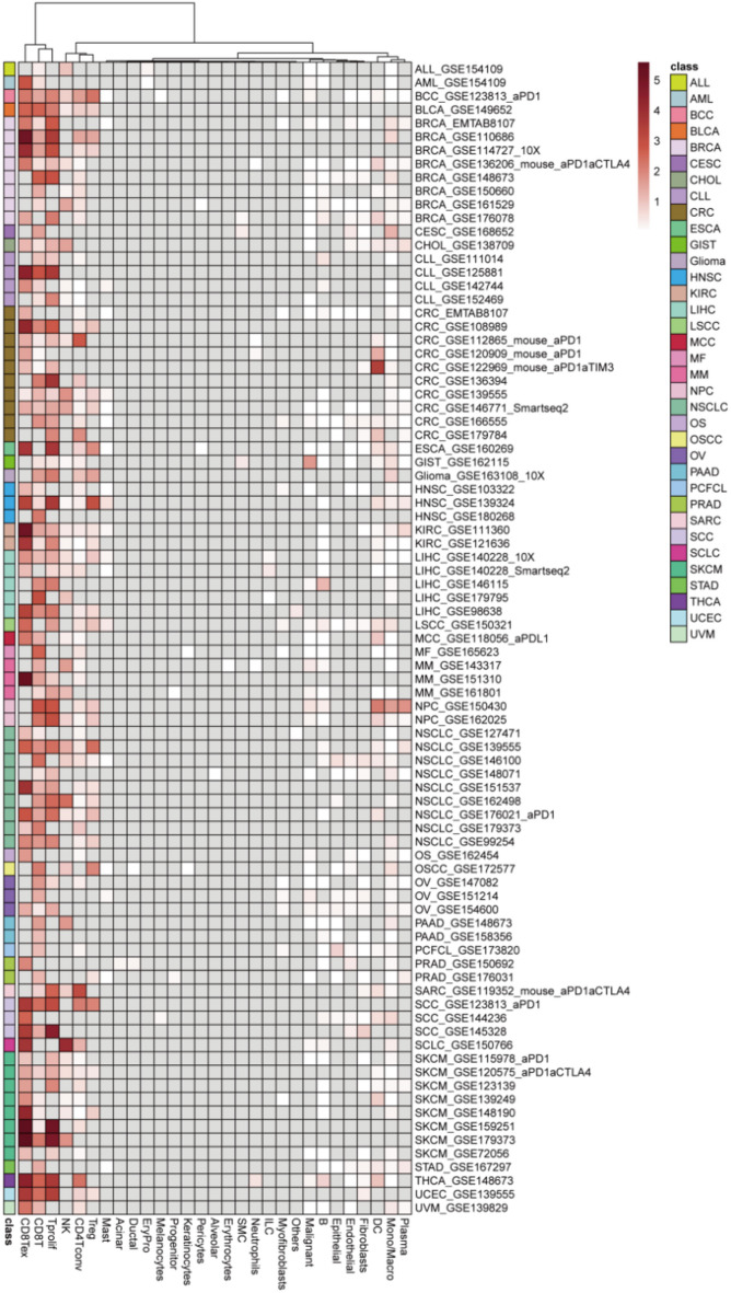
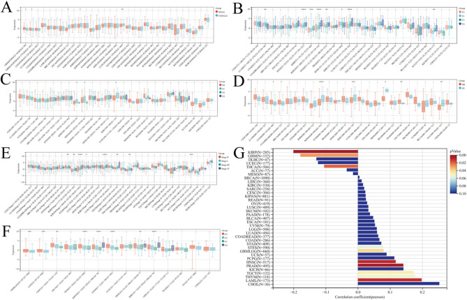
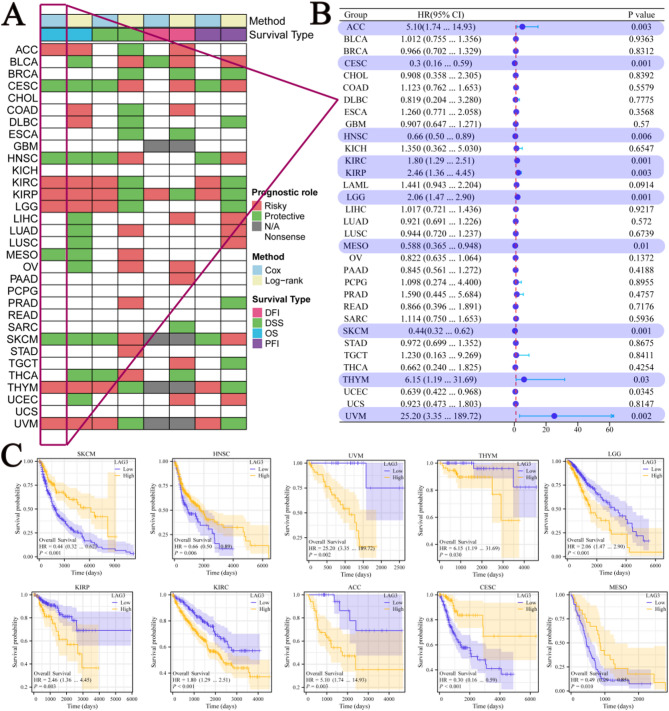
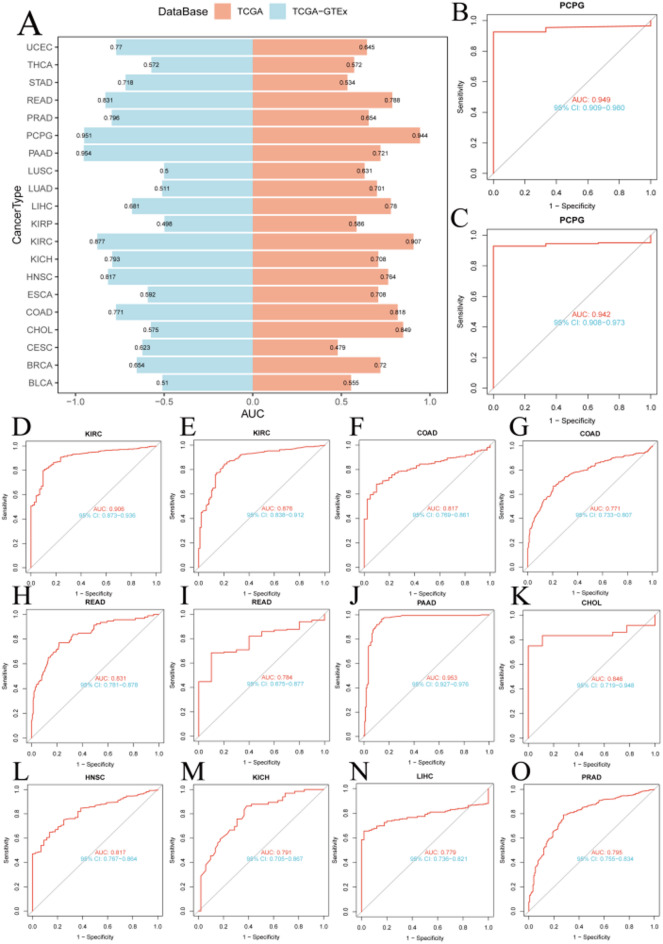
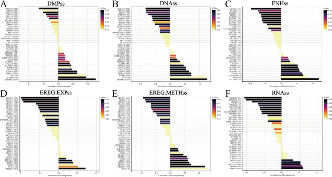
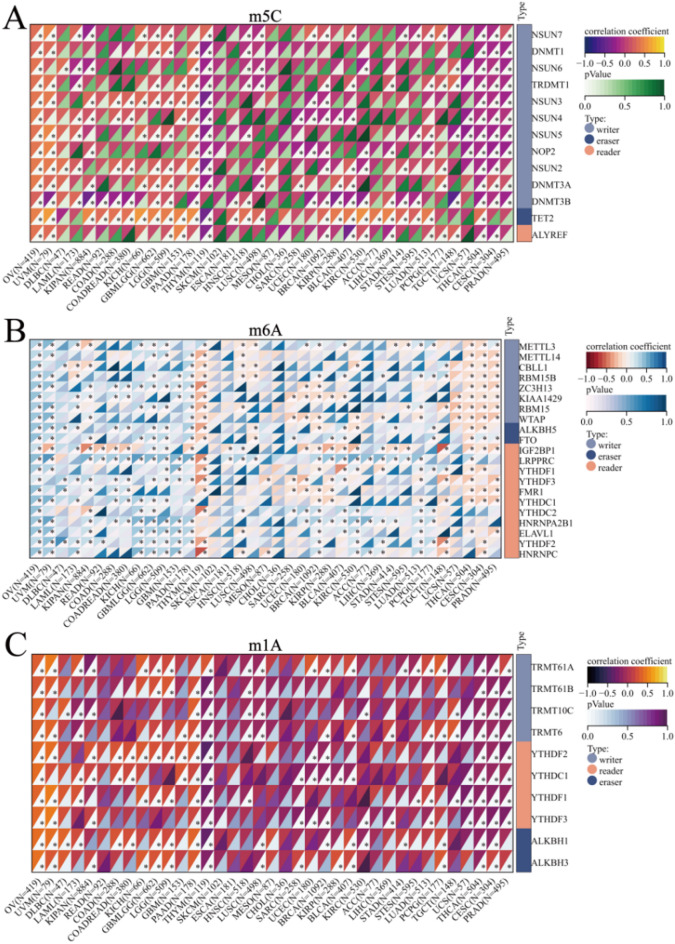
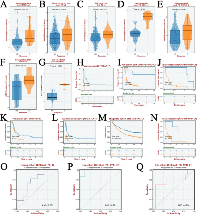
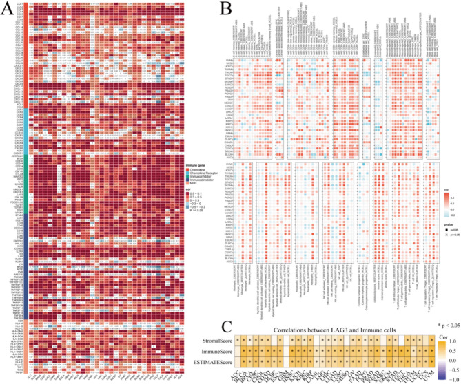
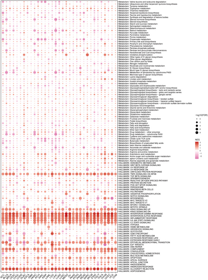
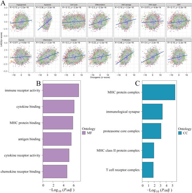
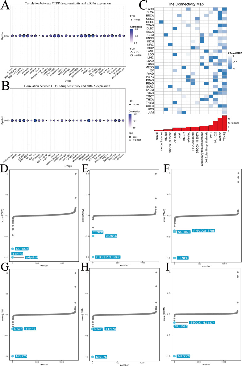
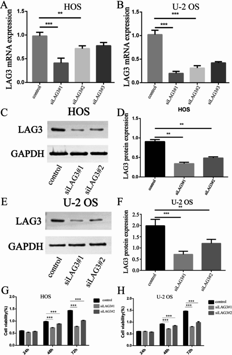
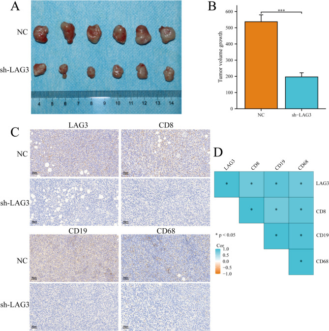
Lymphocyte activation gene 3 (LAG3) is a member of the immunoglobulin superfamily and has been implicated in the development, growth, and progression of several cancers. However, the biological role of LAG3 has not been investigated in pan-cancer datasets. We sought to perform a comprehensive bioinformatics analysis of pan-cancer datasets to determine the relationship of LAG3 with patient survival prognosis, tumor microenvironment, immunotherapy responsiveness, and mechanisms regulating tumorigenesis. We used TCGA, GTEx, TIMER2, GDSC, CTRP, and TISCH databases and online websites to extract data on LAG3 in a variety of cancers, and analyzed pan-cancer patient datasets to explore not only the correlation between LAG3 expression and clinical stage, diagnosis, and prognosis of cancers, but also the correlation between LAG3 expression, gene variants, methylation status, tumor stemness, and tumor immunity. The biological functions of LAG3 in osteosarcoma cells were determined by in vitro CCK-8, wound healing and transwell assays. Finally, through in vivo experiments, the study preliminarily explored the impact of LAG3 on osteosarcoma and its correlation with immune genes. Pan-cancer analysis showed that LAG3 expression was up-regulated in a variety of cancers, and the expression of LAG3 was closely related to the clinical stage, diagnosis and prognosis of cancers. GO and KEGG enrichment analyses showed that LAG3 was enriched in inflammatory, metabolic, and immune signaling pathways in a variety of cancers. Meanwhile, LAG3 expression not only has an impact on patient immunotherapy prognosis and immunotherapy response, but also has a significant effect on drug sensitivity. In vitro experiments have shown that LAG3 promotes the proliferation, migration and invasion of osteosarcoma cells. In vivo xenotransplantation experiments further confirmed that LAG3 promotes the growth of osteosarcoma, and the expression of LAG3 is positively correlated with CD8, CD19, and CD68. Our study suggests that LAG3 is a promising marker for cancer diagnosis, prognosis, and treatment.

Keywords: Diagnosis; Immune; LAG3; Pan-cancer; Prognosis.


Conflict of interest statement

Declarations. Competing interests: The authors declare no competing interests.

Figures

Fig. 1
Fig. 1
Flowchart of data analysis process.
Fig. 2
Fig. 2
LAG3 expression and mutation. (A) LAG3mRNA expression between tumors and normal tissues. (B) The expression of LAG3 in tumors and paired adjacent normal tissues. (C) Expressions and distributions of LAG3 between tumor and normal tissues in various organs. (D) Mutation sites of LAG3. (E) The mutation frequency and corresponding mutation types of LAG3 in different cancers. (F) Sites and numbers of LAG3 genetic alterations across cancers. (*P < 0.05, **P < 0.01, ***P < 0.001, and ****P < 0.0001).
Fig. 3
Fig. 3
LAG3 expression in immune cells infiltrated in cancers.
Fig. 4
Fig. 4
Association between LAG3 expression and clinical features in the pan-cancer setting. (A) The correlation between sex and LAG3 expression in pan-cancer. (B) The correlation between LAG3 expression and T stage in pan-cancer. (C) The correlation between LAG3 expression and N stage in pan-cancer. (D) The correlation between LAG3 expression and M stage in pan-cancer. (E) The correlation between LAG3 expression and staging in pan-cancer. (F) The correlation between LAG3 expression and staging in pan-cancer. (G) The correlation between age and LAG3 expression in pan-cancer. (*P < 0.05, **P < 0.01, ***P < 0.001, and ****P < 0.0001).
Fig. 5
Fig. 5
Survival profiles of LAG3 in pan-cancers. (A) Correlation between LAG3 expression and overall survival (OS), disease-specific survival (SS), disease-free interval (DFI) and progression-free interval (PFI). (B) Forest plot of the relationship between pan-cancer LAG3 expression and overall survival (p < 0.05). (C) Overall survival analysis of LAG3 expression in patients with UVM, THYM, LGG, ACC, KIRC, KIRP, CESC, HNS, MESO and SKCM.
Fig. 6
Fig. 6
Correlation of LAG3 expression with the diagnosis of pan-cancer. (A) Diagnostic value of LAG3 in pan-cancer. (BO) ROC curves of LAG3 in PCPG, KIRC, COAD, READ, CHOL, HNSC, KICH, LIHC and PRAD.
Fig. 7
Fig. 7
Correlation of LAG3 expression with tumor stemness. (A) DMPs. (B) DNAss. (C) ENHss. (D) EREG.EXPss. (E) EREG.METHss. (F) RNAss.
Fig. 8
Fig. 8
Correlation analysis of LAG3 expression with mRNA modification methylation regulators. (A) m5C. (B) m6A. (C) m1A. (*P < 0.05, **P < 0.01, and ***P < 0.001).
Fig. 9
Fig. 9
Correlation between immunotherapy and LAG3 expression. (AG).LAG3 expression in responding and non-responding patients in different immunotherapy cohorts. (HN) Correlation between LAG3 expression and survival in different immunotherapy cohorts. (OQ) Diagnostic value of LAG3 in different immunotherapies.
Fig. 10
Fig. 10
Association of LAG3 expressions with immune infiltration. (A) The heatmap shows correlations between LAG3 mRNA expressions and expressions of chemokine, chemokine receptor, immune-inhibitor, immune-stimulatory, and MHC genes. (B) Correlation between LAG3 expression and cancer immune infiltration. (C) Correlation between LAG3 and immune cells. (*P < 0.05, **P < 0.01, and ***P < 0.001).
Fig. 11
Fig. 11
Enrichment differences of LAG3 in 50 HALLMARK and 83 metabolism gene sets.
Fig. 12
Fig. 12
Functional mechanism analysis. (A) The LAG3 mRNA expression was highly correlated with 14 malignant features of all tumors. (B) Major molecular functions of LAG3. (C) The major cellular component of LAG3.
Fig. 13
Fig. 13
Chemotherapeutic drug resistance analysis. Correlation between LAG3 expression and drug sensitivity studied using two different databases. (A) CTRP. (B) GDSC. (C) Identification of LAG3-targeting compounds through cMap analysis. (DI) LAG3-targeting compounds of clinical importance.
Fig. 14
Fig. 14
Biological function of LAG3 in osteosarcoma. (AF).The mRNA and protein expression levels of LAG3 in HOS and U-2 OS cell lines. (G, H) CCK 8 assay for detecting the proliferative capacity of HOS and U-2 OS cells. (*P < 0.05, **P < 0.01, and ***P < 0.001).
Fig. 15
Fig. 15
(AL) The transwell assay and wound healing were used to assess the migratory potential of HOS and U-2 OS cells. (*P < 0.05, **P < 0.01, ***P < 0.001, and ****P < 0.0001).
Fig. 16
Fig. 16
Knockdown of LAG3 inhibits the malignant growth of osteosarcoma cells. (A) Size of the xenografted tumor tissues. (B) Weight of the xenografted tumor tissues. (C) Immunohistochemical staining analysis of the expression of LAG3, CD8, CD19, and CD68 in tumor tissues from different groups. (D) Correlation of the expression of LAG3, CD8, CD19, and CD68. (*P < 0.05, **P < 0.01, and***P < 0.001).

Similar articles

References

    1. Smith, B. D., Smith, G. L., Hurria, A., Hortobagyi, G. N. & Buchholz, T. A. Future of cancer incidence in the United States: Burdens upon an aging, changing nation. J. Clin. Oncol.27, 2758–2765. 10.1200/jco.2008.20.8983 (2009). - PubMed
    1. Soerjomataram, I. & Bray, F. Planning for tomorrow: global cancer incidence and the role of prevention 2020–2070. Nat. Rev. Clin. Oncol.18, 663–672. 10.1038/s41571-021-00514-z (2021). - PubMed
    1. Zhu, S. et al. Combination strategies to maximize the benefits of cancer immunotherapy. J. Hematol. Oncol.14, 156. 10.1186/s13045-021-01164-5 (2021). - PMC - PubMed
    1. DeVita, V. T. Jr. & Chu, E. A history of cancer chemotherapy. Cancer Res.68, 8643–8653. 10.1158/0008-5472.Can-07-6611 (2008). - PubMed
    1. Chen, H. et al. A pan-cancer analysis of enhancer expression in nearly 9000 patient samples. Cell173, 386-399.e312. 10.1016/j.cell.2018.03.027 (2018). - PMC - PubMed

MeSH terms