Review
. 2025 Aug;487(2):253-273.
doi: 10.1007/s00428-025-04171-w. Epub 2025 Jul 12.

The morphological spectrum of Castleman disease and related disorders: a report from the Lymphoma Workshop of the 22nd Meeting of the European Association of Hematopathology

Affiliations
Review

The morphological spectrum of Castleman disease and related disorders: a report from the Lymphoma Workshop of the 22nd Meeting of the European Association of Hematopathology

Gorana Gasljevic et al. Virchows Arch. 2025 Aug.

Abstract

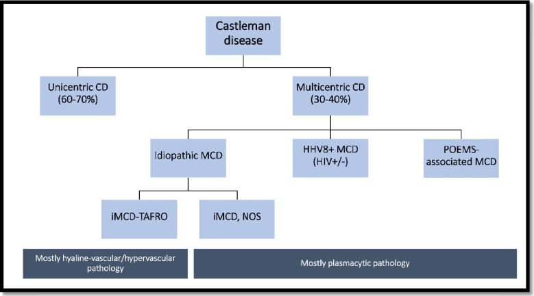
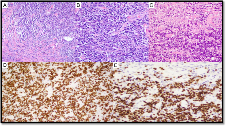
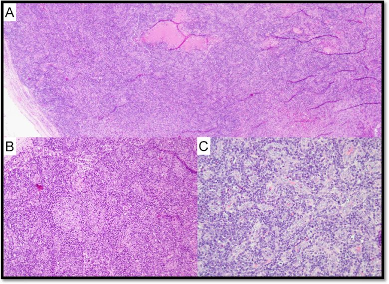
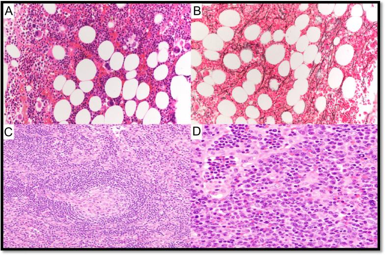
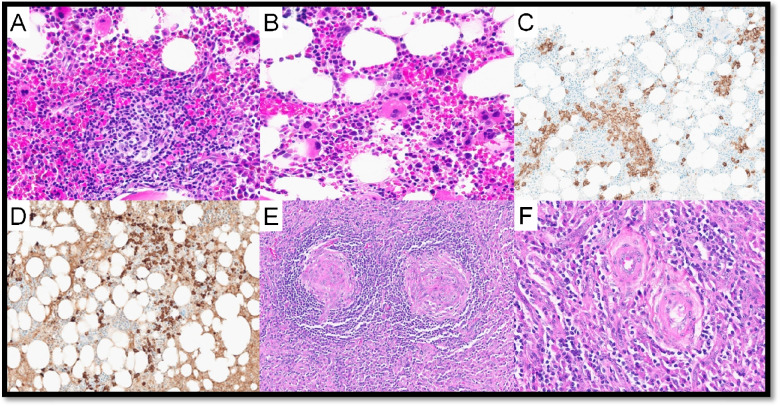
Castleman disease (CD) is an intriguing and complicated group of local and systemic disorders mainly affecting lymph nodes with heterogeneous presentation and therapeutic needs. These disorders were the topic of Session 1 of the Lymphoma Workshop at the 2024 EA4HP in Dubrovnik, Croatia. In this report, we summarize the features of the 85 submitted cases and review the differential diagnosis, pitfalls, and advances for all CD subtypes. Specifically, the molecular landscape of unicentric CD and its relationship with follicular dendritic cell proliferations and indolent T-lymphoblastic proliferation will be discussed. The spectrum of idiopathic multicentric CD (MCD), TAFRO syndrome, as well as the clinical and histopathological peculiarities of POEMS-CD, is reviewed. Cases of Kaposi sarcoma-associated herpesvirus/human herpesvirus 8 (KSHV/HHV8) + MCD were the most complicated and well demonstrated the difficulties and overlaps in the differential diagnosis of KSHV/HHV8 + lymphoproliferative disorders. Finally, the important topic of CD mimickers will be addressed, demonstrating how the integration of clinical, laboratory, histopathological, and molecular data is mandatory to confirm a diagnosis of CD and how to distinguish it from the many neoplastic, autoimmune, and infective mimickers.

Keywords: CD-mimickers; Castleman disease; Idiopathic multicentric Castleman disease; KSHV/HHV8-associated lymphoproliferative disorders; POEMS syndrome; TAFRO syndrome.


Conflict of interest statement

Declarations. Conflict of interest: The authors declare no competing interests.

Figures

Fig. 1
Fig. 1
Clinical classification of CD, associated syndromes, and histological features
Fig. 2
Fig. 2
Stroma-rich variant of HV-CD. Case LyWS-101. Courtesy of R. King. A, B H&E; small follicles with regressively transformed germinal centers, concentric mantle zones, and numerous interfollicular hyalinizing small vessels that occasionally penetrate germinal centers. C Interfollicular stroma replaced by proliferation of hyalinized small vessels and abundant spindled, fibrous stroma. D CD31 highlights the density of small vessels in the stroma
Fig. 3
Fig. 3
Unicentric Castleman disease with atypical stromal cell proliferation. Case LyWS-369. Courtesy of J. Bosch-Schips. A Hyaline vascular features: extensive proliferation of high endothelial venules with perivascular hyalinization, twinning of mantle zones and atretic germinal centers. B Prominent interfollicular stromal spindle cell proliferation. C Focal moderate-to-marked dysplastic features in stromal spindle cell proliferation. D Interfollicular spindle cells are positive for SMA. Interfollicular spindle cells are weakly positive for E CD68, F CD31, and G EMA
Fig. 4
Fig. 4
Follicular dendritic cell sarcoma associated with Castleman disease. Case LyWS-73. Courtesy of S-B. Ng. A, B Tumour composed of spindle to ovoid cells in sheets, fascicles, and storiform whorls. C Neoplastic cells with dispersed chromatin and small nucleoli. Scattered lymphocytes and plasma cells. D Proliferation of FDCs in residual follicles with hyperplastic/dysplastic features. Tumor cells are E CD21 and F CD35 positive
Fig. 5
Fig. 5
Castleman disease, mixed hyaline vascular variant and plasma cell variant, stroma-rich with iT-LBP. Case LyWS-159. Courtesy of J. Li. A Plasma cell and stroma-rich area. B Plasma cell-rich area. C Blastoid small lymphocytic clusters positive for D CD3 and E TdT
Fig. 6
Fig. 6
Idiopathic multicentric Castleman disease with massive infiltration of IgG4-positive plasma cells. Case LyWS-20. Courtesy of K. Karube. A Regressed follicles and expanded paracortex. B Diffuse sheets of plasma cells. Hemosiderin deposition is evident (arrows). No eosinophil infiltration. C IgG and D IgG4—IgG4/IgG ratio is about 50%. E Kappa light chains and F Lambda light chains—no light chain restriction
Fig. 7
Fig. 7
Hypervascular (HvV) morphology in TAFRO syndrome. Case LyWS-169. Courtesy of H. Shao. A Lymph node with respected architecture. B Marked vascular proliferation, atrophy of the germinal centers, and “onion layers” appearance around the follicles. C Marked vascular proliferation. EBV and HHV8 stainings were negative (not shown), there was no restriction of the light chains (not shown), and IgG/IgG4 ratio was below 40% (not shown)
Fig. 8
Fig. 8
Bone marrow and lymph node changes in TAFRO syndrome. Case LyWS-153. Courtesy of M. Nichols. A The bone marrow shows maturing trilineage hematopoiesis with slight megakaryocytic hyperplasia without morphologic atypia. B Reticulin fibrosis is slightly increased (grade 1/3). C Lymph node changes in MCD-TAFRO: germinal centers are regressed with “lollipop follicles” and lymphocyte depletion. D Interfollicular plasmacytosis is present. There was no light chain restriction (not shown) and IgG/IgG4 was < 40% (not shown)
Fig. 9
Fig. 9
Bone marrow and lymph node changes in a patient with MCD-POEMS. Case LyWS-326. Courtesy of I. Prisneac. A Cellular bone marrow with a small lymphoid aggregate surrounded by plasma cells. B Mild megakaryocyte hyperplasia. C CD138 highliting rimming of the plasma cells around lymphoid aggregate. D Plasma cells are λ-restricted. E Atretic germinal centers comprised predominantly of follicular dendritic cells, surrounded by layers of mantle-type cells (onion-skinning) with “lolippop sign” and “twinning.” F Parafollicular regions are expanded by an increase in small blood vessels surrounded by small lymphocytes and plasma cells
Fig. 10
Fig. 10
KSHV/HHV8-associated multicentric Castleman disease with plasmablast aggregates and EBV reactivation. Case LyWS-301. Courtesy of M. Donzel. A, B Lymph node architecture is preserved. The germinal centers vary in size; some have slightly “regressive” appearance. The mantle zones are thickened, sometimes with a “single file” arrangement of cells. C Numerous large cells with a plasmablastic appearance in aggregates are present positive for D HHV8 and E lambda, negative for CD20, CD138, and EBER (not shown). F Some germinal centres contain numerous EBV + cells but do not correspond to HHV8 + cells
Fig. 11
Fig. 11
KHSV/HHV-8 and EBV-positive large B-cell lymphoma arising in the background of multicentric Castleman disease and with concurrent Kaposi sarcoma highlighting the complexity of KSHV/HHV-8-associated pathologies. Case LyWS-197. Courtesy of MG. Daniel. While WHO5 allows rare positivity for EBV in DLBCL-HHV8, panel favored the open differential diagnosis between DLBCL-HHV8 and EBV + and EC-PEL since no analysis of somatic hypermutation was performed. A LN with markedly distorted nodal architecture, with a prominent atypical spindle cell proliferation (lower left part)and multifocal macronodular to sheet-like aggregates of large atypical lymphoid cells (upper part). B Large atypical lymphoid cells have plasmablastic cytomorphology; they are positive for C HHV8, D EBER, E IgM, F spindle cell proliferation positive for CD34
Fig. 12
Fig. 12
Follicular lymphoma with hyaline vascular Castleman-like features. LyWS-62. Courtesy of F. Aguirre-Neira. A Numerous regressed germinal centers surrounded by expanded mantle zones forming concentric rings with an “onion skin” pattern. B Germinal center cells are weakly BCL-2. C BCL-6 and D CD10 positive. E BCL2 and BCL6 (not shown) were rearranged by a fluorescence in situ hybridization study. F Clonality testing by BIOMED-2 revealed a clonal rearrangement of the IgH gene. G NGS showed a pathogenic variant of the CXCR4 gene and probable pathogenic variants of the KMT2D and CARD11 genes
Fig. 13
Fig. 13
Diagnostic algorithm to diagnose Castleman disease and to exclude mimickers

References

    1. Castleman B, Iverson L, Menendez VP (1956) Localized mediastinal lymphnode hyperplasia resembling thymoma. Cancer 9(4):822–830. 10.1002/1097-0142(195607/08)9:4%3c822::aid-cncr2820090430%3e3.0.co;2-4 - PubMed
    1. Weisenburger DD (Mar.1988) Multicentric angiofollicular lymph node hyperplasia. Pathology of the spleen. Am J Surg Pathol 12(3):176–181. 10.1097/00000478-198803000-00002 - PubMed
    1. Fajgenbaum DC et al (Mar.2017) International, evidence-based consensus diagnostic criteria for HHV-8-negative/idiopathic multicentric Castleman disease. Blood 129(12):1646–1657. 10.1182/blood-2016-10-746933 - PMC - PubMed
    1. R. Alaggio et al (2022) The 5th edition of the World Health Organization classification of haematolymphoid tumours: lymphoid neoplasms. Leukemia 36:7. 10.1038/s41375-022-01620-2 - PMC - PubMed
    1. Wang L et al (Aug.2020) Predominant stroma-rich feature in hyaline vascular variant of castleman disease is associated with paraneoplastic pemphigus. Am J Clin Pathol 154(3):403–413. 10.1093/ajcp/aqaa053 - PubMed

Supplementary concepts

LinkOut - more resources