. 2025 Jul 11;23(1):781.
doi: 10.1186/s12967-025-06801-y.

Chronic sleep deprivation induces plasma exosome-derived miR-150-5p downregulation as a novel mechanism involved in Parkinson's disease progression by targeting DCLK1

Affiliations

Chronic sleep deprivation induces plasma exosome-derived miR-150-5p downregulation as a novel mechanism involved in Parkinson's disease progression by targeting DCLK1

Li Liu et al. J Transl Med. .

Abstract

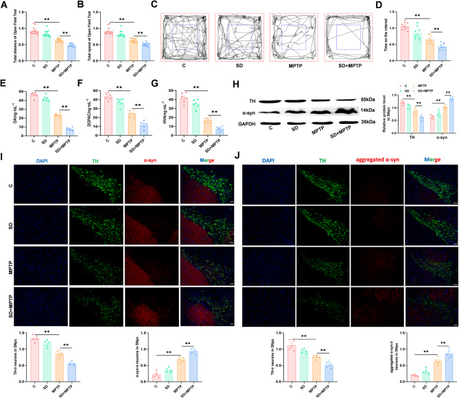
Background: Researches have suggested that chronic sleep deprivation (SD) can lead to neurological dysfunction and facilitate the onset and progression of Parkinson's disease (PD). However, the association between SD and PD remains unclear. Exosome (exo) cargo comprises microRNAs (miRNAs), which are potential regulators of PD. This study focused on assessing the role and related mechanisms of SD on PD.

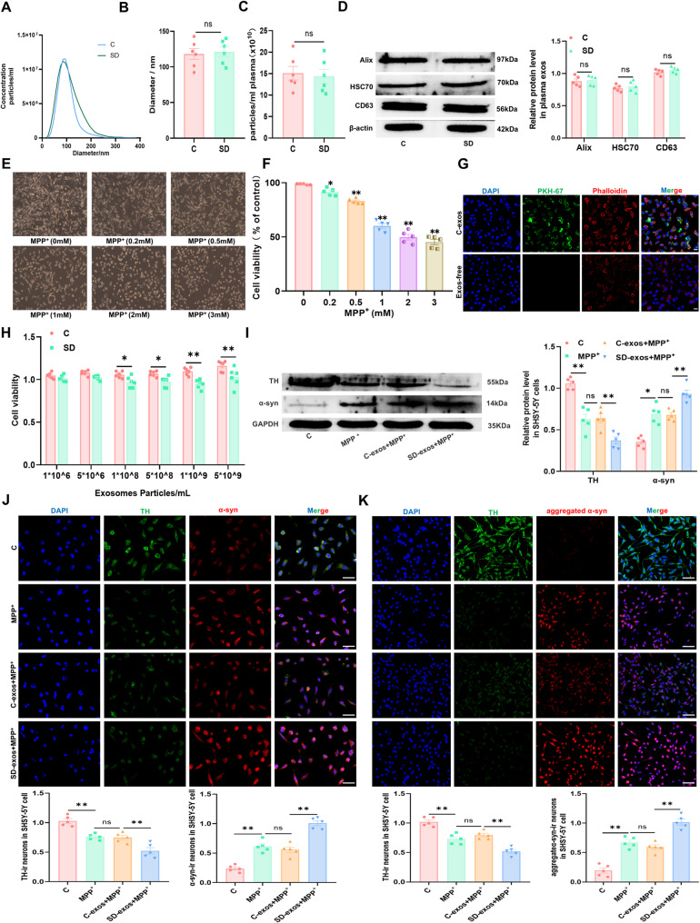
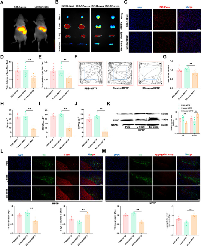
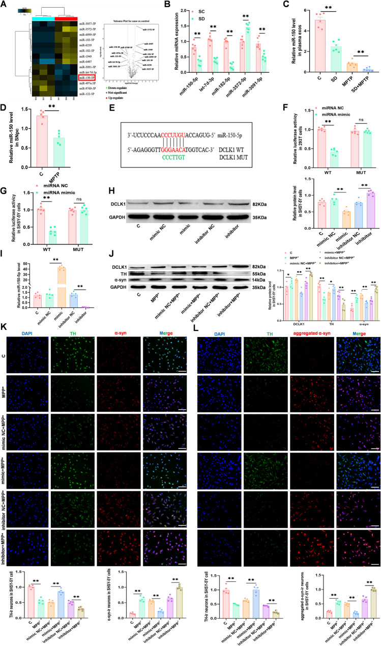
Methods: SD plus 1-methyl-4-phenyl-1,2,3,6-tetrahydropyridine (MPTP)-induced PD mice were used to investigate effects of SD on PD. Exos were extracted from plasma by polymer precipitation method. Impacts of exos on PD were validated through intervention in 1-methyl-4-phenylpyridinium (MPP+)-induced PD cells and MPTP-induced PD mice. Levels of miRNA in exos were analyzed by gene expression profile microarray. Levels of miR-150-5p in exos and substantia nigra pars compacta (SNpc) were further confirmed by reverse transcription quantitative polymerase chain reaction (RT-qPCR). Target genes of miRNAs were predicted by TargetScan and confirmed by Dual-Luciferase Reporter Assay. Mimics and inhibitors of miR-150-5p were transfected into MPP+-induced PD cells, while agomir and antagomir of miR-150-5p were stereotaxic intracranial injected into SNpc of SD + MPTP-induced PD mice, enabling the determination of specific molecular mechanisms affecting PD.

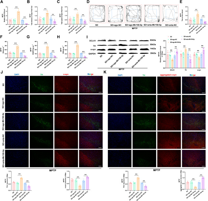
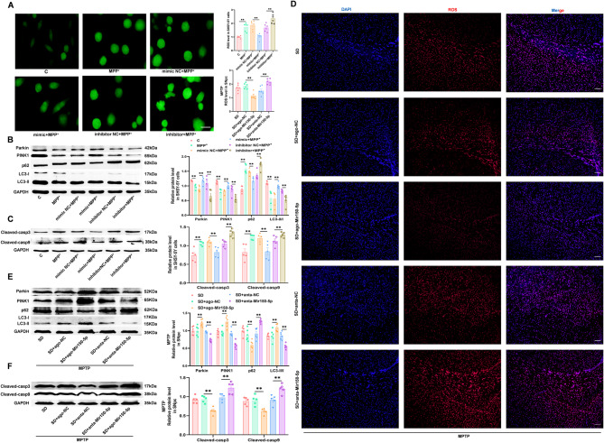
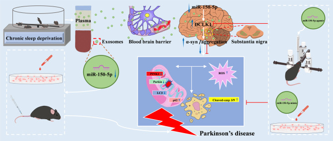
Results: We found that SD and SD-derived exos aggravated PD-related damage. SD-derived exos were identified as potent inducers of PD. MiR-150-5p was recognized as a key element in SD-derived exos, and doublecortin-like kinase 1 (DCLK1) was confirmed as its target gene. Supplementing miR-150-5p alleviated PD damage by inhibiting DCLK1 and abnormal α-synuclein (α-syn) expression, decreasing reactive oxygen species (ROS), p62, cleaved-caspase-3 and cleaved-caspase-9 levels, and increasing Parkin and PINK1 levels and the LC3II/I ratio.

Conclusion: These findings suggested that miR-150-5p-dependent downregulation in SD-derived exos could aggravate the progression of PD via the DCLK1/α-syn pathway. MiR-150-5p decreased ROS levels, promoted mitophagy, and inhibited apoptosis, thus mitigating PD-related damage. These findings indicated that plasma-derived exos and their miRNA cargo might serve as therapeutic targets for PD, providing insights into a mechanism that links SD-related deterioration to the progression of PD.

Keywords: Chronic sleep deprivation; DCLK1; Exosome; Parkinson’s disease; microRNA-150-5p.


Conflict of interest statement

Declarations. Ethics approval and consent to participate: All experiments were approved by the Animal Use and Care Committee of Shandong First Medical University and followed national guidelines for animal care and use. Consent for publication: All authors consent for publication. Competing interests: The authors declare that they have no competing interests.

Figures

Fig. 1
Fig. 1
SD aggravated motor dysfunction and damage to the SNpc and striatum in MPTP-induced PD mice. In the open field test, movement, including (A) motor distance, (B) speed, and (C) trajectory, was significantly decreased in the mice of the SD + MPTP group. (D) Rotarod test results. The duration spent on the rotarod was significantly shorter in the SD + MPTP group than in the control group. The levels of (E) DA, (F) DOPAC, and (G) HVA were significantly decreased in the mice of the SD + MPTP group. (H) Significantly decreased levels of TH and increased levels of α-syn in the SNpc were observed in the SD + MPTP mice. The data are presented as the ratio of TH or α-syn to GAPDH. (I) A significantly lower number of TH-ir neurons and a higher number of α-syn-ir neurons in the SNpc were observed in the SD + MPTP mice. (J) A significantly lower number of TH-ir neurons and a higher number of aggregated α-syn-ir neurons in the SNpc were observed in the SD + MPTP mice. All data were compared to those of the MPTP group. The data are presented as the mean ± SEM, n = 5–14. Bar = 50 μm; *P < 0.05 and **P < 0.01
Fig. 2
Fig. 2
SD-exos aggravated damage in MPP+-induced PD cells. (A) The size distribution and concentration of SD-exos and C-exos were determined by NTA and quantified by determining their (B) diameter and (C) concentration, respectively. (D) No significant differences in exo-specific proteins, including Alix, HSC70, and CD63, were detected between the SD-exos and C-exos. The data are presented as the ratios of Alix, HSC70, or CD63 to β-actin. (E) Changes in the morphology of SHSY-5Y cells, such as rounding, fragmentation, atrophy, and decreased cell density, with different concentrations of MPP+. (F) The optimal concentration of MPP+ (0.5 mM) was determined by cell viability. (G) PKH67-labeled exos were effectively internalized by the SHSY-5Y cells. (H) An appropriate concentration of SD-exos (5 × 108 particles/mL) was used by determination of cell viability. (I) Significantly lower levels of TH and higher levels of α-syn were observed in the SHSY-5Y cells of the SD-exos + MPP+ group. The data are presented as the ratio of TH or α-syn to GAPDH. (J) A significantly lower number of TH-ir neurons and a higher number of α-syn-ir neurons were found in the SHSY-5Y cells of the SD-exos + MPP+ group. (K) A significantly lower number of TH-ir neurons and a higher number of aggregated α-syn-ir neurons were found in the SHSY-5Y cells of the SD-exos + MPP+ group. All data were compared to those of the C-exos + MPP+ group. Each value is represented as the mean ± SEM, n = 5–6. Bar = 50 μm; *P < 0.05 and **P < 0.01
Fig. 3
Fig. 3
SD-exos aggravated damage in MPTP-induced PD mice. (A) The delivery efficiency of DiR-C-exos and DiR-SD-exos in vivo was determined using a small animal imaging system. (B) The distribution of DiR-exos in isolated organs was detected by fluorescence imaging analysis. (C) The distribution of DiR-exos in brain tissue slices was detected via fluorescence microscopy. In the open field test, movement, including (D) motor distance, (E) speed, and (F) trajectory, was significantly decreased in the SD-exos + MPTP group. (G) Rotarod test results. The duration spent on the rotarod was significantly shorter in the SD-exos + MPTP group than in the control group. The levels of (H) DA, (I) DOPAC, and (J) HVA were significantly lower in the SD-exos + MPTP group. (K) Significantly lower levels of TH and higher levels of α-syn in the SNpc were observed in the SD-exos + MPTP group. The data are presented as the ratio of TH or α-syn to GAPDH. (L) A significantly lower number of TH-ir neurons and a higher number of α-syn-ir neurons in the SNpc were observed in the SD-exos + MPTP group. (M) A significantly lower number of TH-ir neurons and a higher number of aggregated α-syn-ir neurons in the SNpc were found in the SD-exos + MPTP group. All data were compared to those of the C-exos + MPTP group. All data are presented as the mean ± SEM, n = 5–14. Bar = 50 μm; *P < 0.05 and **P < 0.01
Fig. 4
Fig. 4
MiR-150-5p alleviated damage in MPP+-induced PD cells by downregulating DCLK1. (A) The levels of miR-150-5p were significantly lower in SD-exos, as determined by a miRNA profiling assay. Changes of miRNAs in the volcano plot. (B) RT-qPCR results validating differential expression levels of miR-150-5p, let-7d-3p, miR-182-5p, miR-3572-5p, and miR-3091-5p in SC-exos compared to SD-exos. (C) The levels of miR-150-5p in exos were significantly lower in the SD group and the SD + MPTP group, as determined by qPCR. (D) The level of miR-150-5p significantly decreased in the SNpc of MPTP-treated mice, as determined by qPCR. All data were compared to those of the C or MPTP group. (E) Binding sites between miR-150-5p and WT as well as mutants of DCLK1. (F) MiR-150-5p decreased the luciferase activity of DCLK1 wild-type 3’UTR constructs in 293T cells. (G) MiR-150-5p decreased the luciferase activity of DCLK1 wild-type 3’UTR constructs in SHSY-5Y cells. (H) DCLK1 levels decreased significantly after the cells were transfected with miR-150-5p mimics and increased significantly after the cells were transfected with miR-150-5p inhibitors. The data are presented as the ratio of DCLK1 to GAPDH. All data were compared to those of the miR-150-5p mimic NC or inhibitor NC groups. (I) Transfection of SHSY-5Y cells with miR-150-5p mimics significantly increased miR-150-5p levels, whereas miR-150-5p inhibitors significantly decreased miR-150-5p levels. (J) Transfection with miR-150-5p mimics significantly decreased the levels of DCLK1 and α-syn while increasing TH levels, whereas miR-150-5p inhibitors significantly increased DCLK1 and α-syn levels while decreasing TH levels. The data are presented as the ratios of DCLK1, α-syn, or TH to GAPDH. (K) Transfection with miR-150-5p mimic significantly increased the number of TH-ir neurons and decreased the number of α-syn-ir neurons, whereas miR-150-5p inhibitors significantly decreased the number of TH-ir neurons and increased the number of α-syn-ir neurons. (L) Transfection with miR-150-5p mimics significantly increased the number of TH-ir neurons and decreased the number of aggregated α-syn-ir neurons, whereas miR-150-5p inhibitors significantly decreased the number of TH-ir neurons and increased the number of aggregated α-syn-ir neurons. All data were compared to those of the corresponding mimic NC or inhibitor NC. All data are presented as the mean ± SEM, n = 5–6. Bar = 50 μm; *P < 0.05 and **P < 0.01
Fig. 5
Fig. 5
MiR-150-5p alleviated the aggravated damage caused by SD in MPTP-induced PD mice through the downregulation of DCLK1. (A) Levels of miR-150-5p increased significantly after miR-150-5p agomir was injected and decreased significantly after miR-150-5p antagomir was injected into the SNpc. The results of the open field test revealed that movement, including (B) distance, (C) speed, and (D) trajectory, increased significantly after miR-150-5p agomir was injected and decreased significantly after miR-150-5p antagomir was injected into the SNpc. (E) Rotarod test results showed that the duration spent on the rotarod increased significantly after the injection of miR-150-5p agomir and decreased significantly after the injection of miR-150-5p antagomir into the SNpc. The levels of (F) DA, (G) DOPAC, and (H) HVA increased significantly after injection of miR-150-5p agomir and decreased significantly after injection of miR-150-5p antagomir into the SNpc. (I) Injection of miR-150-5p agomir into the SNpc decreased DCLK1 and α-syn levels but increased TH levels, whereas miR-150-5p antagomir increased DCLK1 and α-syn levels and decreased TH levels. The data are presented as the ratio of DCLK1, α-syn, or TH to GAPDH. (J) Injection of miR-150-5p agomir into the SNpc significantly increased the number of TH-ir neurons and decreased the number of α-syn-ir neurons, whereas miR-150-5p antagomir significantly decreased the number of TH-ir neurons and increased the number of α-syn-ir neurons. (K) Injection of miR-150-5p agomir into the SNpc significantly increased the number of TH-ir neurons and decreased the number of aggregated α-syn-ir neurons, whereas miR-150-5p antagomir significantly decreased the number of TH-ir neurons and increased the number of aggregated α-syn-ir neurons. All data were compared to the corresponding agomir NC or antagomir NC. All data are presented as the mean ± SEM, n = 5–6. Bar = 50 μm; *P < 0.05 and **P < 0.01
Fig. 6
Fig. 6
MiR-150-5p increased the level of mitophagy and reduced the levels of ROS and apoptosis. (A) ROS levels decreased significantly after transfection with miR-150-5p mimics and increased significantly after transfection with miR-150-5p inhibitors. (B) Transfection with miR-150-5p mimics significantly increased the levels of Parkin and PINK1 and the LC3 II/I ratio but decreased p62 levels. Conversely, transfection with miR-150-5p inhibitors significantly decreased the levels of Parkin and PINK1 and the LC3 II/I ratio while increasing p62 levels. The data are presented as the ratios of Parkin, PINK1, LC3 II/I, and p62 to GAPDH. (C) The levels of cleaved casp3 and cleaved casp9 decreased significantly after transfection with miR-150-5p mimics and increased significantly after transfection with miR-150-5p inhibitors. The data are presented as the ratio of cleaved casp3 or cleaved casp9 to GAPDH. (D) ROS levels decreased significantly after injection of miR-150-5p agomir and increased significantly after injection of miR-150-5p antagomir. (E) Injection of miR-150-5p agomir significantly increased the levels of Parkin and PINK1 and the LC3 II/I ratio but decreased p62 levels. Conversely, injection of miR-150-5p antagomir significantly decreased the levels of Parkin and PINK1 and the LC3 II/I ratio while increasing p62 levels. The data are presented as the ratios of Parkin, PINK1, LC3 II/I, and p62 to GAPDH. (F) Injection of miR-150-5p agomir significantly decreased the levels of cleaved casp3 and cleaved casp9, whereas miR-150-5p antagomir significantly increased their levels. The data are presented as the ratio of cleaved casp3 or cleaved casp9 to GAPDH. All data were compared to those of the corresponding mimic NC or agomir NC and inhibitor NC or antagomir NC. Each value is represented as the mean ± SEM, n = 5–6. Bar = 50 μm; *P < 0.05 and **P < 0.01
Fig. 7
Fig. 7
Schematic representation of the experiment and proposed mechanisms. Decreased levels of miR-150-5p in SD-exos of mice were confirmed. SD-exos can cross the blood-brain barrier. SD-exos, as well as miR-150-5p mimics or inhibitors, were transfected into MPP+-induced PD cells to investigate the mechanisms by which SD affects PD. Tail vein injection of SD-exos and brain stereotactic injection of miR-150-5p agomir or antagomir were performed to investigate the mechanisms by which SD affects PD. Downregulation of miR-150-5p intensifies damage of PD models both in vitro and in vivo by DCLK1/α-syn pathway, exacerbating α-syn aggregation and toxicity, increasing ROS levels, inhibiting mitophagy, and promoting cell apoptosis. Supplementation of miR-150-5p can alleviate these pathological changes

Similar articles

References

    1. Lees AJ, Hardy J, Revesz T. Parkinson’s disease. Lancet. 2009;373:2055–66. - PubMed
    1. Zhou Y, Liu Y, Kang Z, Yao H, Song N, Wang M, Song C, Zhang K, Ding J, Tang J, Hu G, Lu M. CircEPS15, as a sponge of MIR24-3p ameliorates neuronal damage in Parkinson disease through boosting PINK1-PRKN-mediated mitophagy. Autophagy. 2023;19:2520–37. - PMC - PubMed
    1. Mahul-Mellier AL, Burtscher J, Maharjan N, Weerens L, Croisier M, Kuttler F, Leleu M, Knott GW, Lashuel HA. The process of lewy body formation, rather than simply alpha-synuclein fibrillization, is one of the major drivers of neurodegeneration. Proc Natl Acad Sci U S A. 2020;117:4971–82. - PMC - PubMed
    1. Tao Y, Sun Y, Lv S, Xia W, Zhao K, Xu Q, Zhao Q, He L, Le W, Wang Y, Liu C, Li D. Heparin induces alpha-synuclein to form new fibril polymorphs with attenuated neuropathology. Nat Commun. 2022;13:4226. - PMC - PubMed
    1. Sohrabi T, Mirzaei-Behbahani B, Zadali R, Pirhaghi M, Morozova-Roche LA, Meratan AA. Common mechanisms underlying alpha-Synuclein-Induced mitochondrial dysfunction in Parkinson’s disease. J Mol Biol. 2023;435:167992. - PubMed

MeSH terms

Substances