. 2025 Jul 11;44(1):201.
doi: 10.1186/s13046-025-03456-x.

WDR3 undergoes phase separation to mediate the therapeutic mechanism of Nilotinib against osteosarcoma

Affiliations

WDR3 undergoes phase separation to mediate the therapeutic mechanism of Nilotinib against osteosarcoma

Minglei Li et al. J Exp Clin Cancer Res. .

Abstract

Background: Osteosarcoma is highly invasive with a poor prognosis. The phenomenon of liquid-liquid phase separation (LLPS) can promote the formation of biomolecules and participate in the tumor regulation mechanism. Therefore, mining prognostic markers related to LLPS could allow patients to benefit from targeted therapies.

Method: Microarray analysis was performed to identify LLPS-related biomarkers, followed by the validation of binding interactions between genes and drugs via molecular docking analysis. Functions of key genes were investigated in U2-OS cells and xenograft mice. LLPS of WDR3 were observed by the droplet formation assay and fluorescence recovery after photobleaching. The intrinsically disordered region (IDR) of WDR3 was mutated to disrupt LLPS, which was then rescued by the fusion of hnRNAP1 IDR. Therapeutic mechanism of Nilotinib mediated by LLPS was explored in vitro and in vivo.

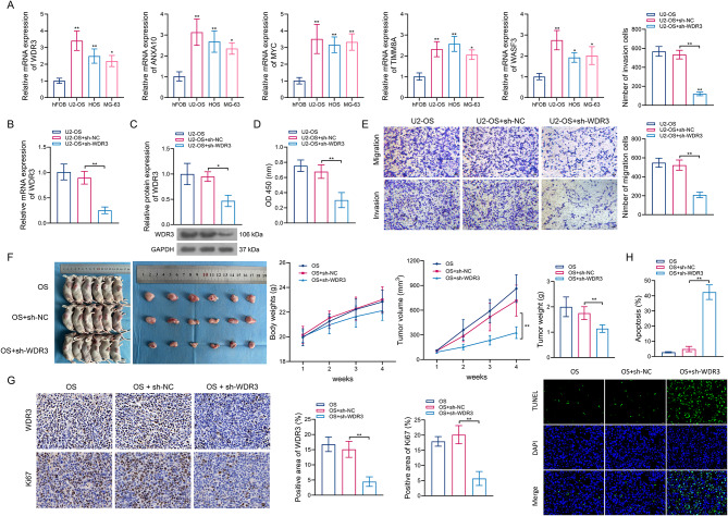
Results: Five LLPS-related biomarkers were screened by bioinformatics analyses to predict the osteosarcoma prognosis. These prognostic genes were significantly associated with the immune cell infiltration, tumor immune escape and drug sensitivity. Among them, WDR3 was a prognostic risk factor for osteosarcoma and stably bound to Nilotinib in the molecular docking model. In transfected U2-OS cells and xenograft mice, the downregulation of WDR3 significantly inhibited the malignant progression of osteosarcoma. More importantly, WDR3 could form droplets in U2-OS cells and restore the fluorescence intensity of WDR3 condensates with liquid-like behavior after photobleaching. The mutation in IDR impaired the phase separation ability of WDR3, whereas the fusion with hnRNAP1 IDR rescued the phase separation abnormality caused by WDR3 mutation. Moreover, the treatment with Nilotinib improved the progression of osteosarcoma in vivo and in vitro, while inhibiting the production of WDR3 phase-separated condensates.

Conclusion: WDR3 phase separation involves in the therapeutic mechanism of Nilotinib against osteosarcoma, and thus may serve as a potent biomarker to ameliorate adverse events following osteosarcoma treatment.

Keywords: IDR mutation; Liquid-liquid phase separation; Nilotinib; Osteosarcoma; Prognostic biomarker; WDR3.


Conflict of interest statement

Declarations. Ethics approval and consent to participate: All animals were carried out under the approval of the Experimental Animal Ethics Committee of Yangzhou University (No.202312013). Consent for publication: Not applicable. Competing interests: The authors declare no competing interests.

Figures

Fig. 1
Fig. 1
Screening of prognostic biomarkers to predict osteosarcoma survival based on a risk score-based model. (A) The volcano plot (left panel) and heatmap (right panel) showing 1497 DEGs between osteosarcoma and normal controls. (B) The Venn diagram depicts 473 intersected genes between DEGs and LLPS-related genes. (C) PCA plots before (top panel) and after (bottom panel) batch effect removal. (D) The univariate Cox regression analyses screened out 15 genes related to osteosarcoma prognosis. (E) Coefficients of the LASSO analysis (left panel) and partial likelihood deviance analysis (right panel) on 15 genes. (FI) The KM (left panel) and ROC (right panel) curves of the combined internal training set (F), combined internal validation set (G), external validation cohort 1 (H), and external validation cohort 2 (I)
Fig. 2
Fig. 2
Characteristic evaluation of prognostic biomarkers. (A) Consensus matrix heatmap defining two clusters (k = 2) and their correlation areas. (B) KM curves showing the survival difference between high- and low-risk groups. (C) Sankey diagram exhibiting the distribution of two clusters in risk groups. (DG) Differences in GSVA pathway scores (D), immune cell infiltrations (E), TIDE scores (F), and drug IC50 values (G) between high- and low-risk groups. *P < 0.05; ****P < 0.0001. (H) Correlations between prognostic biomarkers and drug IC50 values. (I) Molecular docking showing the interaction of WDR3 and Nilotinib
Fig. 3
Fig. 3
Effect of WDR3 downregulation on the progression of osteosarcoma in vitro and in vivo. (A) Expression validation of five prognostic biomarkers using qPCR. (BC) The mRNA (B) and protein (C) expression of WDR3 in U2-OS cells transfected with sh-NC or sh-WDR3. (DE) Effects of WDR3 downregulation on the cell proliferation (D), as well as cell migration and invasion (E). (F) Body weight, tumor value and tumor weight of mice injected with sh-NC- or sh-WDR3-transfected U2-OS cells. (GH) Effects of WDR3 downregulation on Ki67 expression (G) and tumor cell apoptosis (H). *P < 0.05; **P < 0.01
Fig. 4
Fig. 4
WDR3 exhibited phase-separated condensates with liquid-like behavior in OS cells. (AB) The droplet formation of WDR3 under different concentrations of NaCl (A) and WDR3-GFP recombinant protein (B). *P < 0.05; **P < 0.01. (C) FRAP of WDR3-GFP droplets. (D) The droplet formation of endogenous WDR3 in U2-OS cells. (E) Phase-separated condensates of exogenous WDR3-GFP protein in U2-OS cells. (F) FRAP of WDR3-GFP protein exogenously transfected in U2-OS cells. (G) IDR of WDR3 predicted using the IUPred2A online tool. (H) Domain structure of WDR3-WT and three WDR3 IDR mutants. (I) Effect of three WDR3 IDR mutants on the number of intracellular WDR3 droplets. **P < 0.01 versus WDR3-WT. (J) Effect of three WDR3 IDR mutants on the recovery of WDR3 condensate after photobleaching
Fig. 5
Fig. 5
Phase separation of WDR3 expedited OS metastasis in vitro. (A) Schematic illustration of WDR3 mutation and rescue of phase separation. (B) The droplet formation of WDR3-WT, WDR3-MUT, and MUT-IDR. (C) FRAP of MUT-IDR. (D) The droplet formation of WDR3 in U2-OS cells transfected with WDR3-WT, WDR3-MUT, and MUT-IDR. (E) FRAP of WDR3-MUT and MUT-IDR in U2-OS cells. (FG) The proliferation (F) and metastatic ability (G) of U2-OS cells transfected with WDR3-WT, WDR3-MUT, and MUT-IDR. *P < 0.05; **P < 0.01
Fig. 6
Fig. 6
Effect of Nilotinib treatment on WDR3 phase separation and tumor metastasis in vitro. (A) Changes of cell proliferation under the treatment of Nilotinib with different concentrations. (BC) Effects of Nilotinib treatment on WDR3 mRNA (B) and protein (C) expression. (D) The droplet formation of WDR3 in U2-OS cells treated with Nilotinib. (E) FRAP of WDR3 droplets in U2-OS cells treated with Nilotinib. (F) Effect of Nilotinib treatment on migration and invasion of U2-OS cells. *P < 0.05; **P < 0.01
Fig. 7
Fig. 7
Effects of Nilotinib treatment on WDR3 phase separation and tumor progression in vivo. (A) The schedule for animal experiments. (B) Body weight, tumor volume, and tumor weight in xenograft mice of osteosarcoma with or without Nilotinib treatment. (CD) The protein (C) and mRNA (D) expression of WDR3 in mice treated with/without Nilotinib. (E) The positive area of WDR3 in tissues of mice treated with/without Nilotinib. (F) Effects of in vivo treatment of Nilotinib on the number of WDR3 condensates. (G) The number of pulmonary metastatic nodules in each group of mice. *P < 0.05; **P < 0.01
Fig. 8
Fig. 8
Schematic illustration for the mediation of WDR3 phase separation in the treatment of Nilotinib against osteosarcoma metastasis

Similar articles

References

    1. Sun C, Li S, Ding J. Biomaterials-Boosted immunotherapy for osteosarcoma. Adv Healthc Mater. 2024;13:e2400864. - PubMed
    1. Shi Q, Xu J, Chen C, Hu X, Wang B, Zeng F, et al. Direct contact between tumor cells and platelets initiates a FAK-dependent F3/TGF-β positive feedback loop that promotes tumor progression and EMT in osteosarcoma. Cancer Lett. 2024;591:216902. - PubMed
    1. Cascini C, Ratti C, Botti L, Parma B, Cancila V, Salvaggio A, et al. Rewiring innate and adaptive immunity with TLR9 agonist to treat osteosarcoma. J Experimental Clin cancer Research: CR. 2023;42:154. - PMC - PubMed
    1. Yin C, Chokkakula S, Li J, Li W, Yang W, Chong S, et al. Unveiling research trends in the prognosis of osteosarcoma: A bibliometric analysis from 2000 to 2022. Heliyon. 2024;10:e27566. - PMC - PubMed
    1. Mohr A, Marques Da Costa ME, Fromigue O, Audinot B, Balde T, Droit R, et al. From biology to personalized medicine: recent knowledge in osteosarcoma. Eur J Med Genet. 2024;69:104941. - PubMed

MeSH terms

LinkOut - more resources