. 2025 Jul 11;23(1):784.
doi: 10.1186/s12967-025-06831-6.

PDGFR-β/Cav1-induced autophagy via mTOR/FIP200/ATG13 activation in cancer-associated fibroblasts promotes the malignant progression of breast cancer

Affiliations

PDGFR-β/Cav1-induced autophagy via mTOR/FIP200/ATG13 activation in cancer-associated fibroblasts promotes the malignant progression of breast cancer

Lifang Zhang et al. J Transl Med. .

Abstract

Background: Breast cancer incidence rates have been increasing globally. Cancer-associated fibroblasts (CAFs), key stromal components of the tumor microenvironment (TME), play crucial roles in tumor growth by dynamically interacting with cancer cells. Autophagy has been extensively studied in multiple stages of the metastatic cascade. However, the roles of two key membrane proteins, platelet-derived growth factor receptor-β (PDGFR-β) and caveolin-1 (Cav-1), in regulating autophagy in CAFs and their effects on cancer cell invasion and migration remain unclear.

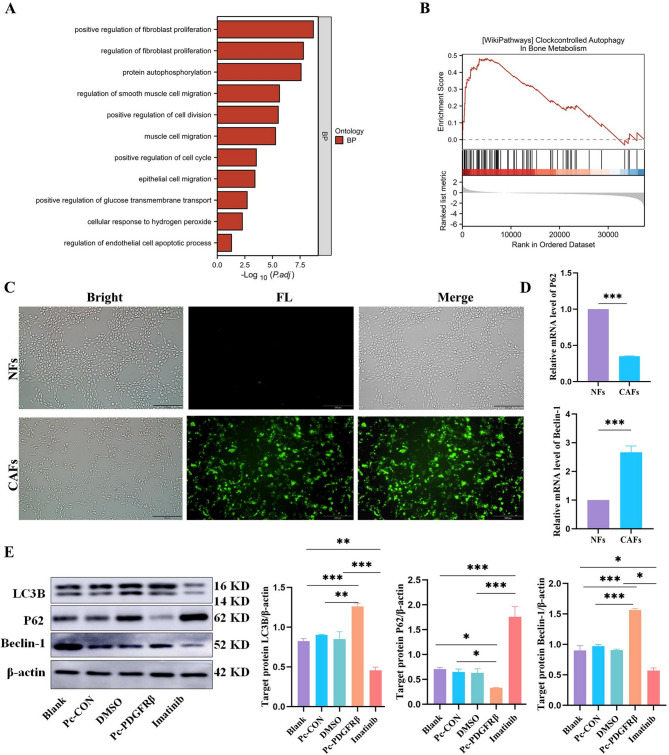
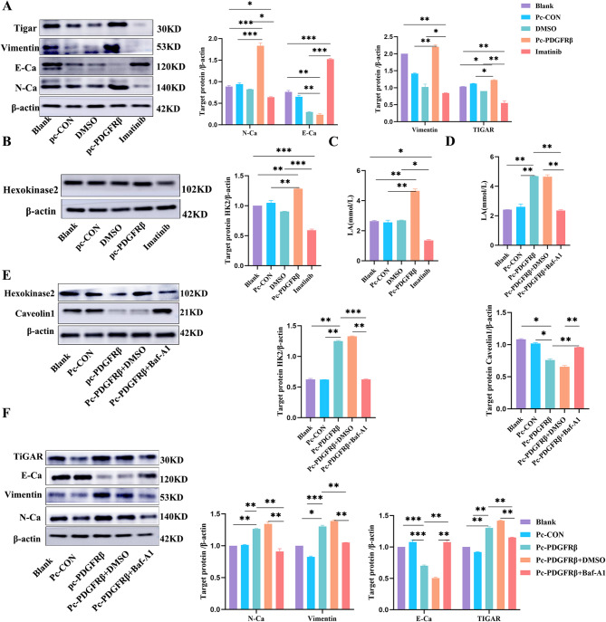
Methods: The association between PDGFR-β expression and clinical features in breast cancer patients was analyzed using TCGA databases. PDGFR-β was either overexpressed or pharmacologically inhibited in cancer cells. Autophagy-related markers and signaling proteins were analyzed by Western blot and RT-qPCR, while lactate secretion and ROS levels were quantified. Breast cancer cell migration and invasion were evaluated through wound healing and transwell assays, and PDGFR-β/Cav1 interactions were verified by immunofluorescence and co-immunoprecipitation (Co-IP). A breast cancer mouse model was employed to assess tumor progression and autophagy modulation in vivo.

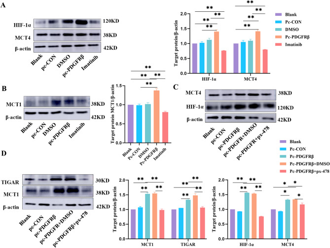
Results: The study demonstrated that PDGFR-β promotes autophagy in CAFs through the mTOR/FIP200/ATG13 signaling. PDGFR-β/Cav-1 enhanced glycolysis in CAFs via autophagy-mediated metabolic reprogramming, resulting in increased lactate export that promoted breast cancer cell growth. Furthermore, CAFs autophagy regulated breast cancer cell invasion and migration via the HIF-1α/MCT4/MCT1 signaling pathway. These findings reveal that PDGFR-β/Cav-1-mediated autophagy in CAFs enhances breast cancer cell invasion, migration, and epithelial-mesenchymal transition (EMT), collectively highlighting the crucial role of CAFs autophagy in facilitating breast cancer progression.

Conclusions: The study elucidates the mechanism by which PDGFR-β/Cav-1 promotes breast cancer progression through autophagy regulation in CAFs, These findings provide a theoretical basis for potential therapeutic method for treating breast cancer.

Keywords: Autophagy; Breast cancer; Cancer-associated fibroblasts; Caveolin-1; PDGFR-β.


Conflict of interest statement

Declarations. Ethical approval and consent to participate: The Nanchang University Animal Care and Use Ethics Committee accepted the protocols used in this work, which adhered to the regulations and guidelines of the Nanchang University Laboratory Animal Center. Consent for publication: All authors have reviewed and approved the final version of the manuscript and agreed to its publication in Journal of Translational Medicine. Competing interests: The authors report no potential conflicts of interest.

Figures

None
In breast cancer, PDGFR-β regulates Cav-1 to induce autophagy in CAFs via mTOR/FIP200/ATG13. This enhances glycolysis and lactate efflux from CAFs to the TME, facilitating breast cancer development
Fig. 1
Fig. 1
Expression of PDGFR-β in breast cancer cell lines and its effect on migration and invasion in breast cancer. (A) PDGFR-β expression in different breast cancer cell lines. (B) PDGFR-β transfection efficiency. (C) The concentration of imatinib was determined with a CCK8 assay. (D, E) Effects of the overexpression and inhibition of PDGFR-β in CAFs on the migration and invasion ability of breast cancer cells
Fig. 2
Fig. 2
PDGFR-β induces autophagy in CAFs. (A) BP analysis of PDGFR-β. (B) GSEA of PDGFR-β. (C) A ROS kit was used to measure the level of ROS in the coculture system (CAFs group) and alone (NF group). (D) RT‒qPCR was used to detect the expression of autophagy-related genes in each group. (E) Western blot analysis of the expression of LC3B, Beclin1, and P62 in CAFs after overexpression or inhibition of PDGFR-β
Fig. 3
Fig. 3
PDGFR-β/Cav1-induced autophagy in CAFs affects the invasion and migration of tumor cells. (A) Immunofluorescence was used to observe the relationship between PDGFR-β and Cav-1 expression. (B) Co-IP analysis of the interaction between PDGFR-β and Cav-1. (C) Western blot analysis of Cav-1 expression after overexpression or inhibition of PDGFR-β. (D) Western blot analysis of Cav-1 expression in CAFs after treatment with the mTOR activator MHY1485. (E) The expression of the autophagy pathway proteins mTOR, FIP200, and ATG13 in the CAFs of each group. (F) Expression levels of autophagy pathway proteins in CAFs after MHY1485 treatment. (G) The expression of autophagy-related proteins in CAFs after the addition of the mTOR activator MHY1485
Fig. 4
Fig. 4
PDGFR-β/Cav-1-mediated autophagy affects glycolysis and lactate efflux from CAFs to the TME and thus affects EMT in tumors. (A, B) The expression of EMT-related proteins, as well as TIGAR and HK2, in MCF-7 cells after overexpression and inhibition of PDGFR-β. (C, D) An LA kit was used to detect the lactate content in each group. (E, F) Effects of autophagy on glycolysis in CAFs, Cav-1, and EMT in tumor cells
Fig. 5
Fig. 5
Autophagy in CAFs promotes glycolysis and lactate efflux to the TME and EMT in tumor cells via HIF-1α/MCT4/MCT1. (A, B) The expression of HIF-1α and MCT4 in CAFs after PDGFR-β overexpression inhibition and MCT1 expression in MCF-7 cells. (C) Expression of HIF-1α and MCT4 in CAFs. (D) MCT1 expression in MCF-7 cells after px-478
Fig. 6
Fig. 6
PDGFR-β promotes breast cancer development in vivo. (A) Breast cancer models in which MDA-MB-231 cells were treated. (B) Immunohistochemistry of PDGFR-β, Caveolin1, LC3 in breast cancer. (C, D) Immunofluorescence of PDGFR-β and LC3 in breast cancer

References

    1. Siegel RL, Giaquinto AN, Jemal A. Cancer statistics, 2024. CA Cancer J Clin. 2024;74(1):12–49. - PubMed
    1. Kong J, et al. Extracellular vesicles of carcinoma-associated fibroblasts creates a pre-metastatic niche in the lung through activating fibroblasts. Mol Cancer. 2019;18(1):175. - PMC - PubMed
    1. Li Y, et al. Pre-metastatic niche: from revealing the molecular and cellular mechanisms to the clinical applications in breast cancer metastasis. Theranostics. 2023;13(7):2301–18. - PMC - PubMed
    1. Xiao Y, Yu D. Tumor microenvironment as a therapeutic target in cancer. Pharmacol Ther. 2021;221:107753. - PMC - PubMed
    1. Giraldo NA, et al. The clinical role of the TME in solid cancer. Br J Cancer. 2019;120(1):45–53. - PMC - PubMed

MeSH terms

LinkOut - more resources