

Endophthalmitis caused by Abiotrophia defectiva with initial presentation as retinal vasculitis: a case report
- PMID: 40646639
- PMCID: PMC12247376
- DOI: 10.1186/s13256-025-05358-0
Endophthalmitis caused by Abiotrophia defectiva with initial presentation as retinal vasculitis: a case report
Abstract
Background: Abiotrophia defectiva is primarily known for its association with endocarditis rather than intraocular infections. We reported a case of endophthalmitis caused by Abiotrophia defectiva, presenting as retinal vasculitis in its early stages, a phenomenon rarely documented in literature.
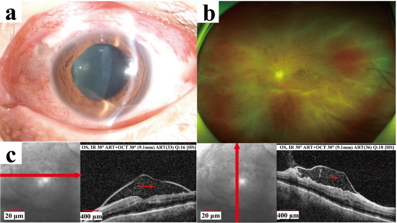
Case presentation: A 50-year-old Han Chinese man presented to the hospital with decreased vision in his left eye. Examination revealed mild vitritis, papilledema, retinal hemorrhages, and peripheral vascular sheathing in the left eye, raising suspicion of retinal vasculitis. In the following hours, his condition worsened dramatically, with the development of hypopyon and severe vitritis obscuring the visualization of the fundus, suggesting endophthalmitis. He subsequently underwent urgent anterior chamber irrigation, vitreous tap, and intravitreal injection. As the symptoms did not improve, a vitrectomy was performed. The culture results identified the presence of Abiotrophia defectiva. Following prompt and effective treatment, the patient's visual acuity showed improvement.
Conclusion: This report delineates a rare case of endophthalmitis caused by Abiotrophia defectiva initially presenting as retinal vasculitis. It emphasizes the need for prompt recognition and treatment of atypical pathogens in postoperative ocular infections to enhance visual outcomes.
Keywords: Abiotrophia defectiva; Case report; Endophthalmitis; Glaucoma; Retinal vasculitis.
© 2025. The Author(s).
Conflict of interest statement
Declarations. Ethics approval and consent to participate: Ethical approval was not applicable for this study, as it involved no novel interventions, and all treatments were standard procedures for Abiotrophia defectiva endophthalmitis. The patient provided written, informed consent for the publication of his case and any accompanying images. Consent for publication: Written informed consent was obtained from the patient for publication of this case report and any accompanying images. A copy of the written consent is available for review by the Editor-in-Chief of this journal. Competing interests: There are no competing interests in this publication.
Figures


References
-
- Kuang TM, Lin YC, Liu CJ, Hsu WM, Chou CK. Early and late endophthalmitis following trabeculectomy in a Chinese population. Eur J Ophthalmol. 2008;18(1):66–70. 10.1177/112067210801800111. - PubMed
-
- Yamamoto T, Sawada A, Mayama C, Araie M, Ohkubo S, Sugiyama K, Kuwayama Y. The 5-year incidence of bleb-related infection and its risk factors after filtering surgeries with adjunctive mitomycin C: collaborative bleb-related infection incidence and treatment study 2. Ophthalmology. 2014;121(5):1001–6. 10.1016/j.ophtha.2013.11.025. - PubMed
-
- Abry F, Sauer A, Riegel P, Saleh M, Gaucher D, Speeg-Schatz C, Bourcier T. Infectious crystalline keratopathy caused by Streptococcus Abiotrophia defectiva. Cornea. 2010;29(8):934–6. 10.1097/ICO.0b013e3181ca2e8f. - PubMed
Publication types
MeSH terms
Substances
Grants and funding
LinkOut - more resources
Full Text Sources
Research Materials
Miscellaneous
