. 2025 Sep;70(5):1704-1720.
doi: 10.1111/1556-4029.70131. Epub 2025 Jul 11.

Evaluating novel and conventional cell-separation techniques for sexual assault investigations

Affiliations

Evaluating novel and conventional cell-separation techniques for sexual assault investigations

Janine Schulte et al. J Forensic Sci. 2025 Sep.

Abstract

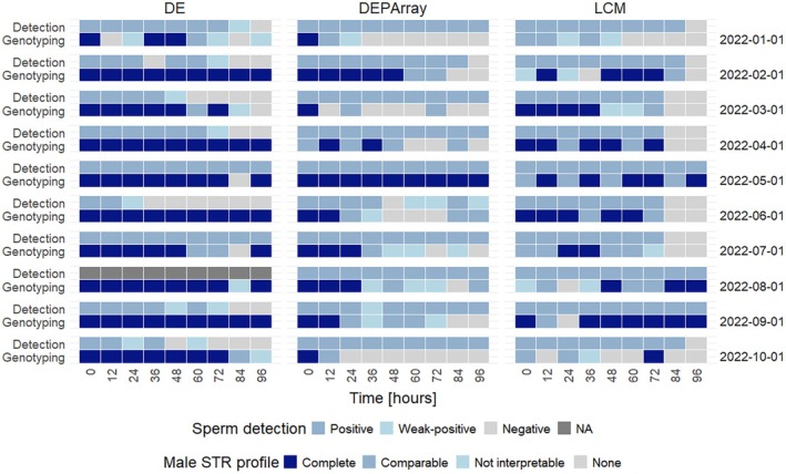
Biological evidence from sexual assaults frequently includes few male cells (i.e., spermatozoa) and numerous female cells (i.e., epithelial cells). In practice, their genetic analysis typically involves separating the victim's cells from the perpetrator's sperm using conventional differential extraction or advanced cell enrichment/capturing techniques. A descriptive study on simulated sexual assault samples was carried out by the recruitment of 10 heterosexual, monogamous couples. Post-coital swabs were collected before and after consensual sexual intercourse, with a sampling period of up to 96 h, and subjected to analysis to detect, quantify, and genotype adhering sperm by three distinct cell-separation techniques: differential extraction, laser capture microdissection, and DEPArray™. Methods differed in sperm detection and genotyping efficacy, while foreign DNA was identifiable up to 96 h. Time since intercourse and individuals were statistically significant factors (p ≤ 0.05) on male DNA yields, while hygienic behavior was not. Prior sperm enrichment was pivotal for cell capture technologies to counteract the abundance of epithelial cells, achieved by a prior mild digestion step for laser microdissection. Evaluating the advantages and disadvantages of standard and advanced methods provided a novel, comprehensive understanding of their merits, postulating that modern applications can assist conventional ones in challenging crime samples.

Keywords: DEPArray™; cell‐separation methods; differential extraction; laser capture microdissection; prostate‐specific antigen; sexual assaults.


Conflict of interest statement

The authors declare no competing financial and non‐financial interests.

Figures

FIGURE 1
FIGURE 1
DE workflow—microscopy and PSA results. Line plot illustrating sperm cell counts of nine subject couples within study cohort over a period of 96 h with respective prostate‐specific antigen (PSA) test results (red = negative, orange = weak positive, black = positive). Couple 2022‐08‐01 is not available (i.e., PSA positive for entire study duration but no microscopy).
FIGURE 2
FIGURE 2
DE workflow—DNA yield estimation by real‐time PCR. Boxplots depicting autosomal and Y chromosomal DNA yield of sperm cell and epithelial fractions at different time points for all subjects.
FIGURE 3
FIGURE 3
LCM workflowmicroscopic image gallery. HE‐stained slide containing sperm cells with (left) and without (right) prior proteinase K digestion in 2022‐06‐01 (above) and 2022‐10‐01 (below).
FIGURE 4
FIGURE 4
DEPArray™ workflowreleased cellular material from swab heads. Box and scatter plot depicting total cell counts (absolute counts/μL) counted using Countess3 (n = 88 for 10 participants (study cohort) and 9 sampling time points (TSI)). For 2022‐02‐01 T96 and 2022‐07‐01 T0, no cell count was conducted, as no large cell pellet was visible. Kruskal–Wallis test revealed no statistical significance (p > 0.05).
FIGURE 5
FIGURE 5
DEPArray™ workflowdetected events. Scatter plot illustrating detected events by CellBrowser™ software after DEPArray™ chip scan. Besides cells, events can include cell‐free nuclei or false‐positive signals. Dotted lines mark the anticipated cell load range from 3000 to the recommended maximum number of 6000 cells that should be analyzed per run. These criteria were met in 51% of the runs (n = 90 for 10 participants (study cohort) and 9 sampling time points (TSI)).
FIGURE 6
FIGURE 6
All methodssummarized sperm detection and genotyping outcome. Heat map comparing the sperm detection limits of the different methods with negative (no sperm), weak positive (1–2 sperm cells) and positive (≥3 sperm cells) as well as their respective genotyping outcomes. Solely male contribution was evaluated. Profiles were generally classified as single‐source or profiles obtained from mixed sources with male major or minor components. The latter was further categorized as “(nearly) complete,” “comparable,” and “not interpretable.” In 2022‐08‐01, microscopy could not be performed due to incorrect sample processing (NA, marked in gray).

References

    1. Inci F, Ozen MO, Saylan Y, Miansari M, Cimen D, Dhara R, et al. A novel on‐chip method for differential extraction of sperm in forensic cases. Adv Sci. 2018;5(9):1800121. 10.1002/advs.201800121 - DOI - PMC - PubMed
    1. Nakagawa T, Doi M, Nishi K, Sugahara T. Advantages of filtration method for sperm‐DNA genotyping in sexual assault cases. Leg Med. 2022;54:101988. 10.1016/j.legalmed.2021.101988 - DOI - PubMed
    1. Gill P, Jeffreys AJ, Werrett DJ. Forensic application of DNA “fingerprints”. Nature. 1985;318:577–579. 10.1038/318577a0 - DOI - PubMed
    1. van den Berge M, Sijen T. Development of a combined differential DNA/RNA co‐extraction protocol and its application in forensic casework. Forensic Sci Int Rep. 2022;5:100261. 10.1016/j.fsir.2022.100261 - DOI
    1. Clark C, Turiello R, Cotton R, Landers JP. Analytical approaches to differential extraction for sexual assault evidence. Anal Chim Acta. 2021;1141:230–245. 10.1016/j.aca.2020.07.059 - DOI - PubMed

Grants and funding