Review
. 2025 Jun 25;17(13):2137.
doi: 10.3390/cancers17132137.

Advances and Challenges in Prostate Cancer Diagnosis: A Comprehensive Review

Affiliations
Review

Advances and Challenges in Prostate Cancer Diagnosis: A Comprehensive Review

Emil Kania et al. Cancers (Basel). .

Abstract

Prostate cancer is the most commonly diagnosed malignancy in men and continues to be a leading cause of cancer-related mortality. Accurate and timely diagnosis is essential for distinguishing clinically significant tumors from indolent lesions and for informing treatment decisions. Multiparametric magnetic resonance imaging (mpMRI) has revolutionized prostate cancer detection by enabling precise lesion localization, risk stratification, and improved biopsy targeting. Fusion biopsy, which combines mpMRI findings with real-time transrectal ultrasonography (TRUS), has emerged as a highly effective method for sampling suspicious lesions. This review provides an integrated anatomical, epidemiological, technical, and clinical overview that highlights the evolving role of fusion biopsy in contemporary prostate cancer diagnostics. We also explore emerging strategies such as penumbra-targeted sampling, discuss ongoing clinical challenges, and examine the impact of biopsy underestimation and lack of standardization. Compared to conventional systematic biopsy, mpMRI-TRUS fusion biopsy improves the detection of clinically significant prostate cancer while reducing the overdiagnosis of low-risk tumors. To our knowledge, few recent reviews have comprehensively synthesized current clinical guidelines, emerging biopsy techniques, and future directions within a single narrative. mpMRI-TRUS-guided fusion biopsy represents a major advancement in the prostate cancer diagnostic pathway, promoting precision oncology by reducing overtreatment and facilitating individualized patient care. This review aims to assist clinicians in adopting biopsy innovations that enhance diagnostic accuracy and improve patient outcomes.

Keywords: PI-RADS; combined fusion biopsy; early detection; fusion biopsy; multiparametric magnetic resonance imaging (mpMRI); prostate biopsy; prostate cancer; prostate-specific antigen (PSA); targeted biopsy; transrectal ultrasonography (TRUS).


Conflict of interest statement

The authors declare no conflicts of interest.

Figures

Figure 1
Figure 1
Zonal anatomy of the prostate gland on axial multiparametric magnetic resonance imaging (mpMRI). Adapted from the authors’ own materials, based on Yacoub and Oto [7]. a: anterior; AS: anterior [fibromuscular] stroma; p: posterior; pl: posterior lateral; pm: posterior medial; PZ: peripheral zone; TZ: transition zone.
Figure 2
Figure 2
Histopathological classification of prostate cancer (PCa). Modified according to Linkon et al. [31] under Creative Commons Attribution (CC BY) 4.0 International License available at https://creativecommons.org/licenses/by/4.0/. GS: Gleason score; ISUP: International Society of Urological Pathology.
Figure 3
Figure 3
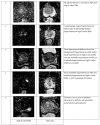
Representative images of peripheral zone lesions of the prostate gland with various Prostate Imaging—Reporting and Data System (PI-RADS) scores. Reprinted from PI-RADS v2.1 [91] under a Creative Commons Attribution-NoDerivatives (CC BY-ND) 4.0 International License available at https://creativecommons.org/licenses/by-nd/4.0/. ADC: apparent diffusion coefficient; DWI: diffusion-weighted imaging. Black arrows indicate lesions in the respective PI-RADS scores.
Figure 4
Figure 4
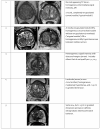
Representative images of transition zone lesions of the prostate gland with various Prostate Imaging—Reporting and Data System (PI-RADS) scores. Reprinted from PI-RADS v2.1 [91] under a Creative Commons Attribution-NoDerivatives (CC BY-ND) 4.0 International License available at https://creativecommons.org/licenses/by-nd/4.0/. TZ: transition zone.
Figure 5
Figure 5
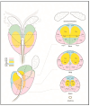
Sector map of the prostate gland illustrating a standardized localization of suspicious lesions on multiparametric magnetic resonance imaging (mpMRI). Reprinted from PI-RADS v2.1 [91] under a Creative Commons Attribution-NoDerivatives (CC BY-ND) 4.0 International available at https://creativecommons.org/licenses/by-nd/4.0/. a: anterior; AFS: anterior fibromuscular stroma; CZ: central zone; p: posterior; pl: posterior lateral; pm: posterior medial; PZ: peripheral zone; US: [external] urethral sphincter; TZ: transition zone.
Figure 6
Figure 6
Transperineal and transrectal access routes for prostate biopsy visualized on sagittal multiparametric magnetic resonance imaging (mpMRI). The image illustrates needle trajectories through the perineum (transperineal approach) and rectum (transrectal approach) directed toward the prostate gland. The transrectal route is commonly used due to its simplicity and familiarity but carries a higher risk of infection. In contrast, the transperineal approach provides improved access to anterior and apical zones of the prostate and has a lower risk of sepsis. Adapted from the authors’ own materials, based on Chang et al. [118].
Figure 7
Figure 7
Discordance in Gleason grading between prostate biopsy cores and whole-gland specimens obtained after radical prostatectomy. Own work based on Goel et al. [140]. GS: Gleason score.
Figure 8
Figure 8
(A) Combined fusion biopsy (ComBx): targeted cores obtained from mpMRI-visible lesions, along with systematic mapping biopsies; (B) Targeted biopsy with additional samples from the 10 mm penumbra zone. Own work based on Brisbane et al. [145]. In both sections of the figure, the orange area represents the lesion, while the blue dots indicate the locations where biopsy cores were taken.

References

    1. Wang L., Lu B., He M., Wang Y., Wang Z., Du L. Prostate Cancer Incidence and Mortality: Global Status and Temporal Trends in 89 Countries From 2000 to 2019. Front. Public Health. 2022;10:811044. doi: 10.3389/fpubh.2022.811044. - DOI - PMC - PubMed
    1. Raporty | Krajowy Rejestr Nowotworów. [(accessed on 19 April 2025)]. Available online: https://onkologia.org.pl/pl/raporty.
    1. Merriel S.W.D., Funston G., Hamilton W. Prostate Cancer in Primary Care. Adv. Ther. 2018;35:1285–1294. doi: 10.1007/s12325-018-0766-1. - DOI - PMC - PubMed
    1. Gnanapragasam V.J., Greenberg D., Burnet N. Urinary symptoms and prostate cancer—The misconception that may be preventing earlier presentation and better survival outcomes. BMC Med. 2022;20:264. doi: 10.1186/s12916-022-02453-7. - DOI - PMC - PubMed
    1. Aaron L., Franco O.E., Hayward S.W. Review of Prostate Anatomy and Embryology and the Etiology of Benign Prostatic Hyperplasia. Urol. Clin. N. Am. 2016;43:279–288. doi: 10.1016/j.ucl.2016.04.012. - DOI - PMC - PubMed

LinkOut - more resources