. 2025 Jul 2;17(13):2219.
doi: 10.3390/cancers17132219.

A Histopathological and Surgical Analysis of Gastric Cancer: A Two-Year Experience in a Single Center

Affiliations

A Histopathological and Surgical Analysis of Gastric Cancer: A Two-Year Experience in a Single Center

Cătălin Prodan-Bărbulescu et al. Cancers (Basel). .

Abstract

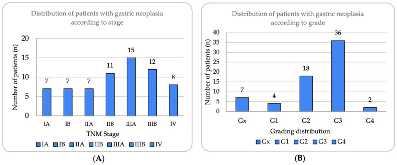
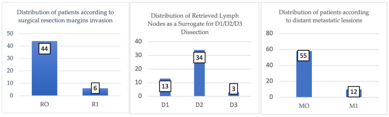
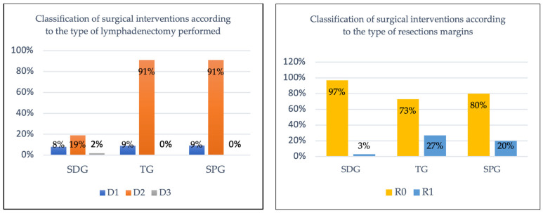
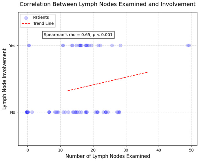
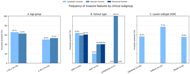
Background: Gastric neoplasms remain pathologies of the malignant spectrum with high incidence and prevalence, with their management requiring a precise histopathological characterization for optimal treatment planning. Methods: The present study is a retrospective analysis that included 67 histopathologically confirmed gastric neoplasia subjects and was performed at a single surgical center from January 2020 to December 2021. Demographics, tumor characteristics, surgical procedures, and oncologic outcomes were included, filtered, and subsequently analyzed using SPSS Statistics 29.0. Results: This study involved 67 patients (mean age 65.7 years, 56.7% men), with adenocarcinoma being the most common histologic type (91.0%) and most tumors being diagnosed directly as Stage III (40.3%). Lauren classification revealed the intestinal type as the most common (49.2%), followed by diffuse (36.1%) and mixed (14.8%). Poorly differentiated tumors (G3) accounted for 53.7% of cases. The surgical team performed curative resection in 75% (n = 50) of patients, achieving R0 margins in 88% of these cases. Subtotal gastrectomy with D2 lymphadenectomy yielded the highest curative success rate with 96.6% R0 resection. Statistically, we identified two significant correlations between age and tumor grade (rho = 0.28; p = 0.021) and between the number of lymph nodes examined and the number of lymph nodes invaded (rho = 0.65, p < 0.001). This study again revealed that adenocarcinomas showed higher rates of lymph node invasion than other tumor types (p = 0.017). Conclusions: The analysis of patients with gastric neoplasms is vital for appropriate therapeutic management. Even though the study period included a pandemic, the analysis remained a complex one with high-quality surgical outcomes, confirming the importance of maintaining oncologic standards during medical crises.

Keywords: COVID-19; Lauren classification; WHO classification; curability parameters; gastric neoplasia; histopathological characteristics; morphopathological analysis; pandemic.


Conflict of interest statement

The authors declare no conflicts of interest.

Figures

Figure 1
Figure 1
Distribution of patients with gastric neoplasia according to stage and histological grade. (A) Tumor stage distribution according to TNM classification (8th edition), showing Stage III as most prevalent (n = 27, 40.3%), followed by Stage II (n = 18, 26.87%), Stage I (n = 14, 20.90%), and Stage IV (n = 8, 11.94%). Substages IIIA and IIIB represented 22.39% and 17.91%, respectively. (B) Histological grade distribution demonstrating predominance of poorly differentiated tumors (G3, n = 36, 53.7%), followed by moderately differentiated (G2, n = 18, 26.87%), well-differentiated (G1, n = 4, 5.97%), undifferentiated (G4, n = 2, 2.99%), and undetermined grade (Gx, n = 7, 10.45%). High prevalence of G3 tumors correlates with advanced stage presentation.
Figure 2
Figure 2
Tubular adenocarcinoma showing well-formed glandular structures with moderate nuclear atypia (H&E, ×200, scale bar = 100 μm).
Figure 3
Figure 3
Poorly cohesive adenocarcinoma with signet ring cell morphology infiltrating the stroma (H&E, ×400, scale bar = 50 μm).
Figure 4
Figure 4
Mucinous adenocarcinoma with abundant extracellular mucin pools surrounding malignant glands (H&E, ×100, scale bar = 200 μm).
Figure 5
Figure 5
Moderately differentiated (G2) adenocarcinoma with irregular glandular architecture and moderate nuclear pleomorphism (H&E, ×200, scale bar = 100 μm).
Figure 6
Figure 6
Poorly differentiated (G3) adenocarcinoma showing solid growth pattern with high nuclear pleomorphism and increased mitotic activity (H&E, ×200, scale bar = 100 μm).
Figure 7
Figure 7
Lymphatic invasion demonstrating tumor cells within lymphatic vessels, supporting our finding of 58.2% lymphatic invasion rate (H&E, ×400, scale bar = 50 μm).
Figure 8
Figure 8
Surgical resection margin status in study cohort (n = 50 curative resections). R0 (negative margins) achieved in 44 cases (88%), indicating complete tumor excision without residual microscopic disease at specimen margins. R1 (positive microscopic margins) found in 6 cases (12%), suggesting remaining tumor cells with implications for adjuvant therapy and prognosis. Chart shows absolute numbers and percentages for each category.
Figure 9
Figure 9
Classification of surgical interventions according to type of lymphadenectomy and status of resectional margins.
Figure 10
Figure 10
Scatterplot correlation between number of lymph nodes examined and number of lymph nodes invaded (Spearman’s rho = 0.65, p < 0.001). Each point represents one patient. Strong positive correlation demonstrates that more extensive lymph node sampling increases detection of nodal metastases. Patients with >20 nodes examined showed 76.0% lymph node involvement versus 48.8% in those with <20 nodes examined. Line represents best-fit trend.
Figure 11
Figure 11
Distribution of invasive features across clinical subgroups. Bar chart comparing lymphatic invasion, vascular invasion, and perineural invasion rates between (A) age groups (<65 vs. ≥65 years), (B) tumor types (adenocarcinoma vs. GIST vs. lymphoma), and (C) Lauren classification subtypes (intestinal vs. diffuse vs. mixed). Error bars represent 95% confidence intervals. Adenocarcinomas showed significantly higher lymphatic invasion rates (63.9%) compared to GIST (20.0%, p = 0.017).
Figure 12
Figure 12
Establishing a set of curability indicators for gastric cancer surgery.

Similar articles

References

    1. Norton E.J., Bateman A.C. Intestinal Type Adenocarcinoma. PathologyOutlines.com Website. [(accessed on 25 April 2025)]. Available online: https://www.pathologyoutlines.com/topic/stomachintestinal.html.
    1. Smyth E.C., Nilsson M., Grabsch H.I., Van Grieken N.C., Lordick F. Gastric Cancer. Lancet. 2020;396:635–648. doi: 10.1016/S0140-6736(20)31288-5. - DOI - PubMed
    1. Sharma R. Burden of Stomach Cancer Incidence, Mortality, Disability-Adjusted Life Years, and Risk Factors in 204 Countries, 1990–2019: An Examination of Global Burden of Disease 2019. J. Gastrointest. Cancer. 2024;55:787–799. doi: 10.1007/s12029-023-01005-3. - DOI - PubMed
    1. Lu B., Lin L., Su X. Global Burden of Depression or Depressive Symptoms in Children and Adolescents: A Systematic Review and Meta-Analysis. J. Affect. Disord. 2024;354:553–562. doi: 10.1016/j.jad.2024.03.074. - DOI - PubMed
    1. Lin J.-L., Lin J.-X., Lin G.-T., Huang C.-M., Zheng C.-H., Xie J.-W., Wang J., Lu J., Chen Q.-Y., Li P. Global Incidence and Mortality Trends of Gastric Cancer and Predicted Mortality of Gastric Cancer by 2035. BMC Public Health. 2024;24:1763. doi: 10.1186/s12889-024-19104-6. - DOI - PMC - PubMed

LinkOut - more resources